

Paper Menu >>
Journal Menu >>
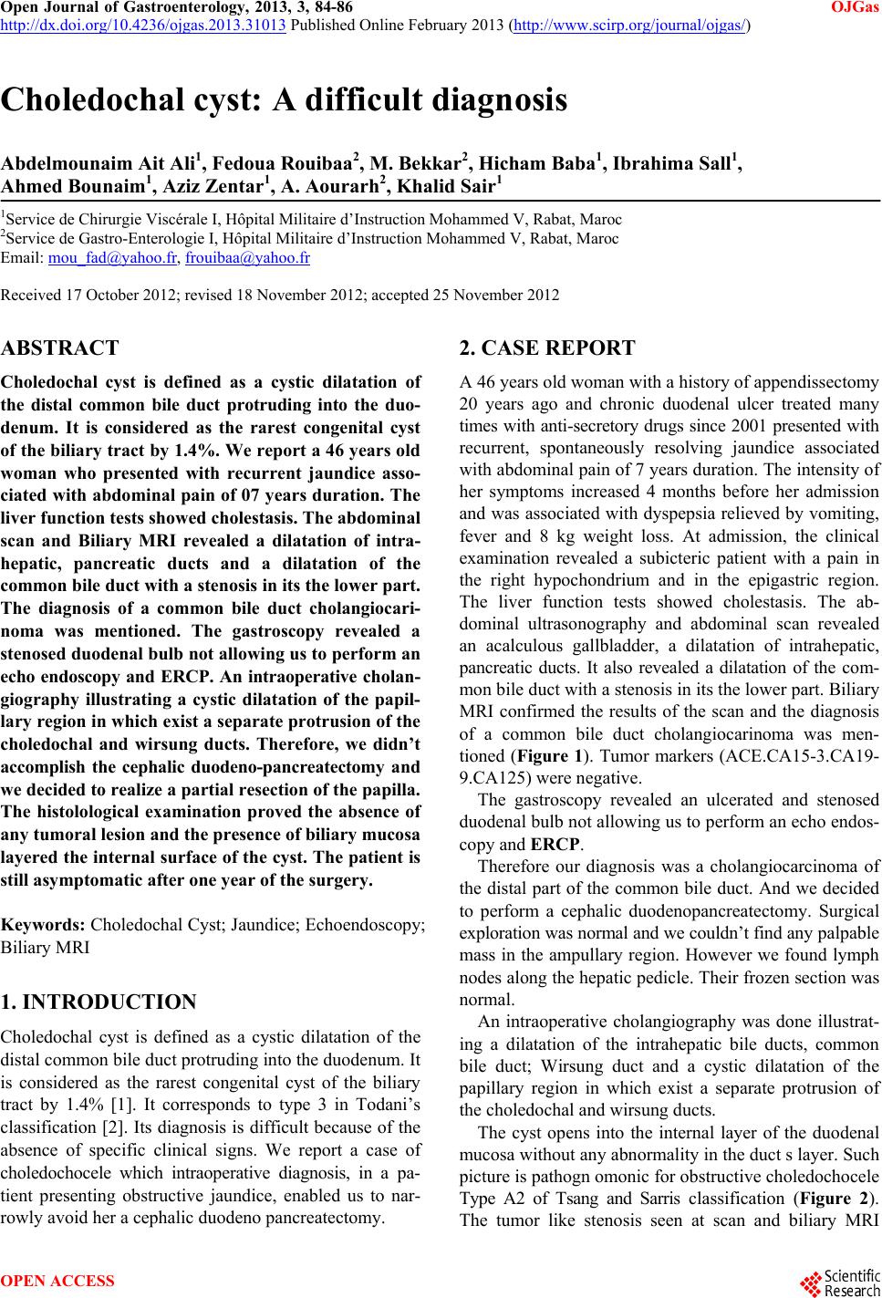 Open Journal of Gastroenterology, 2013, 3, 84-86 OJGas http://dx.doi.org/10.4236/ojgas.2013.31013 Published Online February 2013 (http://www.scirp.org/journal/ojgas/) Choledochal cyst: A difficult diagnosis Abdelmounaim Ait Ali1, Fedoua Rouibaa2, M. Bekkar2, Hicham Baba1, Ibrahima Sall1, Ahmed Bounaim1, Aziz Zentar1, A. Aourarh2, Khalid Sair1 1Service de Chirurgie Viscérale I, Hôpital Militaire d’Instruction Mohammed V, Rabat, Maroc 2Service de Gastro-Enterologie I, Hôpital Militaire d’Instruction Mohammed V, Rabat, Maroc Email: [email protected], [email protected] Received 17 October 2012; revised 18 November 2012; accepted 25 November 2012 ABSTRACT Choledochal cyst is defined as a cystic dilatation of the distal common bile duct protruding into the duo- denum. It is considered as the rarest congenital cyst of the biliary tract by 1.4%. We report a 46 y ears old woman who presented with recurrent jaundice asso- ciated with abdominal pain of 07 years duration. The liver function tests showed cholestasis. The abdominal scan and Biliary MRI revealed a dilatation of intra- hepatic, pancreatic ducts and a dilatation of the common bile duct with a stenosis in its the lower part. The diagnosis of a common bile duct cholangiocari- noma was mentioned. The gastroscopy revealed a stenosed duodenal bulb not allowing us to perform an echo endoscopy and ERCP. An intraoperative cholan- giography illustrating a cystic dilatation of the papil- lary region in which exist a sepa rate protrusion o f the choledochal and wirsung ducts. Therefore, we didn’t accomplish the cephalic duodeno-pancreatectomy and we decided to realize a partial resection of the papilla . The histolological examination proved the absence of any tumoral lesion and the presence of biliary mucosa layered the internal surface of the cyst. The patient is still asymptomatic after one year of the surgery. Keywords: Choledochal Cyst; Jaundice; Echoendoscopy; Biliary MRI 1. INTRODUCTION Choledochal cyst is defined as a cystic dilatation of the distal common bile duct protruding into the duodenum. It is considered as the rarest congenital cyst of the biliary tract by 1.4% [1]. It corresponds to type 3 in Todani’s classification [2]. Its diagnosis is difficult because of the absence of specific clinical signs. We report a case of choledochocele which intraoperative diagnosis, in a pa- tient presenting obstructive jaundice, enabled us to nar- rowly avoid her a cephalic duodeno pancreatectomy. 2. CASE REPORT A 46 years old woman with a history of appendissectomy 20 years ago and chronic duodenal ulcer treated many times with anti-secretory drugs since 2001 presented with recurrent, spontaneously resolving jaundice associated with abdominal pain of 7 years duration. The intensity of her symptoms increased 4 months before her admission and was associated with dyspepsia relieved by vomiting, fever and 8 kg weight loss. At admission, the clinical examination revealed a subicteric patient with a pain in the right hypochondrium and in the epigastric region. The liver function tests showed cholestasis. The ab- dominal ultrasonography and abdominal scan revealed an acalculous gallbladder, a dilatation of intrahepatic, pancreatic ducts. It also revealed a dilatation of the com- mon bile duct with a stenosis in its the lo wer part. Biliary MRI confirmed the results of the scan and the diagnosis of a common bile duct cholangiocarinoma was men- tioned (Figure 1). Tumor markers (ACE.CA15-3.CA19- 9.CA125) we r e ne gat ive. The gastroscopy revealed an ulcerated and stenosed duodenal bulb not allowing us to perform an echo endos- copy and ERCP. Therefore our diagnosis was a cholangiocarcinoma of the distal part of the common bile duct. And we decided to perform a cephalic duodenopancreatectomy. Surgical exploration was normal and we couldn’t find any p a lp ab l e mass in the ampullary region. However we found lymph nodes along the hepatic pedicle. Their frozen section was normal. An intraoperative cholangiography was done illustrat- ing a dilatation of the intrahepatic bile ducts, common bile duct; Wirsung duct and a cystic dilatation of the papillary region in which exist a separate protrusion of the choledochal and wirsung ducts. The cyst opens into the internal layer of the duodenal mucosa without any abnormality in th e duct s layer. Su ch picture is pathogn omonic for obstructive choledochocele Type A2 of Tsang and Sarris classification (Figure 2). The tumor like stenosis seen at scan and biliary MRI OPEN ACCESS 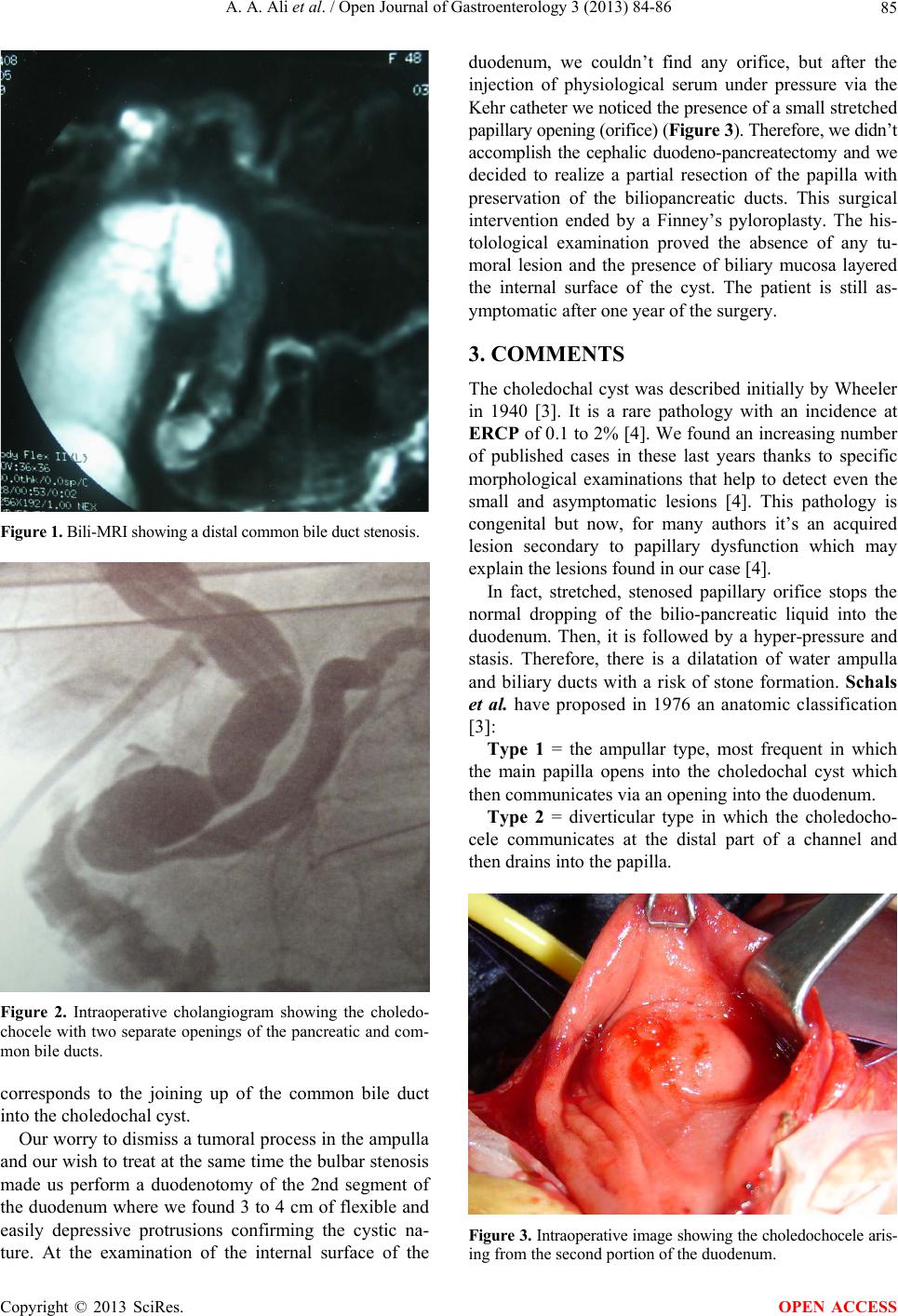 A. A. Ali et al. / Open Journal of Gastroenterology 3 (2013) 84-86 85 Figure 1. Bili-MRI showing a distal common bile duct stenosis. Figure 2. Intraoperative cholangiogram showing the choledo- chocele with two separate openings of the pancreatic and com- mon bile ducts. corresponds to the joining up of the common bile duct into the choledo c hal cyst. Our worry to dismiss a tumoral process in the ampulla and our wish to treat at the sa me time the bulbar stenosis made us perform a duodenotomy of the 2nd segment of the duodenum where we found 3 to 4 cm of flexible and easily depressive protrusions confirming the cystic na- ture. At the examination of the internal surface of the duodenum, we couldn’t find any orifice, but after the injection of physiological serum under pressure via the Kehr catheter we noticed the presence of a smal l s t r e t ch e d papillary opening (orifice) (Figure 3). Therefore, we didn’t accomplish the cephalic duodeno-pancreatectomy and we decided to realize a partial resection of the papilla with preservation of the biliopancreatic ducts. This surgical intervention ended by a Finney’s pyloroplasty. The his- tolological examination proved the absence of any tu- moral lesion and the presence of biliary mucosa layered the internal surface of the cyst. The patient is still as- ymptomatic after one year of the surgery. 3. COMMENTS The choledochal cyst was described initially by Wheeler in 1940 [3]. It is a rare pathology with an incidence at ERCP of 0.1 to 2% [4]. We found an increasing number of published cases in these last years thanks to specific morphological examinations that help to detect even the small and asymptomatic lesions [4]. This pathology is congenital but now, for many authors it’s an acquired lesion secondary to papillary dysfunction which may explain the lesions found in our case [4]. In fact, stretched, stenosed papillary orifice stops the normal dropping of the bilio-pancreatic liquid into the duodenum. Then, it is followed by a hyper-pressure and stasis. Therefore, there is a dilatation of water ampulla and biliary ducts with a risk of stone formation. Schals et al. have proposed in 1976 an anatomic classification [3]: Type 1 = the ampullar type, most frequent in which the main papilla opens into the choledochal cyst which then communicates via an opening into the duodenum. Type 2 = diverticular type in which the choledocho- cele communicates at the distal part of a channel and then drains into the papilla. Figure 3. Intraoperative image showing the chole dochocele aris- ing from the second portion of the duodenum. Copyright © 2013 SciRes. OPEN ACCESS 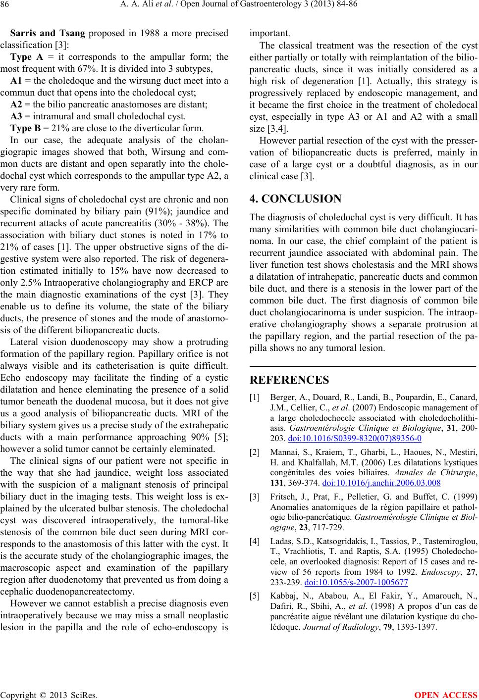 A. A. Ali et al. / Open Journal of Gastroenterology 3 (2013) 84-86 Copyright © 2013 SciRes. 86 Sarris and Tsang proposed in 1988 a more precised classification [3]: OPEN ACCESS Type A = it corresponds to the ampullar form; the most frequent wit h 67 %. It is divi ded into 3 subty pes, A1 = the choled oque and the wirsung duct meet into a commun duct that opens into the choledocal cyst; A2 = the bilio pancreatic anastomoses are distant; A3 = intramural and small choledochal cyst. Type B = 21% are close to the diverticular form. In our case, the adequate analysis of the cholan- giograpic images showed that both, Wirsung and com- mon ducts are distant and open separatly into the chole- dochal cyst which corr esp onds to the ampu llar typ e A2 , a very rare f orm. Clinical signs of choledochal cyst are chronic and non specific dominated by biliary pain (91%); jaundice and recurrent attacks of acute pancreatitis (30% - 38%). The association with biliary duct stones is noted in 17% to 21% of cases [1]. The upper obstructive signs of the di- gestive system were also reported. The risk of degenera- tion estimated initially to 15% have now decreased to only 2.5% Intraoperative cholangiography and ERCP are the main diagnostic examinations of the cyst [3]. They enable us to define its volume, the state of the biliary ducts, the presence of stones and the mode of anastomo- sis of the different biliopancreatic d ucts. Lateral vision duodenoscopy may show a protruding formation of the papillary region. Papillary orifice is not always visible and its catheterisation is quite difficult. Echo endoscopy may facilitate the finding of a cystic dilatation and hence eleminating the presence of a solid tumor beneath the duodenal mucosa, but it does not give us a good analysis of biliopancreatic ducts. MRI of the biliary system gives us a precise study of the extrahepatic ducts with a main performance approaching 90% [5]; however a solid tumor cannot be certainly eleminated. The clinical signs of our patient were not specific in the way that she had jaundice, weight loss associated with the suspicion of a malignant stenosis of principal biliary duct in the imaging tests. This weight loss is ex- plained by the ulcerated bulbar stenosis. The choledochal cyst was discovered intraoperatively, the tumoral-like stenosis of the common bile duct seen during MRI cor- responds to the anastomosis of this latter with the cyst. It is the accurate study of the cholangiographic images, the macroscopic aspect and examination of the papillary region after duodenotomy that prevented us from doing a cephalic duodenopancreatectomy. However we cannot establish a precise diagnosis even intraoperatively because we may miss a small neoplastic lesion in the papilla and the role of echo-endoscopy is important. The classical treatment was the resection of the cyst either partially or totally with reimplantatio n of the bilio- pancreatic ducts, since it was initially considered as a high risk of degeneration [1]. Actually, this strategy is progressively replaced by endoscopic management, and it became the first choice in the treatment of choledocal cyst, especially in type A3 or A1 and A2 with a small size [3,4]. However partial resection of the cyst with the presser- vation of biliopancreatic ducts is preferred, mainly in case of a large cyst or a doubtful diagnosis, as in our clinical case [3]. 4. CONCLUSION The diagnosis of choledochal cyst is very difficult. It has many similarities with common bile duct cholangiocari- noma. In our case, the chief complaint of the patient is recurrent jaundice associated with abdominal pain. The liver function test shows cholestasis and the MRI shows a dilatation of intrah epatic, pancreatic ducts and common bile duct, and there is a stenosis in the lower part of the common bile duct. The first diagnosis of common bile duct cholangiocarinoma is under suspicion. The intraop- erative cholangiography shows a separate protrusion at the papillary region, and the partial resection of the pa- pilla shows no any tumoral lesion. REFERENCES [1] Berger, A., Douard, R., Landi, B., Poupardin, E., Canard, J.M., Cellier, C., et al. (2007) Endoscopic management of a large choledochocele associated with choledocholithi- asis. Gastroentérologie Clinique et Biologique, 31, 200- 203. doi:10.1016/S0399-8320(07)89356-0 [2] Mannai, S., Kraiem, T., Gharbi, L., Haoues, N., Mestiri, H. and Khalfallah, M.T. (2006) Les dilatations kystiques congénitales des voies biliaires. Annales de Chirurgie, 131, 369-374. doi:10.1016/j.anchir.2006.03.008 [3] Fritsch, J., Prat, F., Pelletier, G. and Buffet, C. (1999) Anomalies anatomiques de la région papillaire et pathol- ogie bilio-pancré atique. Gastroentéro logie Clinique et Biol- ogique, 23, 717-729. [4] Ladas, S.D., Katsogridakis, I., Tassios, P., Tastemiroglou, T., Vrachliotis, T. and Raptis, S.A. (1995) Choledocho- cele, an overlooked diagnosis: Report of 15 cases and re- view of 56 reports from 1984 to 1992. Endoscopy, 27, 233-239. doi:10.1055/s-2007-1005677 [5] Kabbaj, N., Ababou, A., El Fakir, Y., Amarouch, N., Dafiri, R., Sbihi, A., et al. (1998) A propos d’un cas de pancréatite aigue révélant une dilatation kystique du cho- lédoque. Journal of Radiology, 79, 1393-1397. |