

Paper Menu >>
Journal Menu >>
 Advances in Anthropology 2013. Vol.3, No.1, 10-15 Published Online February 2013 in SciRes (http://www.scirp.org/journal/aa) http://dx.doi.org/10.4236/aa.2013.31002 Copyright © 2013 SciRes. 10 Sexual Dimorphism in Prehispanic Populations of the Cochabamba Valleys, Bolivia* José A. Cocilovo1#, María L. Fuchs1, Tyler G. O’Brien2, Héctor H. Varela1 1Departamento de Ciencias Naturales, Facultad de Ciencias Exactas, Físico-Química y Naturales, Universidad Nacional de Río Cuarto, Río Cuarto, Argentina 2Department of Sociology, Anthropology and Criminology, University of Northern Iowa, Cedar Falls, USA Email: #[email protected] Received November 17th, 2012; revised December 21st, 2012; accepted January 1st, 2013 The expression of sexual dimorphism may vary across time and space, as well as within and between populations depending on genetic and environmental factors that influence growth and development. The objective of the present work is to contribute to the knowledge of factors that determine the physical cha- racteristics of ancient human groups in the eastern valleys of Cochabamba—a key region for cultural development, inter-regional interaction with northern Chile and northwest Argentina, and their noticeable role in the settlement of the south central Andean region. This paper analyzes the differences between males and females crania representing ancient human groups that inhabited the eastern valleys of Co- chabamba, Bolivia. Thirty-one craniometric variables are analyzed from a sample of 234 individuals. Differences are evaluated using univariate analysis by ANOVA and multivariate discriminant analysis. The results indicate the existence of higher mean values in males compared to females in most cranial measurements (mean difference 5.3%). The discriminant analysis also reveals a significant morphological difference exists between sexes. Furthermore, by means of the discriminant function, the reclassification of correct sex was 99% of cases. This information indicates that members of this population could live in optimal conditions, with adequate resources to ensure growth and development and normal expression of the phenotype of each sex. Keywords: Sexual Dimorphism; Growth and Development; Population Biology; Craniometrics Introduction Sexual dimorphism is the result of the complex action of a series of genetic and environmental factors linked to the growth and development process of individuals. Although this charac- teristic is already seen in the fetal development, this is much more evident in the puberal stage of the vital cycle, affecting the anatomy, physiology and behavior. In current populations, normal growth tends to conclude with the development of the secondary sexual characteristics around 16 years old in the female and about 18 years old in the males, with the most con- spicuous change being the overall difference in size. This diffe- rence is produced by differential growth in regards to duration, intensity and velocity. A complete synthesis of this theme, from an evolutionary perspective, can be found in work by Frayer and Wolpoff (1985). Additional work by Guntupalli and Baten (2009) evaluates stature differences between both sexes from historic and socioeconomic points of view. Sexual differences manifest themselves at various levels in respect to size and form of the tissue and organ. In particular, the acquisition in the male of greater muscular mass also brings about a greater development of the skeletal system. The bone structure reflects these differences in conjunction with anatomic traits. Such characteristics are currently employed in the dis- crimination of sex in skeletal remains (Saunders, 1992). The magnitude of sexual dimorphism constitutes a topic of great interest because of its association with growth problems produced by disease and malnutrition. In fact, in situations of chronic stress a reduction of sexual dimorphism is expected but at the expense of normal masculine growth. Males are more ecosensitive and experience a much more marked response to prolonged nutritional shortages (Frayer & Wolpoff, 1985; Stinson, 1985). Three dramatic examples of the effects on sexual dimorphism by way of nutritional shortages and ineffi- cient sanitary conditions are evident in the cases of Heliconia, Colombia (Stini, 1969), of the Villa IAPI, a poor village outside of Buenos Aires, Argentina (Pucciarelli et al., 1993) and an- other case in the Republic of Mali, Africa (Dettwyler, 1992). However, in contrast, the Tehuelches and Mapuches, two in- digenous groups of Chubut, Argentina, express normal rates of sexual dimorphism in supposedly unfavorable economic and environmental conditions because of the governmental assis- tance they receive and the social cooperation that permits them to reach an appropriate nutritional state (Pinotti et al., 2005). This feature was evaluated in current native groups from la Puna in Jujuy in a sample of 231 individuals, by means of 21 somatometric measurements. All the variables showed signifi- cant differences between both sexes, and the male mean values were higher than those of the females. This result was very precise as the measurements were performed independently of *The research for this work was supported by grants from Consejo Nacional de Investigaciones Científicas y Técnicas (PIP 2405/08), Universidad Na- cional de Río Cuarto (C18/328), Agencia Nacional de Promoción Científica y Técnica (PICT 02210/07) of Argentina; Faculty Fellowship, University o f N orthern Iowa; and Nacional Science Foundation’s doctoral dissertation improvement grant (SBR 9903631). #Corresponding author. 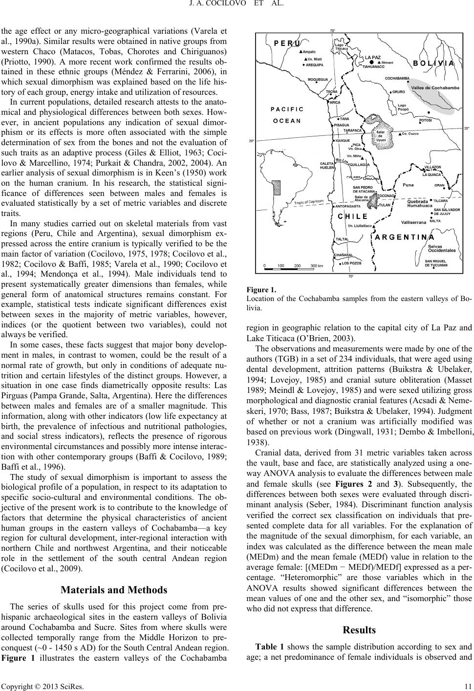 J. A. COCILOVO ET AL. the age effect or any micro-geographical variations (Varela et al., 1990a). Similar results were obtained in native groups from western Chaco (Matacos, Tobas, Chorotes and Chiriguanos) (Priotto, 1990). A more recent work confirmed the results ob- tained in these ethnic groups (Méndez & Ferrarini, 2006), in which sexual dimorphism was explained based on the life his- tory of each group, energy intake and utilization of resources. In current populations, detailed research attests to the anato- mical and physiological differences between both sexes. How- ever, in ancient populations any indication of sexual dimor- phism or its effects is more often associated with the simple determination of sex from the bones and not the evaluation of such traits as an adaptive process (Giles & Elliot, 1963; Coci- lovo & Marcellino, 1974; Purkait & Chandra, 2002, 2004). An earlier analysis of sexual dimorphism is in Keen’s (1950) work on the human cranium. In his research, the statistical signi- ficance of differences seen between males and females is evaluated statistically by a set of metric variables and discrete traits. In many studies carried out on skeletal materials from vast regions (Peru, Chile and Argentina), sexual dimorphism ex- pressed across the entire cranium is typically verified to be the main factor of variation (Cocilovo, 1975, 1978; Cocilovo et al., 1982; Cocilovo & Baffi, 1985; Varela et al., 1990; Cocilovo et al., 1994; Mendonça et al., 1994). Male individuals tend to present systematically greater dimensions than females, while general form of anatomical structures remains constant. For example, statistical tests indicate significant differences exist between sexes in the majority of metric variables, however, indices (or the quotient between two variables), could not always be verified. In some cases, these facts suggest that major bony develop- ment in males, in contrast to women, could be the result of a normal rate of growth, but only in conditions of adequate nu- trition and certain lifestyles of the distinct groups. However, a situation in one case finds diametrically opposite results: Las Pirguas (Pampa Grande, Salta, Argentina). Here the differences between males and females are of a smaller magnitude. This information, along with other indicators (low life expectancy at birth, the prevalence of infectious and nutritional pathologies, and social stress indicators), reflects the presence of rigorous environmental circumstances and possibly more intense interac- tion with other contemporary groups (Baffi & Cocilovo, 1989; Baffi et al., 1996). The study of sexual dimorphism is important to assess the biological profile of a population, in respect to its adaptation to specific socio-cultural and environmental conditions. The ob- jective of the present work is to contribute to the knowledge of factors that determine the physical characteristics of ancient human groups in the eastern valleys of Cochabamba—a key region for cultural development, inter-regional interaction with northern Chile and northwest Argentina, and their noticeable role in the settlement of the south central Andean region (Cocilovo et al., 2009). Materials and Methods The series of skulls used for this project come from pre- hispanic archaeological sites in the eastern valleys of Bolivia around Cochabamba and Sucre. Sites from where skulls were collected temporally range from the Middle Horizon to pre- conquest (~0 - 1450 s AD) for the South Central Andean region. Figure 1 illustrates the eastern valleys of the Cochabamba Figure 1. Location of the Cochabamba samples from the eastern valleys of Bo- livia. region in geographic relation to the capital city of La Paz and Lake Titicaca (O’Brien, 2003). The observations and measurements were made by one of the authors (TGB) in a set of 234 individuals, that were aged using dental development, attrition patterns (Buikstra & Ubelaker, 1994; Lovejoy, 1985) and cranial suture obliteration (Masset 1989; Meindl & Lovejoy, 1985) and were sexed utilizing gross morphological and diagnostic cranial features (Acsadi & Neme- skeri, 1970; Bass, 1987; Buikstra & Ubelaker, 1994). Judgment of whether or not a cranium was artificially modified was based on previous work (Dingwall, 1931; Dembo & Imbelloni, 1938). Cranial data, derived from 31 metric variables taken across the vault, base and face, are statistically analyzed using a one- way ANOVA analysis to evaluate the differences between male and female skulls (see Figures 2 and 3). Subsequently, the differences between both sexes were evaluated through discri- minant analysis (Seber, 1984). Discriminant function analysis verified the correct sex classification on individuals that pre- sented complete data for all variables. For the explanation of the magnitude of the sexual dimorphism, for each variable, an index was calculated as the difference between the mean male (MEDm) and the mean female (MEDf) value in relation to the average female: [(MEDm − MEDf)/MEDf] expressed as a per- centage. “Heteromorphic” are those variables which in the ANOVA results showed significant differences between the mean values of one and the other sex, and “isomorphic” those who did not express that difference. Results Table 1 shows the sample distribution according to sex and age; a net predominance of female individuals is observed and Copyright © 2013 SciRes. 11  J. A. COCILOVO ET AL. Figure 2. Example of adult male from the Mojocoya site, Cochabamba. Figure 3. Example of adult female from the Mojocoya site, Cochabamba. Table 1. Distribution of the Cochabamba sample by age and sex. Sex Age Male Female Total Adult 43 (39%) 67 (61%) 110 Mature 50 (52%) 46 (48%) 96 Senile 7 (25%) 21 (75%) 28 Total 100 134 234 few senile individuals. Table 2 shows the distribution of both sexes according to artificial cranial deformation. The majority consists of non-deformed individuals (76%). The other types: tabular erect (TE), tabular oblique (TO) and circular oblique (CO) represent 24% of all cases. Table 3 displays each variable separated by sex, number of cases (N), mean ( x ), standard error (s.e.), and the test for sex differences between means (F). All but three of the craniometric variables express a sig- nificant difference between the sexes (p < 0.01). The three exceptions are nasal breadth, orbit height and occipital chord. Overall, males tend to present greater mean values than females (see Figures 4-6). This difference, measured with regard to the female value, is always positive and is distributed with a mean of 4.9% (s.d. = 0.06%) in a range with a low of 2.3% (for the frontal chord) and a high of 19.4% (for mastoid length). The magnitude of the difference is smaller in measurements of the Table 2. Composition of the Cochabamba sample by sex and artificial defor- mation type*. Artificial Deformation Sex TE TO CO ND Total Male 9 (9%) 8 (8%) 5 (5%) 78 (78%) 100 Female10 (7%)9 (7%) 16 (12%) 99 (74%) 134 Total 19 (8%)17 (7%)21 (9%) 176 (76%) 234 Note: row percentages. *TE = Tabular erect; TO = Tabular oblique; CO = Circular oblique; ND = no deformation. vault (mean = 4.4%) and greater in facial skull measurements (mean = 5.4%). In general, the adult male tends to possess greater growth and development in the majority of the anatomical substructures of the skull. The greater dimensions are most evident in the vault, neurocranial base, foramen magnum, size of the face and fron- tal, malar and palatine bones, wider orbits and longer nasal length. Upon considering all the variables together, the results of the discriminant analysis indicate that there is a significant difference between sexes (Wilks’ Lambda = 0.21343, approxi- mate F (31 and 47 df) = 5.5876, p < 0.0001). The reclassifica- tion of individuals into the correct sex was 99% (Table 4). The distributions of male and female agrees with the first canonical discriminant variable accounting for 100% of the variance (see Figure 7). Discussion The magnitude of sexual dimorphism depends on genetic variation and local conditions such as the availability of food resources to ensure adequate nutrition, in order to guarantee normal growth and development. Such effects are often evident in a metric examination of the skeleton. Results from the uni- variate and multivariate tests, carried out on the crania in the Cochabamba population, clearly demonstrate a difference exists between the male and females phenotypes. In males, during the process of body tissue differentiation, major skeletal develop- ment enables greater and more functional muscle mass to form than in females. This characteristic, constituting an important property of the biological profile of the population, is charac- terized by the notable constancy of the sexual dimorphic dif- ferences across the cranial dimensions. Incidentally, in only 10% of the variables was it not possible to show this effect. These facts are consistent with a normal growth and a later development in male individuals in an environment with exten- sive availability of nutritional resources. Undoubtedly, the living conditions in prehispanic Cocha- bamba surpassed that of other localities, where it was found that the proportion of isomorphic variables was higher, ranging between 13% and 62% of all measurements. In comparison, values for pre-ceramic populations in northern Chile are 19% in Morro de Arica and 13% in agro pastoral group of San Pedro de Atacama. More extreme values come from the site of Pisagua where the absence of sexual dimorphism is determined from 38% of the metric variables analyzed. Similarly, in Caleta Huelén 42, 45% of the variables were unaffected. In late ceramic groups of northwest Argentina, like La Paya, in the Calchaqui Valleys, 27% of the variables do not manifest sexual dimorphic diffe- Copyright © 2013 SciRes. 12  J. A. COCILOVO ET AL. Copyright © 2013 SciRes. 13 Table 3. Cochabamba, statistics for each variable and test the difference between mean values (ANOVA) by sex. Sex Male Female Variable N x se N x se F1 df Maximum Cranial Length 100 175.52 0.66 133 168.64 0.64 ** 231 Maximum Cranial Breadth 98 137.08 0.70 133 134.56 0.53 ** 229 Maximum Cranial Height 93 132.49 0.55 125 129.4 0.53 ** 216 Minimum Frontal Breadth 98 92.02 0.46 131 89.10 0.33 ** 227 Maximum Frontal Breadth 96 113.92 0.57 127 110.67 0.51 ** 221 Upper Facial Breadth 100 103.78 0.45 131 99.83 0.34 ** 229 Upper Facial Height 87 72.33 0.64 115 67.63 0.69 ** 200 Bizygomatic Breadth 89 137.26 0.63 117 128.39 0.56 ** 204 External Palatal Length 65 53.2 0.33 82 50.29 0.35 ** 145 External Palatal Breadth 58 65.31 0.40 77 62.10 0.34 ** 133 Internal Palatal Length 64 47.55 0.37 87 44.87 0.30 ** 149 Internal Palatal Breadth 55 42.47 0.41 78 40.42 0.28 ** 131 Nasal Height 89 51.38 0.34 117 48.68 0.29 ** 204 Nasal Breadth 87 25.39 0.21 117 25.02 0.16 ns 202 Orbit Height 91 35.68 0.28 121 35.55 0.21 ns 210 Orbit Breadth 90 37.87 0.20 122 36.85 0.15 ** 210 Interorbit Breadth 89 23.42 0.24 127 21.94 0.20 ** 214 Biorbital Breadth 100 95.91 0.39 128 92.41 0.33 ** 226 Malar Length. Inferior 81 32.91 0.33 105 30.67 0.35 ** 184 Malar Length. Maximum 80 52.26 0.32 107 48.33 0.42 ** 185 Cheek Height. Minimum 91 24.35 0.29 121 22.10 0.22 ** 210 Biauricular Breadth 97 126.57 0.56 130 120.38 0.42 ** 225 Basion Prosthion Length 79 96.19 0.55 104 91.80 0.45 ** 181 Cranial Base Length 92 96.85 0.37 124 92.25 0.35 ** 214 Foramen Magnum Length 89 35.56 0.29 120 34.21 0.25 ** 207 Foramen Magnum Breadth 93 29.74 0.23 120 28.87 0.21 ** 211 Biasterion Breadth 97 107.10 0.51 132 103.71 0.47 ** 227 Mastoid Length 97 29.03 0.29 128 24.32 0.28 ** 223 Frontal Chord 97 111.24 0.57 133 108.75 0.57 ** 228 Parietal Chord 99 105.12 0.64 133 102.31 0.55 ** 230 Occipital Chord 91 100.66 0.70 120 99.13 0.69 ns 209 Age 100 39.30 0.81 134 37.65 0.89 ns 232 Note: F: test sex differences; **p < 0.01; ns = p > 0.05; df: denominator degree of freedom. Numerator degrees of freedom equal to 1. rences (Cocilovo & Baffi, 1985). Also in Doncellas (Puna), a cemetery of the Late Period, 26% of the variables did not differ between sex (Mendonça et al., 1994). Finally, in Trelew, a hunter and gatherer site in Patagonia, 15% of 41 variables do not exhibit sexual dimorphism (Cocilovo, 1978). Cochabamba unlike, Pampa Grande (Northwest Argentina), represents a situation in which the differences between males and females reach a minimum expression with 62% of isomor- phic variables from 37 measurements. This fact clearly demon- strates the life conditions based on a subsistence economy with insufficient nutritional resources, which was already seen pre- viously (Baffi & Cocilovo, 1989; Cocilovo et al., 1994). The difficulty to establish morphometric differences between both sexes for some variables is certainly caused by problems related to experimental design and the quantity of observations available in each case. Furthermore, assuming an adequate design, the early cessation of growth in a particular region of the skull, like the anatomical units that protect and contain the visual and olfactory systems, can limit the expression of the differences between sexes. For example, this could explain the absence of sexual dimorphism in the dimensions of the orbit and nose in several samples from northern Chile: Morro de 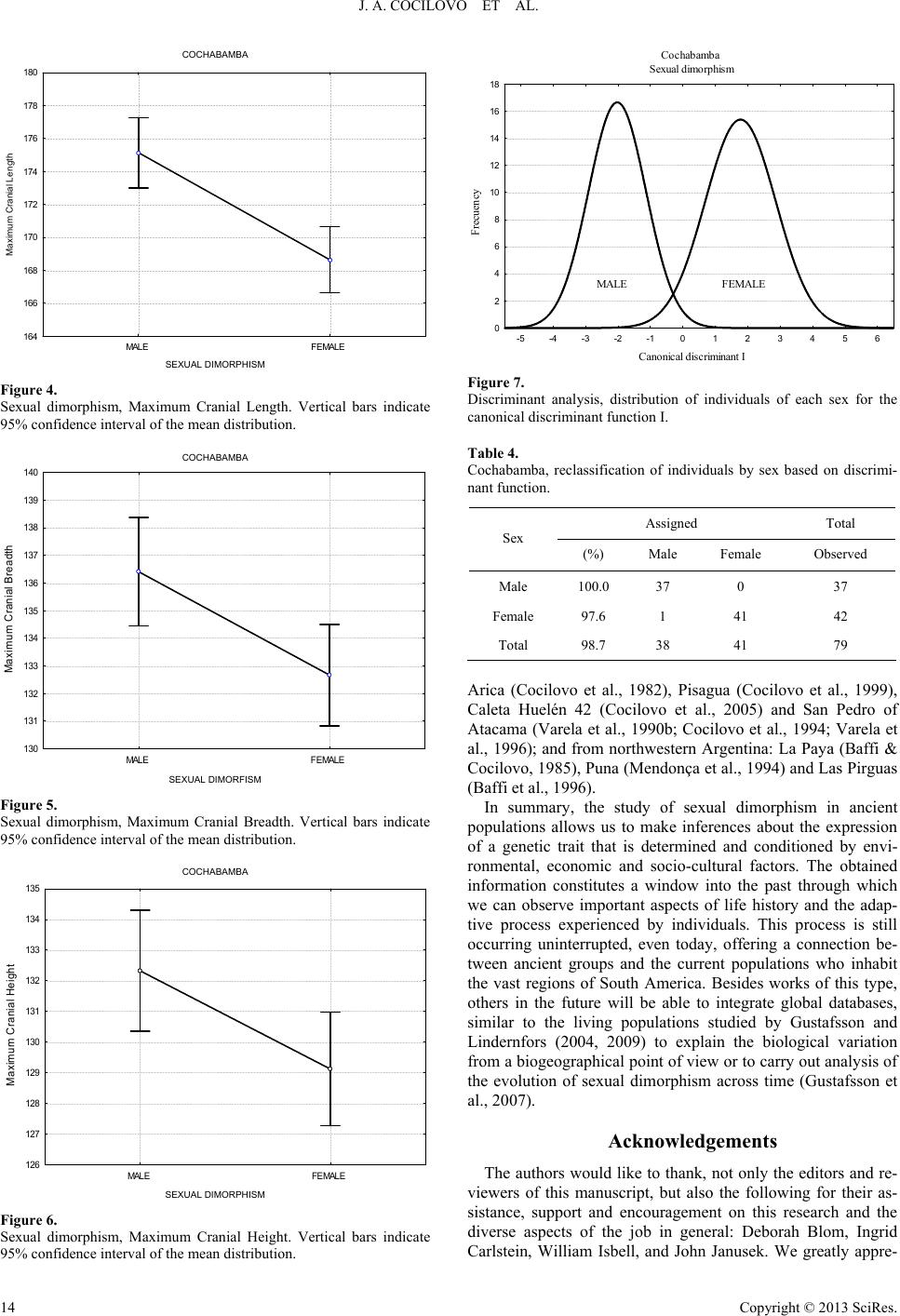 J. A. COCILOVO ET AL. COCHABAMBA MALE FEMALE 164 166 168 170 172 174 176 178 180 Maxim um Cr ania l Length SEXUAL DIMORPHISM Figure 4. Sexual dimorphism, Maximum Cranial Length. Vertical bars indicate 95% confidence interval of the mean distribution. COCHABAMBA MALE FEMALE 130 131 132 133 134 135 136 137 138 139 140 Maximum Cranial Breadth SEXUAL DIMORFISM Figure 5. Sexual dimorphism, Maximum Cranial Breadth. Vertical bars indicate 95% confidence interval of the mean distribution. COCHABAMBA MALE FEMALE 126 127 128 129 130 131 132 133 134 135 Maximum Cranial H eig ht SEXUAL DIMORPHISM Figure 6. Sexual dimorphism, Maximum Cranial Height. Vertical bars indicate 95% confidence interval of the mean distribution. Cochabamba Sexual dimorphism Canonical discriminant I Frecuency -5 -4 -3 -2 -10123456 0 2 4 6 8 10 12 14 16 18 MALE FEMALE Figure 7. Discriminant analysis, distribution of individuals of each sex for the canonical discriminant function I. Table 4. Cochabamba, reclassification of individuals by sex based on discrimi- nant function. Assigned Total Sex (%) Male Female Observed Male 100.0 37 0 37 Female 97.6 1 41 42 Total 98.7 38 41 79 Arica (Cocilovo et al., 1982), Pisagua (Cocilovo et al., 1999), Caleta Huelén 42 (Cocilovo et al., 2005) and San Pedro of Atacama (Varela et al., 1990b; Cocilovo et al., 1994; Varela et al., 1996); and from northwestern Argentina: La Paya (Baffi & Cocilovo, 1985), Puna (Mendonça et al., 1994) and Las Pirguas (Baffi et al., 1996). In summary, the study of sexual dimorphism in ancient populations allows us to make inferences about the expression of a genetic trait that is determined and conditioned by envi- ronmental, economic and socio-cultural factors. The obtained information constitutes a window into the past through which we can observe important aspects of life history and the adap- tive process experienced by individuals. This process is still occurring uninterrupted, even today, offering a connection be- tween ancient groups and the current populations who inhabit the vast regions of South America. Besides works of this type, others in the future will be able to integrate global databases, similar to the living populations studied by Gustafsson and Lindernfors (2004, 2009) to explain the biological variation from a biogeographical point of view or to carry out analysis of the evolution of sexual dimorphism across time (Gustafsson et al., 2007). Acknowledgements The authors would like to thank, not only the editors and re- viewers of this manuscript, but also the following for their as- sistance, support and encouragement on this research and the diverse aspects of the job in general: Deborah Blom, Ingrid Carlstein, William Isbell, and John Janusek. We greatly appre- Copyright © 2013 SciRes. 14 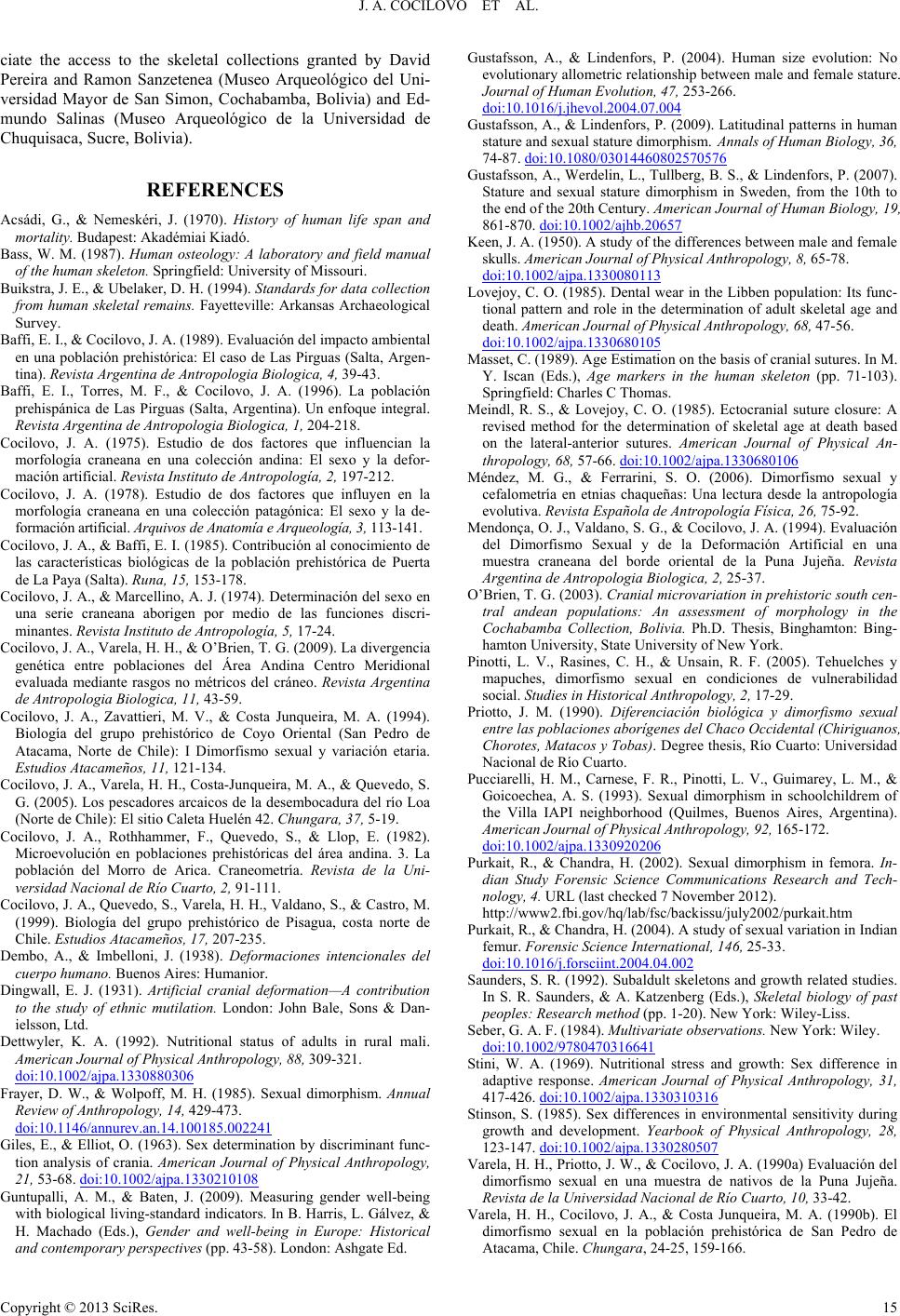 J. A. COCILOVO ET AL. Copyright © 2013 SciRes. 15 ciate the access to the skeletal collections granted by David Pereira and Ramon Sanzetenea (Museo Arqueológico del Uni- versidad Mayor de San Simon, Cochabamba, Bolivia) and Ed- mundo Salinas (Museo Arqueológico de la Universidad de Chuquisaca, Sucre, Bolivia). REFERENCES Acsádi, G., & Nemeskéri, J. (1970). History of human life span and mortality. Budapest: Akadémiai Kiadó. Bass, W. M. (1987). Human osteology: A laboratory and field manual of the human skeleton. Springfield: University of Missouri. Buikstra, J. E., & Ubelaker, D. H. (1994). Standards for data collection from human skeletal remains. Fayetteville: Arkansas Archaeological Survey. Baffi, E. I., & Cocilovo, J. A. (1989). Evaluación del impacto ambiental en una población prehistórica: El caso de Las Pirguas (Salta, Argen- tina). Revista Argentina de Antropologia Biologica, 4, 39-43. Baffi, E. I., Torres, M. F., & Cocilovo, J. A. (1996). La población prehispánica de Las Pirguas (Salta, Argentina). Un enfoque integral. Revista Argentina de Antropologia Biologica, 1, 204-218. Cocilovo, J. A. (1975). Estudio de dos factores que influencian la morfología craneana en una colección andina: El sexo y la defor- mación artificial. Revista Instituto de A nt ro po l og ía , 2 , 197-212. Cocilovo, J. A. (1978). Estudio de dos factores que influyen en la morfología craneana en una colección patagónica: El sexo y la de- formación artificial. Arquivos de Anatomía e Arqueología, 3, 113-141. Cocilovo, J. A., & Baffi, E. I. (1985). Contribución al conocimiento de las características biológicas de la población prehistórica de Puerta de La Paya (Salta). Runa, 15, 153-178. Cocilovo, J. A., & Marcellino, A. J. (1974). Determinación del sexo en una serie craneana aborigen por medio de las funciones discri- minantes. Revista Instituto de Antropología, 5, 17-24. Cocilovo, J. A., Varela, H. H., & O’Brien, T. G. (2009). La divergencia genética entre poblaciones del Área Andina Centro Meridional evaluada mediante rasgos no métricos del cráneo. Revista Argentina de Antropologia Biologica, 11, 43-59. Cocilovo, J. A., Zavattieri, M. V., & Costa Junqueira, M. A. (1994). Biología del grupo prehistórico de Coyo Oriental (San Pedro de Atacama, Norte de Chile): I Dimorfismo sexual y variación etaria. Estudios Atacameños, 11, 121-134. Cocilovo, J. A., Varela, H. H., Costa-Junqueira, M. A., & Quevedo, S. G. (2005). Los pescadores arcaicos de la desembocadura del río Loa (Norte de Chile): El sitio Caleta Huelén 42. Chungara, 37, 5-19. Cocilovo, J. A., Rothhammer, F., Quevedo, S., & Llop, E. (1982). Microevolución en poblaciones prehistóricas del área andina. 3. La población del Morro de Arica. Craneometría. Revista de la Uni- versidad Nacional de Río Cuarto, 2 , 91-111. Cocilovo, J. A., Quevedo, S., Varela, H. H., Valdano, S., & Castro, M. (1999). Biología del grupo prehistórico de Pisagua, costa norte de Chile. Estudios Atacameños, 1 7, 207-235. Dembo, A., & Imbelloni, J. (1938). Deformaciones intencionales del cuerpo humano. Buenos Aires: Humanior. Dingwall, E. J. (1931). Artificial cranial deformation—A contribution to the study of ethnic mutilation. London: John Bale, Sons & Dan- ielsson, Ltd. Dettwyler, K. A. (1992). Nutritional status of adults in rural mali. American Journal of Physical Anthropology, 88, 309-321. doi:10.1002/ajpa.1330880306 Frayer, D. W., & Wolpoff, M. H. (1985). Sexual dimorphism. Annual Review of Anthropology, 14, 429-473. doi:10.1146/annurev.an.14.100185.002241 Giles, E., & Elliot, O. (1963). Sex determination by discriminant func- tion analysis of crania. American Journal of Physical Anthropology, 21, 53-68. doi:10.1002/ajpa.1330210108 Guntupalli, A. M., & Baten, J. (2009). Measuring gender well-being with biological living-standard indicators. In B. Harris, L. Gálvez, & H. Machado (Eds.), Gender and well-being in Europe: Historical and contemporary perspectives (pp. 43-58). London: Ashgate Ed. Gustafsson, A., & Lindenfors, P. (2004). Human size evolution: No evolutionary allometric relationship between male and female stature. Journal of Human Evolution, 4 7, 253-266. doi:10.1016/j.jhevol.2004.07.004 Gustafsson, A., & Lindenfors, P. (2009). Latitudinal patterns in human stature and sexual stature dimorphism. Annals of Human Biology, 36, 74-87. doi:10.1080/03014460802570576 Gustafsson, A., Werdelin, L., Tullberg, B. S., & Lindenfors, P. (2007). Stature and sexual stature dimorphism in Sweden, from the 10th to the end of the 20th Century. American Journal of Human Biology, 19, 861-870. doi:10.1002/ajhb.20657 Keen, J. A. (1950). A study of the differences between male and female skulls. American Journal of Physical Anthropology, 8, 65-78. doi:10.1002/ajpa.1330080113 Lovejoy, C. O. (1985). Dental wear in the Libben population: Its func- tional pattern and role in the determination of adult skeletal age and death. American Journal of P hys ica l Anthropology, 68, 47-56. doi:10.1002/ajpa.1330680105 Masset, C. (1989). Age Estimation on the basis of cranial sutures. In M. Y. Iscan (Eds.), Age markers in the human skeleton (pp. 71-103). Springfield: Charles C Thomas. Meindl, R. S., & Lovejoy, C. O. (1985). Ectocranial suture closure: A revised method for the determination of skeletal age at death based on the lateral-anterior sutures. American Journal of Physical An- thropology, 68, 57-66. doi:10.1002/ajpa.1330680106 Méndez, M. G., & Ferrarini, S. O. (2006). Dimorfismo sexual y cefalometría en etnias chaqueñas: Una lectura desde la antropología evolutiva. Revista Española de Antropología Física, 2 6, 75-92. Mendonça, O. J., Valdano, S. G., & Cocilovo, J. A. (1994). Evaluación del Dimorfismo Sexual y de la Deformación Artificial en una muestra craneana del borde oriental de la Puna Jujeña. Revista Argentina de Antropologia Biologica, 2, 25-37. O’Brien, T. G. (2003). Cranial microvariation in prehistoric south cen- tral andean populations: An assessment of morphology in the Cochabamba Collection, Bolivia. Ph.D. Thesis, Binghamton: Bing- hamton University, State University of New York. Pinotti, L. V., Rasines, C. H., & Unsain, R. F. (2005). Tehuelches y mapuches, dimorfismo sexual en condiciones de vulnerabilidad social. Studies in Historical Anthropology, 2, 17-29. Priotto, J. M. (1990). Diferenciación biológica y dimorfismo sexual entre las poblaciones aborígenes del Chaco Occidental (Chiriguanos, Chorotes, Matacos y Tobas). Degree thesis, Río Cuarto: Universidad Nacional de Río Cuarto. Pucciarelli, H. M., Carnese, F. R., Pinotti, L. V., Guimarey, L. M., & Goicoechea, A. S. (1993). Sexual dimorphism in schoolchildrem of the Villa IAPI neighborhood (Quilmes, Buenos Aires, Argentina). American Journal of Physical Anthropology, 92, 165-172. doi:10.1002/ajpa.1330920206 Purkait, R., & Chandra, H. (2002). Sexual dimorphism in femora. In- dian Study Forensic Science Communications Research and Tech- nology, 4. URL (last checked 7 November 2012). http://www2.fbi.gov/hq/lab/fsc/backissu/july2002/purkait.htm Purkait, R., & Chandra, H. (2004). A study of sexual variation in Indian femur. Forensic Science Inter na t i on a l, 146, 25-33. doi:10.1016/j.forsciint.2004.04.002 Saunders, S. R. (1992). Subaldult skeletons and growth related studies. In S. R. Saunders, & A. Katzenberg (Eds.), Skeletal biology of past peoples: Research method (pp. 1-20). New York: Wiley-Liss. Seber, G. A. F. (1984). Multivariate observations. New York: Wiley. doi:10.1002/9780470316641 Stini, W. A. (1969). Nutritional stress and growth: Sex difference in adaptive response. American Journal of Physical Anthropology, 31, 417-426. doi:10.1002/ajpa.1330310316 Stinson, S. (1985). Sex differences in environmental sensitivity during growth and development. Yearbook of Physical Anthropology, 28, 123-147. doi:10.1002/ajpa.1330280507 Varela, H. H., Priotto, J. W., & Cocilovo, J. A. (1990a) Evaluación del dimorfismo sexual en una muestra de nativos de la Puna Jujeña. Revista de la Universidad Nacional de Río Cuarto, 10, 33-42. Varela, H. H., Cocilovo, J. A., & Costa Junqueira, M. A. (1990b). El dimorfismo sexual en la población prehistórica de San Pedro de Atacama, Chile. Chungara, 24-25, 159-166. |