

Paper Menu >>
Journal Menu >>
 J. Biomedical Science and Engineering, 2013, 6, 109-115 JBiSE http://dx.doi.org/10.4236/jbise.2013.62014 Published Online February 2013 (http://www.scirp.org/journal/jbise/) A study on the activity of dermal multipotent stem cells in initiation of wound repair Jifu Qu1,2, Tianmin Cheng 2, Yongping Su2, Chunmeng Shi2, Wei Sun1 1Trauma Center, Department of Emergency Medicine, Southwest Hospital, Third Military Medical University, Chongqing, China 2State Key Laboratory of Trauma, Burn and Combined Injury, Institute of Combined Injuries of Chinese PLA, Academy of Preven- tive Medicine, Third Military Medical University, Chongqing, China Email: [email protected] Received 17 December 2012; revised 16 January 2013; accepted 24 January 2013 ABSTRACT Background: Wound healing is a process of cell-cell interaction and cell-extracellular matrix interaction. Dermal multipotent stem cells (dMSCs) have the abilities to promote survival and wound healing, but the potential function of dMSCs in wound healing, particularly in the initiation of wound repair, has not been fully understood. Methods: dMSCs and fibro- blasts were isolated from neonatal rat dermis and were further purified and expanded. The cell cycles were determined with flow cytometry, while the ra- diosensitivity was measured by MTT assay. Rats were wounded with a 7-cm incision on the back skin and the wound fluids were collected by inserting two pieces of sterile polyvinyl alcohol sponge (1 cm in di- ameter and 0.4 cm in thickness) subcutaneously into the dorsum of each rat through the midline of incision on the 1st, 2nd, 3rd and 4th day after incision. The ef- fects of wound fluids on the proliferation of dMSCs and fibroblasts were measured with MTT assays. dMSC’s abilities of adhesion and attachment and its migration in response to wound fluids collected on the 1st day after incision were explored by measuring the percentage of floating cells and the cells migrated into wounding area in vitro, respectively. Results: The iso- lated dMSCs were morphologically homogenous and highly proliferative. Most of the cultured dMSCs were quiescent with few apoptotic cells. Compared with fibroblasts, dMSCs were more sensitive to ra- diation and more proliferative in response to wound fluids, especially to the wound fluids collected on the 1st day after wounding. Moreover, their abilities to attach, adhere and migrate were significantly en- hanced with the early-phase wound fluids. Conclu- sions: As primitive stem cells, dMSCs are very re- sponsive to wound fluids, which suggests dMSCs’ important role in wound healing, especially in initiat- ing wound repair. Keywords: Dermal Multipotent Stem Cells; Initiation of Wound Repair; Wound Healing; Wound Fluids 1. INTRODUCTION Stem cells are cells with self-renewal capacity, multipo- tentiality and unparalleled superiority in cell-replacement therapy, gene therapy, developmental biology, pharma- cology and toxicology [1,2]. Mesenchymal stem cells have the potential to differentiate into fibroblasts, vascu- lar endothelial cells and other tissue repairing cells, there- fore playing important roles in tissue repair [3]. Recent studies have suggested that stem cells with different de- velopmental potentials exist in several adult mammalian tissues, especially in newly formed tissues or tissues with rapid renewal potentials [4-6]. Skin, which consists of the epidermis and dermis, is one of the rapidly renewing tissues in adults. Several studies from our group and two other groups have indicated that multipotent cells can be isolated from adult mammalian dermis [4,7,8]. Given their easy accessibility, these cells could be a source of stem cells for cell transplantation and tissue engineering. Particularly, we have found that multipotent stem cells derived from dermal mesenchymal tissue, namely dMSCs, have the abilities to promote survival and wound healing in rats subjected to radiation and wound injury, as well as enhance hematopoietic recovery in sublethally irradiated rats [9,10]. Wound healing is a process of cell-cell interaction and cell-extracellular matrix interaction. Wound environments exert significant effects on cell growth and differentiation during the healing process. Different cell types are acti- vated by the environment and subsequently migrate to and proliferate at the wound site. Studies have indicated that it is the specific tissue environment that determines the plasticity of mesenchymal stem cells in vivo [11-14]. Although activation of epithelial cells, fibroblasts and endothelial cells has been observed after wounding, little has been described about the multipotent cells in the OPEN ACCESS  J. F. Qu et al. / J. Biomedical Science and Engineering 6 (2013) 109-115 110 dermis [15-18]. In our previous study, we investigated the biological effects of the acute wound environment (wound fluids) on dMSCs isolated from newborn rats (neonatal dMSCs), and showed that neonatal suspensions enhanced the initial rate of wound contraction and some cells differentiated from dMSCs were found in the re- generative dermis. We also observed that treatment with acute wound fluid promoted proliferation and migration of dMSCs [4]. These results suggest that neonatal dMSCs could participate in the regeneration of the in- jured dermis and thus may be an alternative choice for cell-based therapies for skin injuries. As stem cells are primitive with self-renewal capacity, it awaits clarification whether the dMSCs are primitive stem cells. Moreover, the potential function of dMSCs in wound healing has also not been fully understood. In the present study, we further characterized the radiosensitiv- ity of dMSCs, as well as their proliferation, attachment, adhesion and migration in response to early-phase wound fluids so as to explore their potential roles in wound healing. 2. MATERIALS AND METHODS 2.1. Cell Culture dMSCs and fibroblasts were isolated from neonatal rat dermis, cultured in IMDM supplemented with 10% (v/v) fetal bovine serum, 100 U/mL penicillin and 100 µg/mL streptomycin (Hyclone). dMSCs were amplified and identified as previously described [9]. Briefly, skin tissue from 1-day-old rat was digested with 0.25% trypsin at 4˚C overnight. The dermal layer (confirmed with histo- logical examination) was dissociated by flushing with D-Hanks’ solution; the suspension was filtered through a nylon mesh and centrifuged to remove cellular debris. The cell pellet was resuspended and cultured in IMDM containing 10% FBS, 100 U/mL penicillin and 100 µg/mL streptomycin at 37˚C in a humidified atmosphere containing 5% CO2. Six hours later, the adherent cells were subcultured at a low density of 5 cells/cm2. The presence of separately adherent single cell was assessed microscopically. Two weeks later, single colonies were isolated by cloning rings under an inverted microscope and expanded. The differentiation characteristics of dMSC were further confirmed in an induction medium contain- ing dexamethasone as reported previously [4]. 2.2. Radiosensitivity Measurement by MTT Assay Radiosensitivities of fibroblasts and the 10th passage of dMSCs were measured by MTT assay. 80% confluent cells were detached with 0.25% trypsin. After washed once with D-Hank’s solution, the cell concentration was adjusted to 2 × 10⁴/mL with IMDM supplemented with 10 ml/L fetal bovine serum. 0.2 ml cells were transferred into sterile penicillin bottles and irradiated with 60Co γ ray at 0, 2, 6, 10, 12 and 15 Gy, respectively. After irradiation, the cells were added into 96-well plate and the numbers of live cells were measured with MTT assay in quadruplicate for each radiation dosage. Specifically, 20 μL 5 g/L MTT solution (Sigma) was added into each well of the 96-well plate and incubated for 4 hours at 37˚C in a humidified atmosphere containing 5% CO2. Then the culture solution was removed and 150 μL DMSO was added into each well and oscillated for 10 minutes. The absorptions at 492 nm were measured with an enzyme immunoassay analyzer (HTS 7000 plus, Perkin Elmer, USA). 2.3. Measurement of Cell Cycle and Apoptotic Rate The changes in cell cycle progression and apoptotic rate of dMSCs at the 3rd passage were assayed with flow cy- tometry. 80% confluent cells were detached with 0.25% trypsin. After washed once with D-Hank’s solution, the cells were fixed for 30 min with 70% alcohol at 4˚C, and then centrifuged at 1500 rpm for 5 min. After discarding the fixing solution, the cells were washed once with cold PBS solution. dMSC cells were incubated with 200 µl of lysis buffer, stained with propidium iodide at a concen- tration of 50 mg/mL for 15 min, and analyzed with the Coulter Epic C flow cytometer to calculate the G0/G1 population and apoptotic rate. 2.4. Collection of Rat Wound Fluids Wistar rats were bred and fed in Animal Center of our university with first class standard. All animal proce- dures were approved by the institution’s animal-use committee and by the Ministry of Justice. 15-week old Wistar rats of mixed sex were wounded with a 7-cm in- cision on the back skin and the wound fluids were col- lected by inserting two pieces of sterile polyvinyl alcohol sponges (1cm in diameter and 0.4 cm in thickness) sub- cutaneously in the dorsum of each animal through the midline incision [10]. The rats were then sacrificed on the 1st, 2nd, 3rd and 4th day after wounding. The wound fluids were collected from the sponge after the cellular components were removed by centrifugation and sterilized with a 0.2 µm filter and then stored at −20˚C until use. 2.5. Effect of Wound Fluids on Proliferation of dMSCs After 48-hour culture in serum-free IMDM [19], cells were plated at 2 × 10⁴/mL to 96-well plate. The wound fluids were added at various concentrations of 10ml/L, Copyright © 2013 SciRes. OPEN ACCESS 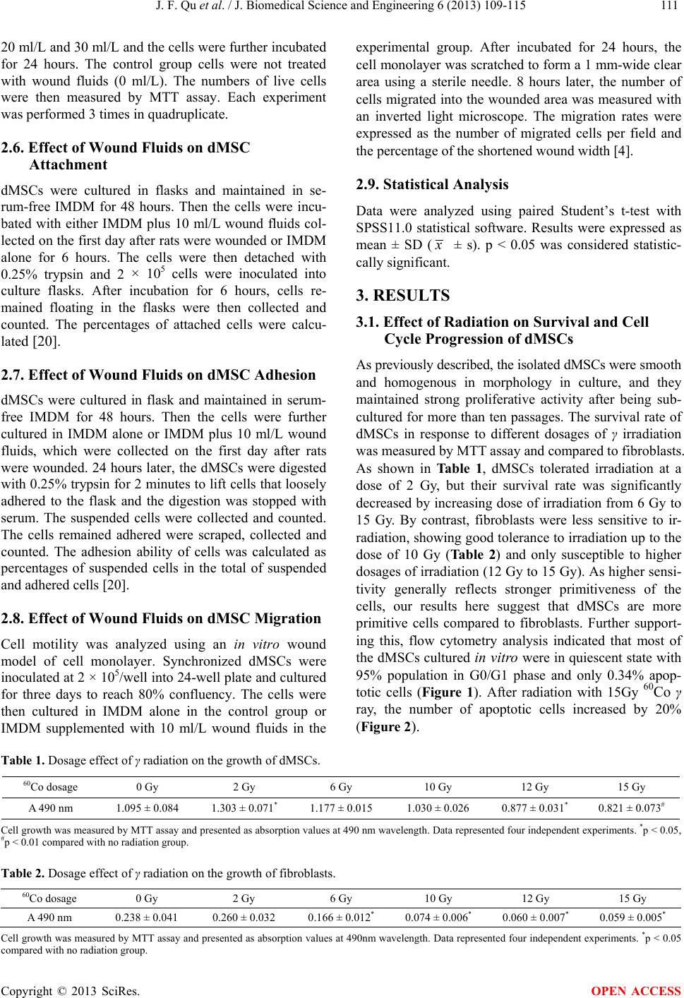 J. F. Qu et al. / J. Biomedical Science and Engineering 6 (2013) 109-115 Copyright © 2013 SciRes. 111 20 ml/L and 30 ml/L and the cells were further incubated for 24 hours. The control group cells were not treated with wound fluids (0 ml/L). The numbers of live cells were then measured by MTT assay. Each experiment was performed 3 times in quadruplicate. experimental group. After incubated for 24 hours, the cell monolayer was scratched to form a 1 mm-wide clear area using a sterile needle. 8 hours later, the number of cells migrated into the wounded area was measured with an inverted light microscope. The migration rates were expressed as the number of migrated cells per field and the percentage of the shortened wound width [4]. 2.6. Effect of Wound Fluids on dMSC Attachment 2.9. Statistical Analysis dMSCs were cultured in flasks and maintained in se- rum-free IMDM for 48 hours. Then the cells were incu- bated with either IMDM plus 10 ml/L wound fluids col- lected on the first day after rats were wounded or IMDM alone for 6 hours. The cells were then detached with 0.25% trypsin and 2 × 10⁵ cells were inoculated into culture flasks. After incubation for 6 hours, cells re- mained floating in the flasks were then collected and counted. The percentages of attached cells were calcu- lated [20]. Data were analyzed using paired Student’s t-test with SPSS11.0 statistical software. Results were expressed as mean ± SD ( x ± s). p < 0.05 was considered statistic- cally significant. 3. RESULTS 3.1. Effect of Radiation on Survival and Cell Cycle Progression of dMSCs As previously described, the isolated dMSCs were smooth and homogenous in morphology in culture, and they maintained strong proliferative activity after being sub- cultured for more than ten passages. The survival rate of dMSCs in response to different dosages of γ irradiation was measured by MTT assay and compared to fibroblasts. As shown in Table 1, dMSCs tolerated irradiation at a dose of 2 Gy, but their survival rate was significantly decreased by increasing dose of irradiation from 6 Gy to 15 Gy. By contrast, fibroblasts were less sensitive to ir- radiation, showing good tolerance to irradiation up to the dose of 10 Gy (Ta b le 2) and only susceptible to higher dosages of irradiation (12 Gy to 15 Gy). As higher sensi- tivity generally reflects stronger primitiveness of the cells, our results here suggest that dMSCs are more primitive cells compared to fibroblasts. Further support- ing this, flow cytometry analysis indicated that most of the dMSCs cultured in vitro were in quiescent state with 95% population in G0/G1 phase and only 0.34% apop- totic cells (Figure 1). After radiation with 15Gy 60Co γ ray, the number of apoptotic cells increased by 20% (Figure 2). 2.7. Effect of Wound Fluids on dMSC Adhesion dMSCs were cultured in flask and maintained in serum- free IMDM for 48 hours. Then the cells were further cultured in IMDM alone or IMDM plus 10 ml/L wound fluids, which were collected on the first day after rats were wounded. 24 hours later, the dMSCs were digested with 0.25% trypsin for 2 minutes to lift cells that loosely adhered to the flask and the digestion was stopped with serum. The suspended cells were collected and counted. The cells remained adhered were scraped, collected and counted. The adhesion ability of cells was calculated as percentages of suspended cells in the total of suspended and adhered cells [20]. 2.8. Effect of Wound Fluids on dMSC Migration Cell motility was analyzed using an in vitro wound model of cell monolayer. Synchronized dMSCs were inoculated at 2 × 10⁵/well into 24-well plate and cultured for three days to reach 80% confluency. The cells were then cultured in IMDM alone in the control group or IMDM supplemented with 10 ml/L wound fluids in the Table 1. Dosage effect of γ radiation on the growth of dMSCs. 60Co dosage 0 Gy 2 Gy 6 Gy 10 Gy 12 Gy 15 Gy A 490 nm 1.095 ± 0.084 1.303 ± 0.071* 1.177 ± 0.015 1.030 ± 0.026 0.877 ± 0.031* 0.821 ± 0.073# Cell growth was measured by MTT assay and presented as absorption values at 490 nm wavelength. Data represented four independent experiments. *p < 0.05, #p < 0.01 compared with no radiation group. Table 2. Dosage effect of γ radiation on the growth of fibroblasts. 60Co dosage 0 Gy 2 Gy 6 Gy 10 Gy 12 Gy 15 Gy A 490 nm 0.238 ± 0.041 0.260 ± 0.032 0.166 ± 0.012* 0.074 ± 0.006* 0.060 ± 0.007* 0.059 ± 0.005* Cell growth was measured by MTT assay and presented as absorption values at 490nm wavelength. Data represented four independent experiments. *p < 0.05 compared with no radiation group. OPEN ACCESS 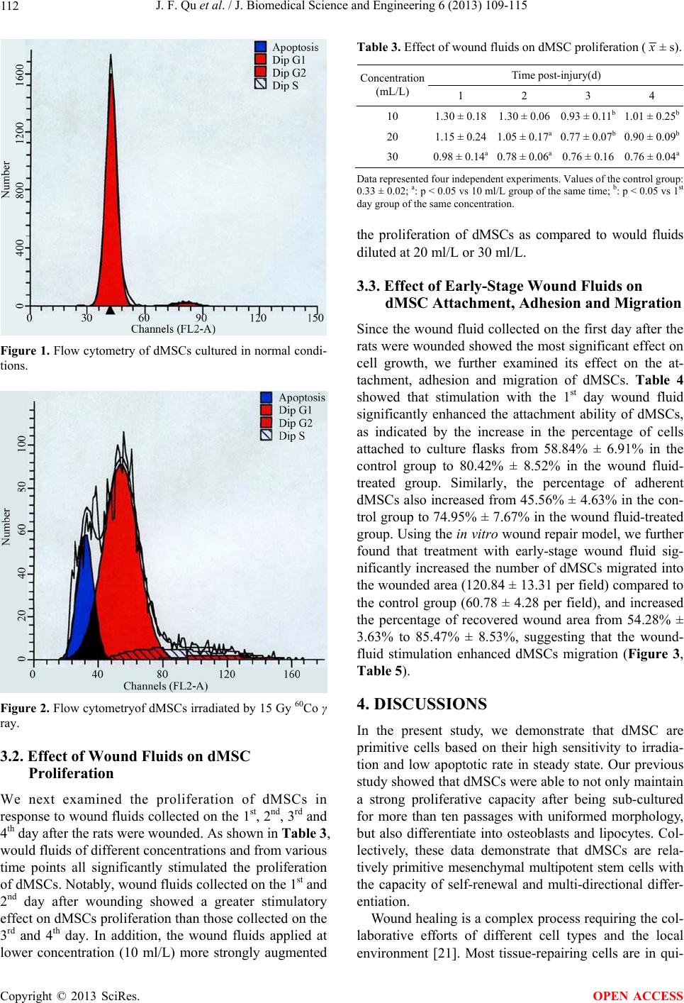 J. F. Qu et al. / J. Biomedical Science and Engineering 6 (2013) 109-115 112 Figure 1. Flow cytometry of dMSCs cultured in normal condi- tions. Figure 2. Flow cytometryof dMSCs irradiated by 15 Gy 60Co γ ray. 3.2. Effect of Wound Fluids on dMSC Proliferation We next examined the proliferation of dMSCs in response to wound fluids collected on the 1st, 2nd, 3rd and 4th day after the rats were wounded. As shown in Tab le 3, would fluids of different concentrations and from various time points all significantly stimulated the proliferation of dMSCs. Notably, wound fluids collected on the 1st and 2nd day after wounding showed a greater stimulatory effect on dMSCs proliferation than those collected on the 3rd and 4th day. In addition, the wound fluids applied at lower concentration (10 ml/L) more strongly augmented Table 3. Effect of wound fluids on dMSC proliferation ( x ± s). Time post-injury(d) Concentration (mL/L) 1 2 3 4 10 1.30 ± 0.181.30 ± 0.06 0.93 ± 0.11b 1.01 ± 0.25b 20 1.15 ± 0.241.05 ± 0.17a 0.77 ± 0.07b 0.90 ± 0.09b 30 0.98 ± 0.14a0.78 ± 0.06a 0.76 ± 0.16 0.76 ± 0.04a Data represented four independent experiments. Values of the control group: 0.33 ± 0.02; a: p < 0.05 vs 10 ml/L group of the same time; b: p < 0.05 vs 1st day group of the same concentration. the proliferation of dMSCs as compared to would fluids diluted at 20 ml/L or 30 ml/L. 3.3. Effect of Early-Stage Wound Fluids on dMSC Attachment, Adhesion and Migration Since the wound fluid collected on the first day after the rats were wounded showed the most significant effect on cell growth, we further examined its effect on the at- tachment, adhesion and migration of dMSCs. Table 4 showed that stimulation with the 1st day wound fluid significantly enhanced the attachment ability of dMSCs, as indicated by the increase in the percentage of cells attached to culture flasks from 58.84% ± 6.91% in the control group to 80.42% ± 8.52% in the wound fluid- treated group. Similarly, the percentage of adherent dMSCs also increased from 45.56% ± 4.63% in the con- trol group to 74.95% ± 7.67% in the wound fluid-treated group. Using the in vitro wound repair model, we further found that treatment with early-stage wound fluid sig- nificantly increased the number of dMSCs migrated into the wounded area (120.84 ± 13.31 per field) compared to the control group (60.78 ± 4.28 per field), and increased the percentage of recovered wound area from 54.28% ± 3.63% to 85.47% ± 8.53%, suggesting that the wound- fluid stimulation enhanced dMSCs migration (Figure 3, Table 5). 4. DISCUSSIONS In the present study, we demonstrate that dMSC are primitive cells based on their high sensitivity to irradia- tion and low apoptotic rate in steady state. Our previous study showed that dMSCs were able to not only maintain a strong proliferative capacity after being sub-cultured for more than ten passages with uniformed morphology, but also differentiate into osteoblasts and lipocytes. Col- lectively, these data demonstrate that dMSCs are rela- tively primitive mesenchymal multipotent stem cells with the capacity of self-renewal and multi-directional differ- entiation. Wound healing is a complex process requiring the col- laborative efforts of different cell types and the local environment [21]. Most tissue-repairing cells are in qui- Copyright © 2013 SciRes. OPEN ACCESS 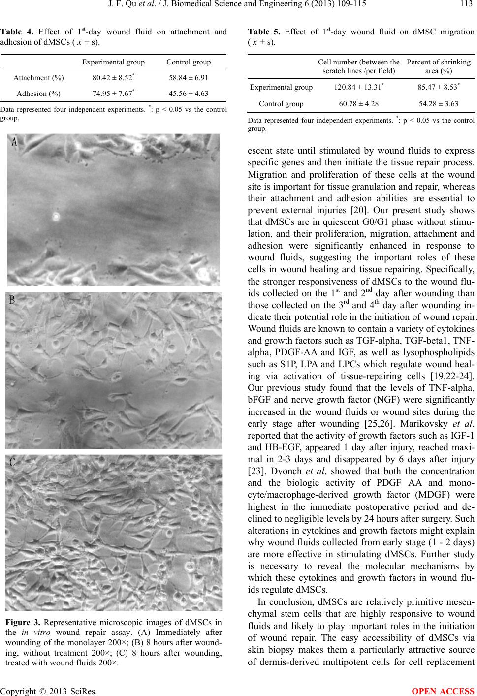 J. F. Qu et al. / J. Biomedical Science and Engineering 6 (2013) 109-115 113 Table 4. Effect of 1st-day wound fluid on attachment and adhesion of dMSCs ( x ± s). Experimental group Control group Attachment (%) 80.42 ± 8.52* 58.84 ± 6.91 Adhesion (%) 74.95 ± 7.67* 45.56 ± 4.63 Data represented four independent experiments. *: p < 0.05 vs the control group. Figure 3. Representative microscopic images of dMSCs in the in vitro wound repair assay. (A) Immediately after wounding of the monolayer 200×; (B) 8 hours after wound- ing, without treatment 200×; (C) 8 hours after wounding, treated with wound fluids 200×. Table 5. Effect of 1st-day wound fluid on dMSC migration ( x ± s). Cell number (between the scratch lines /per field) Percent of shrinking area (%) Experimental group120.84 ± 13.31* 85.47 ± 8.53* Control group 60.78 ± 4.28 54.28 ± 3.63 Data represented four independent experiments. *: p < 0.05 vs the control group. escent state until stimulated by wound fluids to express specific genes and then initiate the tissue repair process. Migration and proliferation of these cells at the wound site is important for tissue granulation and repair, whereas their attachment and adhesion abilities are essential to prevent external injuries [20]. Our present study shows that dMSCs are in quiescent G0/G1 phase without stimu- lation, and their proliferation, migration, attachment and adhesion were significantly enhanced in response to wound fluids, suggesting the important roles of these cells in wound healing and tissue repairing. Specifically, the stronger responsiveness of dMSCs to the wound flu- ids collected on the 1st and 2nd day after wounding than those collected on the 3rd and 4th day after wounding in- dicate their potential role in the initiation of wound repair. Wound fluids are known to contain a variety of cytokines and growth factors such as TGF-alpha, TGF-beta1, TNF- alpha, PDGF-AA and IGF, as well as lysophospholipids such as S1P, LPA and LPCs which regulate wound heal- ing via activation of tissue-repairing cells [19,22-24]. Our previous study found that the levels of TNF-alpha, bFGF and nerve growth factor (NGF) were significantly increased in the wound fluids or wound sites during the early stage after wounding [25,26]. Marikovsky et al. reported that the activity of growth factors such as IGF-1 and HB-EGF, appeared 1 day after injury, reached maxi- mal in 2-3 days and disappeared by 6 days after injury [23]. Dvonch et al. showed that both the concentration and the biologic activity of PDGF AA and mono- cyte/macrophage-derived growth factor (MDGF) were highest in the immediate postoperative period and de- clined to negligible levels by 24 hours after surgery. Such alterations in cytokines and growth factors might explain why wound fluids collected from early stage (1 - 2 days) are more effective in stimulating dMSCs. Further study is necessary to reveal the molecular mechanisms by which these cytokines and growth factors in wound flu- ids regulate dMSCs. In conclusion, dMSCs are relatively primitive mesen- chymal stem cells that are highly responsive to wound fluids and likely to play important roles in the initiation of wound repair. The easy accessibility of dMSCs via skin biopsy makes them a particularly attractive source of dermis-derived multipotent cells for cell replacement Copyright © 2013 SciRes. OPEN ACCESS 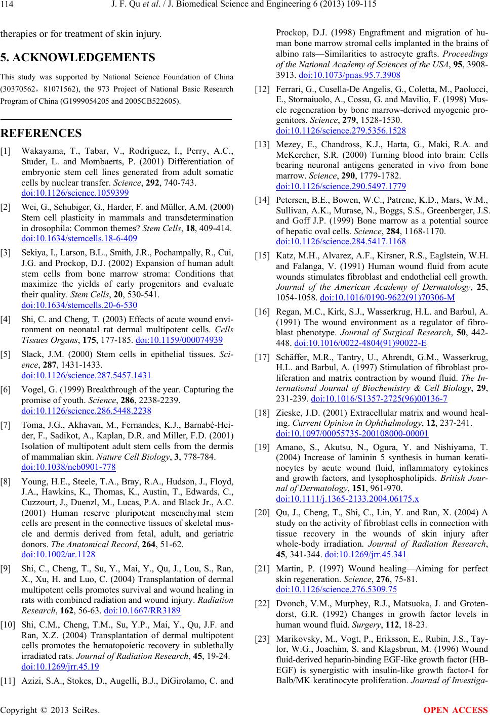 J. F. Qu et al. / J. Biomedical Science and Engineering 6 (2013) 109-115 114 therapies or for treatment of skin injury. 5. ACKNOWLEDGEMENTS This study was supported by National Science Foundation of China (30370562,81071562), the 973 Project of National Basic Research Program of China (G1999054205 and 2005CB522605). REFERENCES [1] Wakayama, T., Tabar, V., Rodriguez, I., Perry, A.C., Studer, L. and Mombaerts, P. (2001) Differentiation of embryonic stem cell lines generated from adult somatic cells by nuclear transfer. Science, 292, 740-743. doi:10.1126/science.1059399 [2] Wei, G., Schubiger, G., Harder, F. and Müller, A.M. (2000) Stem cell plasticity in mammals and transdetermination in drosophila: Common themes? Stem Cells, 18, 409-414. doi:10.1634/stemcells.18-6-409 [3] Sekiya, I., Larson, B.L., Smith, J.R., Pochampally, R., Cui, J.G. and Prockop, D.J. (2002) Expansion of human adult stem cells from bone marrow stroma: Conditions that maximize the yields of early progenitors and evaluate their quality. Stem Cells, 20, 530-541. doi:10.1634/stemcells.20-6-530 [4] Shi, C. and Cheng, T. (2003) Effects of acute wound envi- ronment on neonatal rat dermal multipotent cells. Cells Tissues Organs, 175, 177-185. doi:10.1159/000074939 [5] Slack, J.M. (2000) Stem cells in epithelial tissues. Sci- ence, 287, 1431-1433. doi:10.1126/science.287.5457.1431 [6] Vogel, G. (1999) Breakthrough of the year. Capturing the promise of youth. Science, 286, 2238-2239. doi:10.1126/science.286.5448.2238 [7] Toma, J.G., Akhavan, M., Fernandes, K.J., Barnabé-Hei- der, F., Sadikot, A., Kaplan, D.R. and Miller, F.D. (2001) Isolation of multipotent adult stem cells from the dermis of mammalian skin. Nature Cell Biology, 3, 778-784. doi:10.1038/ncb0901-778 [8] Young, H.E., Steele, T.A., Bray, R.A., Hudson, J., Floyd, J.A., Hawkins, K., Thomas, K., Austin, T., Edwards, C., Cuzzourt, J., Duenzl, M., Lucas, P.A. and Black Jr., A.C. (2001) Human reserve pluripotent mesenchymal stem cells are present in the connective tissues of skeletal mus- cle and dermis derived from fetal, adult, and geriatric donors. The Anatomical Record, 264, 51-62. doi:10.1002/ar.1128 [9] Shi, C., Cheng, T., Su, Y., Mai, Y., Qu, J., Lou, S., Ran, X., Xu, H. and Luo, C. (2004) Transplantation of dermal multipotent cells promotes survival and wound healing in rats with combined radiation and wound injury. Radiation Research, 162, 56-63. doi:10.1667/RR3189 [10] Shi, C.M., Cheng, T.M., Su, Y.P., Mai, Y., Qu, J.F. and Ran, X.Z. (2004) Transplantation of dermal multipotent cells promotes the hematopoietic recovery in sublethally irradiated rats. Journal of Radiation Research, 45, 19-24. doi:10.1269/jrr.45.19 [11] Azizi, S.A., Stokes, D., Augelli, B.J., DiGirolamo, C. and Prockop, D.J. (1998) Engraftment and migration of hu- man bone marrow stromal cells implanted in the brains of albino rats—Similarities to astrocyte grafts. Proceedings of the National Academy of Sciences of the USA, 95, 3908- 3913. doi:10.1073/pnas.95.7.3908 [12] Ferrari, G., Cusella-De Angelis, G., Coletta, M., Paolucci, E., Stornaiuolo, A., Cossu, G. and Mavilio, F. (1998) Mus- cle regeneration by bone marrow-derived myogenic pro- genitors. Science, 279, 1528-1530. doi:10.1126/science.279.5356.1528 [13] Mezey, E., Chandross, K.J., Harta, G., Maki, R.A. and McKercher, S.R. (2000) Turning blood into brain: Cells bearing neuronal antigens generated in vivo from bone marrow. Science, 290, 1779-1782. doi:10.1126/science.290.5497.1779 [14] Petersen, B.E., Bowen, W.C., Patrene, K.D., Mars, W.M., Sullivan, A.K., Murase, N., Boggs, S.S., Greenberger, J.S. and Goff J.P. (1999) Bone marrow as a potential source of hepatic oval cells. Science, 284, 1168-1170. doi:10.1126/science.284.5417.1168 [15] Katz, M.H., Alvarez, A.F., Kirsner, R.S., Eaglstein, W.H. and Falanga, V. (1991) Human wound fluid from acute wounds stimulates fibroblast and endothelial cell growth. Journal of the American Academy of Dermatology, 25, 1054-1058. doi:10.1016/0190-9622(91)70306-M [16] Regan, M.C., Kirk, S.J., Wasserkrug, H.L. and Barbul, A. (1991) The wound environment as a regulator of fibro- blast phenotype. Journal of Surgical Research, 50, 442- 448. doi:10.1016/0022-4804(91)90022-E [17] Schäffer, M.R., Tantry, U., Ahrendt, G.M., Wasserkrug, H.L. and Barbul, A. (1997) Stimulation of fibroblast pro- liferation and matrix contraction by wound fluid. The In- ternational Journal of Biochemistry & Cell Biology, 29, 231-239. doi:10.1016/S1357-2725(96)00136-7 [18] Zieske, J.D. (2001) Extracellular matrix and wound heal- ing. Current Opinion in Ophthalmology, 12, 237-241. doi:10.1097/00055735-200108000-00001 [19] Amano, S., Akutsu, N., Ogura, Y. and Nishiyama, T. (2004) Increase of laminin 5 synthesis in human kerati- nocytes by acute wound fluid, inflammatory cytokines and growth factors, and lysophospholipids. British Jour- nal of Dermatology, 151, 961-970. doi:10.1111/j.1365-2133.2004.06175.x [20] Qu, J., Cheng, T., Shi, C., Lin, Y. and Ran, X. (2004) A study on the activity of fibroblast cells in connection with tissue recovery in the wounds of skin injury after whole-body irradiation. Journal of Radiation Research, 45, 341-344. doi:10.1269/jrr.45.341 [21] Martin, P. (1997) Wound healing—Aiming for perfect skin regeneration. Science, 276, 75-81. doi:10.1126/science.276.5309.75 [22] Dvonch, V.M., Murphey, R.J., Matsuoka, J. and Groten- dorst, G.R. (1992) Changes in growth factor levels in human wound fluid. Surgery, 112, 18-23. [23] Marikovsky, M., Vogt, P., Eriksson, E., Rubin, J.S., Tay- lor, W.G., Joachim, S. and Klagsbrun, M. (1996) Wound fluid-derived heparin-binding EGF-like growth factor (HB- EGF) is synergistic with insulin-like growth factor-I for Balb/MK keratinocyte proliferation. Journal of Investiga- Copyright © 2013 SciRes. OPEN ACCESS  J. F. Qu et al. / J. Biomedical Science and Engineering 6 (2013) 109-115 Copyright © 2013 SciRes. 115 OPEN ACCESS tive Dermatology, 106, 616-621. doi:10.1111/1523-1747.ep12345413 [24] Kroeze, K.L., Jurgens, W.J., Doulabi, B.Z., van Milligen, F.J., Scheper, R.J. and Gibbs, S. (2009) Chemokine-me- diated migration of skin-derived stem cells: Predominant role for CCL5/RANTES. Journal of Investigative Derma- tology, 129, 1569-1581. doi:10.1038/jid.2008.405 [25] Qu, J.F., Cheng, T.M., Shi, C.M., Lin, Y., Yan, G.H. and Ran, X.Z. (2002) Changes of contents of wound bFGF and NGF after combined total body irradiation (TBI) in- jury. Chinese Journal of Radiological Medicine & Pro- tection, 22, 158-160. [26] Song, S.Q., Cheng, T.M. and Lin, Y. (1998) Changes of cytokines in wound fluids and the effects of phenytoin sodium on whole body irradiated rats. Basic Medicine & Clinical, 18, 208-211. |