

Paper Menu >>
Journal Menu >>
 International Journal of Clinical Medicine, 2013, 4, 91-95 http://dx.doi.org/10.4236/ijcm.2013.42017 Published Online February 2013 (http://www.scirp.org/journal/ijcm) National Approach to Premarital Diagnosis of Trait Thalassemia and Silent Carriers Narges Beigom Mirbehbahani1, Azam Rashidbaghan2*, Maryam Mazji2, Nasser Behnampour2 1Golestan University of Medical Sciences, Gorgan, Iran; 2Hematology and Oncology Research Center, Golestan University of Medi- cal Sciences, Gorgan, Iran. Email: *[email protected] Received October 6th, 2012; revised November 7th, 2012; accepted November 15th, 2012 ABSTRACT Background: Major β-thalassemia occurs when impaired genes received by a neonate from the parents. In most of the parents, there are no apparent clinical manifestation and they just show some impairments in hematological indices. This study was designed to determine prevalence of minor and silent carries parents that have children suffering from major β-thalassemia, compare it with national protocol about prevention of thalassemia. Methods: A blood sample was taken from parents of all major thalassemic patients covered by Taleghani Hospital in Gorgan (n = 195), CBC and He- moglobin electrophoresis were done. Data were analyzed using SPSS software. Results: Amongst 196 parents one case have normal level of MCV, MCH, RBC, HbA, HbF and mentzer = 22.05 (0.51%) that diagnosed as alpha triplication/N by real time PCR, RFLP informative. The means of hematological indices were based on the national protocol. Conclu- sion: Present results showed that there are a few cases of thalassemia disorders with normal MCV, MCH, RBC, Mentzer index and Hb electrophoresis which could be missed in routine and pre-marital screening tests, resulted in a thalassemia child that it is possible in every screening test. Generally, indices were according to range of the national guideline. Keywords: Beta-Thalassemia; Silent Carrier; Hemoglobin; Electrophoresis; Hematological Tests; Thalassemia Intermedia 1. Introduction The inherited hemoglobin (Hb) disorders are the most common single gene detect in men. The prevalence of hemoglobinopathies is on the rise worldwide. This is of special importance in developing countries where it in- creases the burden of health care delivery system. The abnormalities can either be quantitative (the thalassemia syndrome) or qualitative (the hemoglobin variants) or a combination of both. Of these, the thalassemia syn- dromes particularly the beta thalassemias and some aloha thalassemias are the major cause of morbidity [1]. Beta-thalassemia is the common hereditary disease all around the world, especially in Iran. It is estimated that 900,000 thalassemia patients will born in the next com- ing 20 years; which 95% of them will be in Asia, India and middle east. Iran Thalassemia Association reported that 18,616 thalassemia patients live in this country that Ma- zandaran and Fars have the most affected population [2]. Reduction or absence of beta globin chains of the hemoglobin results in Beta-thalassemia. The beta globin (HBB) chain gene is located on the short arm of chro- mosome 11. Beta-thalassemia silent carrier, beta thalas- semia trait, thalassemia intermediate and thalassemia major are four recognized category of this condition. Heterozygosis for beta-thalassemia will lead to beta- thalassemia silent carrier and trait [3]. In silent carrier often revealed mild microcytosis or a slight impairment in B-globin synthesis on radial labeling of the globin chains peripheral blood reticulocytes. Characteristically, silent carriers of -thalassemia have normal levels of HbA2 [4]. Also silent carrier is known by normal or small decrease in MCV and MCH and normal hemoglobin electrophoresis [5]. This type will lead to a transfusion- dependent baby if married with a minor thalassemia [6]. In 1969, a silent thalassemia was reported through ex- amination not only of the hematological picture but also of the hemoglobin status and the α/ globin synthesis ratio; the carrier was undoubtedly the father of two tha- lassemia intermedia patients [7] in subsequent years many similar cases were observed that confirmed the existence of varieties of silent thalassemia [8-12]. In these patients increased RBC count, reduced hemo- globin level, MCV and MCH, altered erythrocyte mor- phology and increased HbA2 level imbalance in globin *Corresponding author. Copyright © 2013 SciRes. IJCM  National Approach to Premarital Diagnosis of Trait Thalassemia and Silent Carriers 92 synthesis with the α/ ratio greater than 1 [13]. Also they have microcytic and hypochromic anemia, target cells, basophilic aggregation and eliptocytosis, or can be nor- mal appearance [14]. Hemoglobin ranges from 9 - 11 gr/dl, mean MCV is 68 fl and MCH is around 20 - 22 gr and HbA2 is greater than 3.5 [14-16]. Thalassemia intermediate is a clinical term that de- scribes the transfusion status of the patient. In these pa- tients who also homozygous for beta-thalassemia muta- tions based on family studies, a Hb concentrate of 6 - 10 gr/dl, is maintained without blood transfusions [3]. The most severe form of thalassemia is thalassemia major that sufferer is transfusion-dependent due to severe anemia. High risk populations are detectable in Mediter- ranean region and screening programs are developing there to inform them about the best decision on repro- ductive choices associated with genetic counseling and prenatal diagnosis [17]. National screening program for detecting carrier cou- ples (heterozygotes) was proved in Iran from 1991 to prevent the birth of major thalassemia, as a result of pre- venting the marriage of carriers, informing the couples for active participation in screening programs and genetic counseling [18]. In Iran, the annual prevention cost is constant but an- nual treatment costs rise year by year. National Thalas- semia Prevention Program in the Iranian province of Mazandaran demonstrated that an unbearable financial burden can be prevented [19]. The main goal of the present project was determining MCV, MCH, RBC and hemoglobin electrophoresis in parents with thalassemia major child and evaluating that if these parents were at the time of national screening program could they be diagnosed as silent carriers or not (Figure 1). 2. Materials and Methods This was a cross-sectional descriptive study. Generally, 196 individuals of parents of thalassemia major patients referred to Taleghani hospital of Gorgan, Iran, had been evaluated. This research was done during 2006-2007. All couples were married before the development of national screening program. Invitation letters were distributed for a free-of-charge testing as followings: MCV, MCH, RBC, Mentzer index (MCV/RBC) and hemoglobin electropho- resis. A check list was filled for each person. Finally, all data were coded and entered into SPSS-15 software. Central and distributive parameters were calculated. 3. Results In the present study 196 parents were tested. Mean and other parameters of studied subjects are shown in Tables 1 and 2. Generally, among 196 persons, 194 (98.98%) cases had 3.5% ≤ HbA2 and 2 cases had 3.5% > HbA2. HbF was 1.2% ≤ for 129 (66%) individuals and was 1.2% > for 69 (44%) persons. Just one person (0.51% of population) had 3% ≤ HbF and remaining individuals had 3% > HbF. Mentzer index > 13 was found in 45 persons (23%) and the others had a mentzer index < 13. One of the indi- viduals with mentzer index > 13 had 80 ≤ MCV and about others, it was 80>. MCH for one person was 27 ≤ and for others, it was <27. HbA2 was 3.5% ≤ for 44 per- sons of them and for one of them was 3.5%>. Among these persons, one had 3% ≤ HbF and 3.5% ≤ HbA2 and blood indexes were as fallowing: HbA = 92.8, HbF = 3.4, HbA2 = 3.8, MCH = 22.85, MCV = 67.87 and RBC = 4.42. In fact one of persons with mentzer index > 13 had normal MCH, MCV and 3.5% > HbA2. That diagnosed as alpha triplication/N by real time PCR, RFLP informa- tive. The means of hematological indices were based on the national protocol. Finally, 194 of 196 persons had 3.5% ≤ HbA2 and mentzer index for them was 8.09 - 18.37 with 11.99 as the mean count. This group was thalassemia minor with high HbA2. Just one person had 3% ≤ HbF, 3.5% ≤ HbA2 and mentzer index = 16.97 that was thalassemia minor with high HbA2 and HbF. 2 individuals had 3.5% > HbA2. One of them had 80 > MCV, 27 > MCH and mentzer index = 12.78 that was thalassemia minor with normal HbA2. RT-PCR showed Alpha triplication/N in the affected mother and RFLP showed IVSI-130/N model in her husband, their thalassemic major child had IVSI-130/ Alpha triplication in RT-PCR. 4. Discussion By researching, some studies on parents of patients with thalassemia major were as same as this study. Ehteram et al. (1997) reported that in parents of tha- lassemia major patients, the mean of MCV is 60 fl and for MCH, HbA2 and HbF are 20, 5.2 and 2.4 pg, respec- tively [4]. In comparison to this study, our results had a larger rang of changes. While in our study, 98.98% of population had 3.5 ≤ HbA2 and in 1.02% of them, HbA2 was normal (3.5 > HbA2), Ehteram et al. (1997) demon- strated that in their study, 89.5% of persons have an elec- trophoresis disorder (3.5 < HbA2) and 10.5% of them had normal HbA2. There is markedly note that Ehteram and colleges didn’t find any individuals with normal HbA2, MCV and MCH and all of cases had a disorder in hema- tological index. Probably, all parents were in beta thalas- semia minor with normal HbA2 [4]. But in our investiga- tion one of the persons with normal HbA2 and hemato Copyright © 2013 SciRes. IJCM 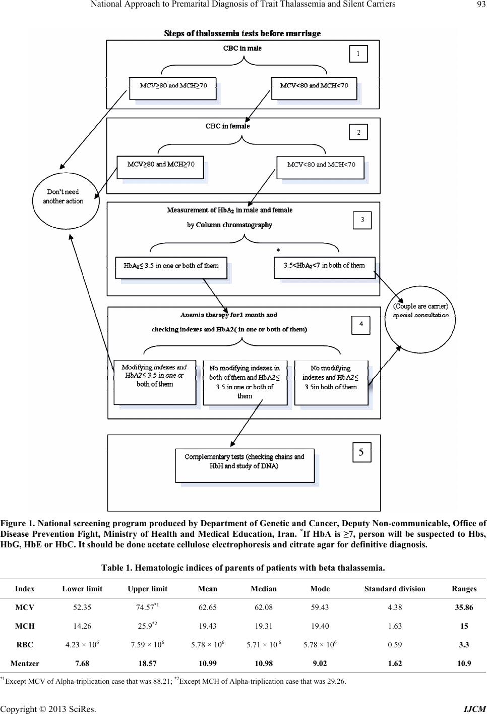 National Approach to Premarital Diagnosis of Trait Thalassemia and Silent Carriers Copyright © 2013 SciRes. IJCM 93 Figure 1. National screening program produced by Department of Genetic and Cancer, Deputy Non-communicable, Office of Disease Prevention Fight, Ministry of Health and Medical Education, Iran. *If HbA is ≥7, person will be suspected to Hbs, HbG, HbE or HbC. It should be done acetate cellulose electrophoresis and citrate agar for definitive diagnosis. Table 1. Hematologic indices of parents of patients with beta thalassemia. Ranges Standard division Mode Median Mean Upper limit Lower limit Index 35.86 4.38 59.43 62.08 62.65 74.57*1 52.35 MCV 15 1.63 19.40 19.31 19.43 25.9*2 14.26 MCH 3.3 0.59 5.78 × 10⁶ 5.71 × 10 6 5.78 × 10⁶ 7.59 × 10⁶ 4.23 × 10⁶ RBC 10.9 1.62 9.02 10.98 10.99 18.57 7.68 Mentzer *1Except MCV of Alpha-triplication case that was 88.21; *2Except MCH of Alpha-triplication case that was 29.26. 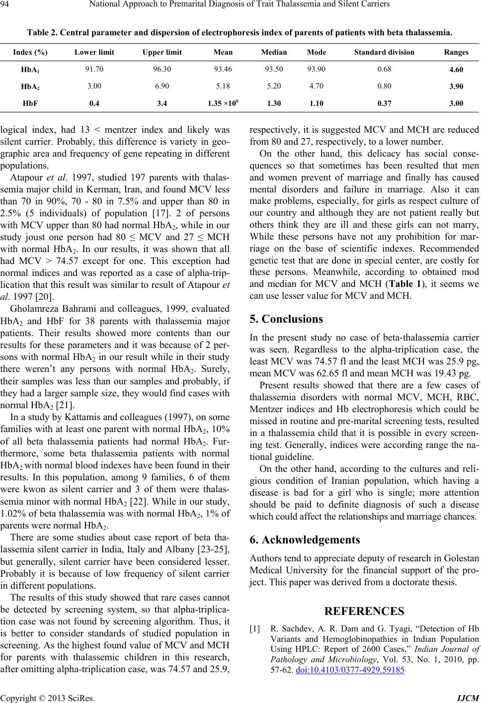 National Approach to Premarital Diagnosis of Trait Thalassemia and Silent Carriers 94 Table 2. Central parameter and dispersion of electrophoresis index of parents of patients with beta thalassemia. Ranges Standard division Mode Median Mean Upper limit Lower limit Index (%) 4.60 0.68 93.90 93.50 93.46 96.30 91.70 HbA1 3.90 0.80 4.70 5.20 5.18 6.90 3.00 HbA2 3.00 0.37 1.10 1.30 1.35 ×10⁶ 3.4 0.4 HbF logical index, had 13 < mentzer index and likely was silent carrier. Probably, this difference is variety in geo- graphic area and frequency of gene repeating in different populations. Atapour et al. 1997, studied 197 parents with thalas- semia major child in Kerman, Iran, and found MCV less than 70 in 90%, 70 - 80 in 7.5% and upper than 80 in 2.5% (5 individuals) of population [17]. 2 of persons with MCV upper than 80 had normal HbA2, while in our study joust one person had 80 ≤ MCV and 27 ≤ MCH with normal HbA2. In our results, it was shown that all had MCV > 74.57 except for one. This exception had normal indices and was reported as a case of alpha-trip- lication that this result was similar to result of Atapour et al. 1997 [20]. Gholamreza Bahrami and colleagues, 1999, evaluated HbA2 and HbF for 38 parents with thalassemia major patients. Their results showed more contents than our results for these parameters and it was because of 2 per- sons with normal HbA2 in our result while in their study there weren’t any persons with normal HbA2. Surely, their samples was less than our samples and probably, if they had a larger sample size, they would find cases with normal HbA2 [21]. In a study by Kattamis and colleagues (1997), on some families with at least one parent with normal HbA2, 10% of all beta thalassemia patients had normal HbA2. Fur- thermore, some beta thalassemia patients with normal HbA2 with normal blood indexes have been found in their results. In this population, among 9 families, 6 of them were kwon as silent carrier and 3 of them were thalas- semia minor with normal HbA2 [22]. While in our study, 1.02% of beta thalassemia was with normal HbA2, 1% of parents were normal HbA2. There are some studies about case report of beta tha- lassemia silent carrier in India, Italy and Albany [23-25], but generally, silent carrier have been considered lesser. Probably it is because of low frequency of silent carrier in different populations. The results of this study showed that rare cases cannot be detected by screening system, so that alpha-triplica- tion case was not found by screening algorithm. Thus, it is better to consider standards of studied population in screening. As the highest found value of MCV and MCH for parents with thalassemic children in this research, after omitting alpha-triplication case, was 74.57 and 25.9, respectively, it is suggested MCV and MCH are reduced from 80 and 27, respectively, to a lower number. On the other hand, this delicacy has social conse- quences so that sometimes has been resulted that men and women prevent of marriage and finally has caused mental disorders and failure in marriage. Also it can make problems, especially, for girls as respect culture of our country and although they are not patient really but others think they are ill and these girls can not marry, While these persons have not any prohibition for mar- riage on the base of scientific indexes. Recommended genetic test that are done in special center, are costly for these persons. Meanwhile, according to obtained mod and median for MCV and MCH (Table 1), it seems we can use lesser value for MCV and MCH. 5. Conclusions In the present study no case of beta-thalassemia carrier was seen. Regardless to the alpha-triplication case, the least MCV was 74.57 fl and the least MCH was 25.9 pg, mean MCV was 62.65 fl and mean MCH was 19.43 pg. Present results showed that there are a few cases of thalassemia disorders with normal MCV, MCH, RBC, Mentzer indices and Hb electrophoresis which could be missed in routine and pre-marital screening tests, resulted in a thalassemia child that it is possible in every screen- ing test. Generally, indices were according range the na- tional guideline. On the other hand, according to the cultures and reli- gious condition of Iranian population, which having a disease is bad for a girl who is single; more attention should be paid to definite diagnosis of such a disease which could affect the relationships and marriage chances. 6. Acknowledgements Authors tend to appreciate deputy of research in Golestan Medical University for the financial support of the pro- ject. This paper was derived from a doctorate thesis. REFERENCES [1] R. Sachdev, A. R. Dam and G. Tyagi, “Detection of Hb Variants and Hemoglobinopathies in Indian Population Using HPLC: Report of 2600 Cases,” Indian Journal of Pathology and Microbiology, Vol. 53, No. 1, 2010, pp. 57-62. doi:10.4103/0377-4929.59185 Copyright © 2013 SciRes. IJCM  National Approach to Premarital Diagnosis of Trait Thalassemia and Silent Carriers 95 [2] M. B. H. Soteh, H. A. Niaki, M. Kowsarian, A. Alias- gharian and A. Banihashemi, “Frequency of Beta-Globin Gene Mutations in Beta-Thalassemia Patients from East of Mazandaran,” Journal of Mazandaran University of Medical Sciences, Vol. 67, No. 18, 2008, pp. 17-25. [3] A. Ahmadi A. R. M. Khalilabadi, M. Noroozi, N. Kohan and M. Ramzi, “Evaluation of Discrimination Indices Va- lidity for Screening of β-Thalassemia Trait,” Journal of Qom University of Medical Sciences, Vol. 3, No. 3, 2009, pp. 31-36. [4] S. H. Oskie and D. G. Nathan, “The Thalassemia,” In: D. G. Nathan, S. H. Oskie, D. Ginsburg and T. Look, Eds., Hematology of Infancy and Childhood, Sunders, Philadel- phia, 2009, p. 1051. [5] H. Ehteram and B. Shafaghi, “Laboratory Findings in Pa- rents with Thalassemic Patients Referred to Imam Hos- sein Hospital 1997,” Feyz Quarterly Journal of Kashan Medical University, Vol. 13, 2000, pp. 81-88. [6] J. N. Luknes, “The Thalassemia and Related Disorders,” In: G. R. Lee, J. Foerester, J. N. Luknes, J. P. P. Greer and G. Rodgers, Eds., Wintrobes Clinical Hematology, Sunders, Philadelphia, 1999, pp. 1421-1430. [7] E. Schwartz, “The Silent Carrier of Beta Thalassemia,” The New England Journal of Medicine, Vol. 281, No. 24, 1969, pp. 1327-1333. doi:10.1056/NEJM196912112812403 [8] M. Aksoy, G. Dincol and S. Erdem, “Different Types of Beta-Thalassemia Intermedia,” Acta Haematologica, Vol. 59, No. 3, 1978, pp. 178-189. doi:10.1159/000207760 [9] C. Kattamis, A. Metaxotou-Mavromati, W. G. Wood, J. R. Nash and D. J. Weatherall, “The Heterogeneity of Normal Hb A2-b Thalassaemia,” British Journal of Haematology, Vol. 42, No. 1, 1979, pp. 109-123. [10] G. Aicardi, A. Naselli, G. V. Sciarratta and G. Sansone, “The Silent Carrier of Beta Thalassemia: Interaction with the Typical Beta Thalassemia Trait,” Blut, Vol. 38, No. 6, 1979, pp. 473-478. doi:10.1007/BF01013508 [11] M. Aksoy, G. Bermek, G. Almis and A. Kutlar, “β-Tha- lassemia Intermedia Homozygous for Normal Hemoglo- bin A2 b Thalassemia. Study in Four Families,” Acta Ha- ematologica, Vol. 67, No. 1, 1982, pp. 57-61. doi:10.1159/000207025 [12] G. V. Sciarratta, M. I. Parodi, S. F. A. Vallerino and G. Sansone, “The Silent Carrier of β-Thalassemia,” Annals of the New York Academy of Sciences, Vol. 445, 1984, pp. 111-118. doi:10.1111/j.1749-6632.1985.tb17180.x [13] I. Bianco, M. P. Cappabianca, E. Foglietta, M. Lerone, G. Deidda, L. Morlupi, et al., “Silent Thalassemias: Geno- types and Phenotypes,” Haematologica, Vol. 82, No. 3, 1997, pp. 269-280. [14] S. H. Oskie and D. G. Nathan, “The Thalassemia,” In: D. G. Nathan, S. H. Oskie, D. Ginsburg and T. Look, Eds., Hematology of Infancy and Childhood, Sunders, Phila- delphia, 2003, pp. 826-883. [15] J. B. Edward, “Hemoglobin Disorders,” In: D. L. Kasper, A. S. Fauci, D. L. Longo, E. Braunwald, S. L. Hauser, J. L. Jameson and J. Loscalzo, Eds., Harrison’s Haematol- ogy and Oncology, Nasle Farda Publication, Tehran, 2005, pp. 105-114. [16] A. Samavat and B. Modell, “Iranian National Thalassae- mia Screening Programme,” British Medical Journal, Vol. 329, No. 7475, 2004, pp. 1134-1137. doi:10.1136/bmj.329.7475.1134 [17] A. Cao and R. Galanello, “Beta-Thalassemia,” Genetics in Medicine, Vol. 12, No. 2, 2010, pp. 61-76. doi:10.1097/GIM.0b013e3181cd68ed [18] M. Kosaryan, A. Okhvatian and F. Babamahmoodi, “How Much We Have Been Successful in Nationwide Preven- tive Program for Beta-Thalassemia in Mazandaran Uni- versity of Medical Sciences in 1992-2002,” Journal of Mazandaran University of Medical Sciences, Vol. 4, No. 13, 2004, pp. 47-54. [19] M. Karimi, S. Johari and N. Cohan, “Attitude toward Prenatal Diagnosis for β-Thalassemia Major and Medical Abortion in Southern Iran,” Hemoglobin, Vol. 34, No. 1, 2010, pp. 49-54. doi:10.3109/03630260903547690 [20] M. Atapour, A. Zohoor, A. Zolali and A. Arjmand, “He- moglobin A2 Percentage in Definite Carriers of Beta Tha- lassemia Major Patients Referred to Especial Disease Center in Kerman,” Journal of Kerman University of Me- dical Sciences, Vol. 10, No. 2, 2003, pp. 99-104. [21] G. H. Bahrami, H. Rahi and H. Dehghan, “Hemoglobin A2 and F Levels and Hematologic Parameters in Patients of Transfusion-Dependent Thalassemia Major Patients,” Journal of Kermanshah University of Medical Sciences, Vol. 5, No. 2, 2001, pp. 6-12. [22] C. Kattamis, A. Metaxotou Mavomati, W. G. Wood and J. R. Nash, “The Heterogeneity of Normal HbA2-Beta Tha- lassemia in Greec,” British Journal of Hematology, Vol. 42, No. 1, 1997, pp. 109-123. doi:10.1111/j.1365-2141.1979.tb03703.x [23] G. Aicardi, A. Naselli and F. Sciarratta, “Silent Carrier of β Thalassemia: Interaction with Typical Beta Thalassemia Trait,” Blut, Vol. 38, No. 6, 1979, pp. 473-478. doi:10.1007/BF01013508 [24] G. L. Semenza, K. Delgross, P. Malladi and Z. Schwart, “The Silent Carrier Allele: Beta Thalassemia a without Mu- tation in the Beta Globin Gene or Its Immediate Flank- ing Regions,” Cell, Vol. 39, No. 1, 1984, pp. 123-128. doi:10.1016/0092-8674(84)90197-1 [25] A. Nad Korin, A. M. Pawar, V. C. Mudera, D. M. Mo- hanty and R. Colah, “Clinical Heterogeneity of Silent Gene β-Thalassemia among India,” Annals of Hematol- ogy, Vol. 70, No. 1, 1995, pp. 61-64. Copyright © 2013 SciRes. IJCM |