

Paper Menu >>
Journal Menu >>
 Journal of Surface Engineered Materials and Advanced Technology, 2013, 3, 49-54 http://dx.doi.org/10.4236/jsemat.2013.31A007 Published Online February 2013 (http://www.scirp.org/journal/jsemat) 49 Advances in Probing Wood-Coating Interface by Microscopy: A Review Adya P. Singh1*, Byung-Dae Park1, Arif Nuryawan1, Menda Kazayawoko2 1Department of Wood Science and Technology, Kyungpook National University, Daegu, South Korea; 2Departement de Gestion des Ressources Naturelles, Faculte des Sciences Agronomiques, Universite de Kinshasa, Kinshasa, Democratic Republic of the Congo. Email: *[email protected] Received December 18th, 2012; revised January 20th, 2013; accepted January 28th, 2013 ABSTRACT Surface coatings provide protection to wood products against weathering and other deteriorating factors, such as mois- ture uptake and microbial invasion. The effectiveness of coatings depends on many factors, including how well the ap- plied coatings adhere to the wood surface. Coating adhesion to wood involves both chemical and physical interactions between the coating and wood tissues in contact, and the particular focus of this mini-review will be on the advances being made in understanding the physical aspects of the interaction by probing wood-coating interface using novel and high resolution imaging techniques, including confocal laser scanning microscopy (CLSM), SEM-backscattered elec- tron imaging and correlative microscopy employing light, confocal and scanning electron microscopy. Keywords: Wood-Coating Interface; Coating Adhesion; Correlative Microscopy; Backscattered Electron Imaging; Confocal Laser Scanning Microscopy 1. Introduction Wood products exposed in service under outdoor condi- tions can deteriorate from the damaging effects of solar radiation, which can cause weathering of cell walls [1- 10], and cell wall degradation by microorganisms [11,12]. Application of suitable coatings to the product surface can significantly extend the service life by preventing solar radiation from reaching the wood surface and or- ganisms and moisture to come in direct contact with the underlying wood tissues. Among the factors that influ- ence coating performance coating adhesion to wood is of major importance [13], which primarily depends on coat- ing penetration into wood [14] and mechanical anchorage through entanglement of coatings within penetrated sur- face and subsurface layers of wood tissues [15,16]. Ex- amining wood-coating interface is therefore important to understand the nature of wood-coating interaction, in- formation that can be helpful in evaluating adhesion and performance of applied coatings and ultimately in opti- mizing formulations and processes to achieve high dura- bility of wood-coating composites. Microscopy has been the most widely applied tool to examine and image wood-coating interface. Whereas the majority microscopic studies have aimed to record in- formation on depth to which the applied coatings can penetrate into wood products made from different wood types as well as those variously surfaced [14,17-24], specialized microscopic techniques have also been de- veloped and employed that can resolve wood-coating interface more clearly, providing improved understand- ing of wood-coating interaction at cellular and sub- cellular levels [15,16,25]. In this mini-review the focus will be on these technical advances in probing wood- coating interface, often involving a combination of dif- ferent types of microscopes that differ in their imaging capabilities and can provide complementary information. The information presented is in a broader context to in- clude places of physical contact of coatings with the sur- faces of wood products to which they are applied as well as all contact interfaces between penetrated coatings and cell walls around cell lumens and lining cell wall cracks within tissue layers underneath the surface. 2. Novel Microscopic Approaches for Imaging Wood-Coating Interface The importance of coating adhesion for coating durabil- ity and performance has been recognized for a long time. Among the factors that influence coating adhesion to wood surface, coating penetration into and entanglement within surface and subsurface tissues are important, as improvements in water repellency result in greater di- mensional stability and less cracking of wood in outdoor exposures, aspects that have been emphasized in several *Corresponding a uthor. Copyright © 2013 SciRes. JSEMAT 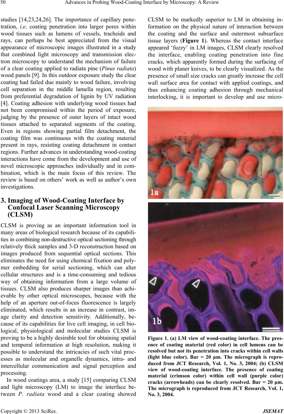 Advances in Probing Wood-Coating Interface by Microscopy: A Review 50 studies [14,23,24,26]. The importance of capillary pene- tration, i.e. coating penetration into larger pores within wood tissues such as lumens of vessels, tracheids and rays, can perhaps be best appreciated from the visual appearance of microscopic images illustrated in a study that combined light microscopy and transmission elec- tron microscopy to understand the mechanism of failure of a clear coating applied to radiata pine (Pinus radiata) wood panels [9]. In this outdoor exposure study the clear coating had failed due mainly to wood failure, involving cell separation in the middle lamella region, resulting from preferential degradation of lignin by UV radiation [4]. Coating adhesion with underlying wood tissues had not been compromised within the period of exposure, judging by the presence of outer layers of intact wood tissues attached to separated segments of the coating. Even in regions showing partial film detachment, the coating film was continuous with the coating material present in rays, resisting coating detachment in contact regions. Further advances in understanding wood-coating interactions have come from the development and use of novel microscopic approaches individually and in com- bination, which is the main focus of this review. The review is based on others’ work as well as author’s own investigations. 3. Imaging of Wood-Coating Interface by Confocal Laser Scanning Microscopy (CLSM) CLSM is proving as an important information tool in many areas of biological research because of its capabili- ties in combining non-destructive optical sectioning through relatively thick samples and 3-D reconstruction based on images produced from sequential optical sections. This eliminates the need for using chemical fixation and poly- mer embedding for serial sectioning, which can alter cellular structures and is a time-consuming and tedious way of obtaining information from a large volume of tissues. CLSM also produces sharper images than achi- evable by other optical microscopes, because with the help of an aperture out-of-focus fluorescence is largely eliminated, which results in an increase in contrast, im- age clarity and detection sensitivity. Additionally, be- cause of its capabilities for live cell imaging, in cell bio- logical, physiological and molecular studies CLSM is proving to be a highly desirable tool for ob taining spatial and temporal information at high resolution, making it possible to understand the intricacies of such vital proc- esses as molecular and organelle dynamics, intra- and intercellular communication and signal perception and processing. In wood coatings area, a study [15] comparing CLSM and light microscopy (LM) to image the interface be- tween P. radiata wood and a clear coating showed CLSM to be markedly superior to LM in obtaining in- formation on the physical nature of interaction between the coating and the surface and outermost subsurface tissue layers (Figure 1). Whereas the contact interface appeared ‘fuzzy’ in LM images, CLSM clearly resolved the interface, enabling coating penetration into fine cracks, which apparently formed during the surfacing of wood with planer knives, to be clearly visualized. As the presence of small size cracks can greatly increase the cell wall surface area for contact with applied coatings, and thus enhancing coating adhesion through mechanical interlocking, it is important to develop and use micro- Figure 1. (a) LM view of wood-coating interface. The pres- ence of coating material (red color) in cell lumens can be resolved but not its penetration into cracks within cell walls (light blue color). Bar = 20 μm. The micrograph is repro- duced from JCT Research, Vol. 1, No. 3, 2004; (b) CLSM view of wood-coating interface. The presence of coating material (crimson color) within cell wall (purple color) cracks (arrowheads) can be clearly resolved. Bar = 20 μm. The micrograph is reproduced from JCT Research, Vol. 1, No. 3, 2004. Copyright © 2013 SciRes. JSEMAT 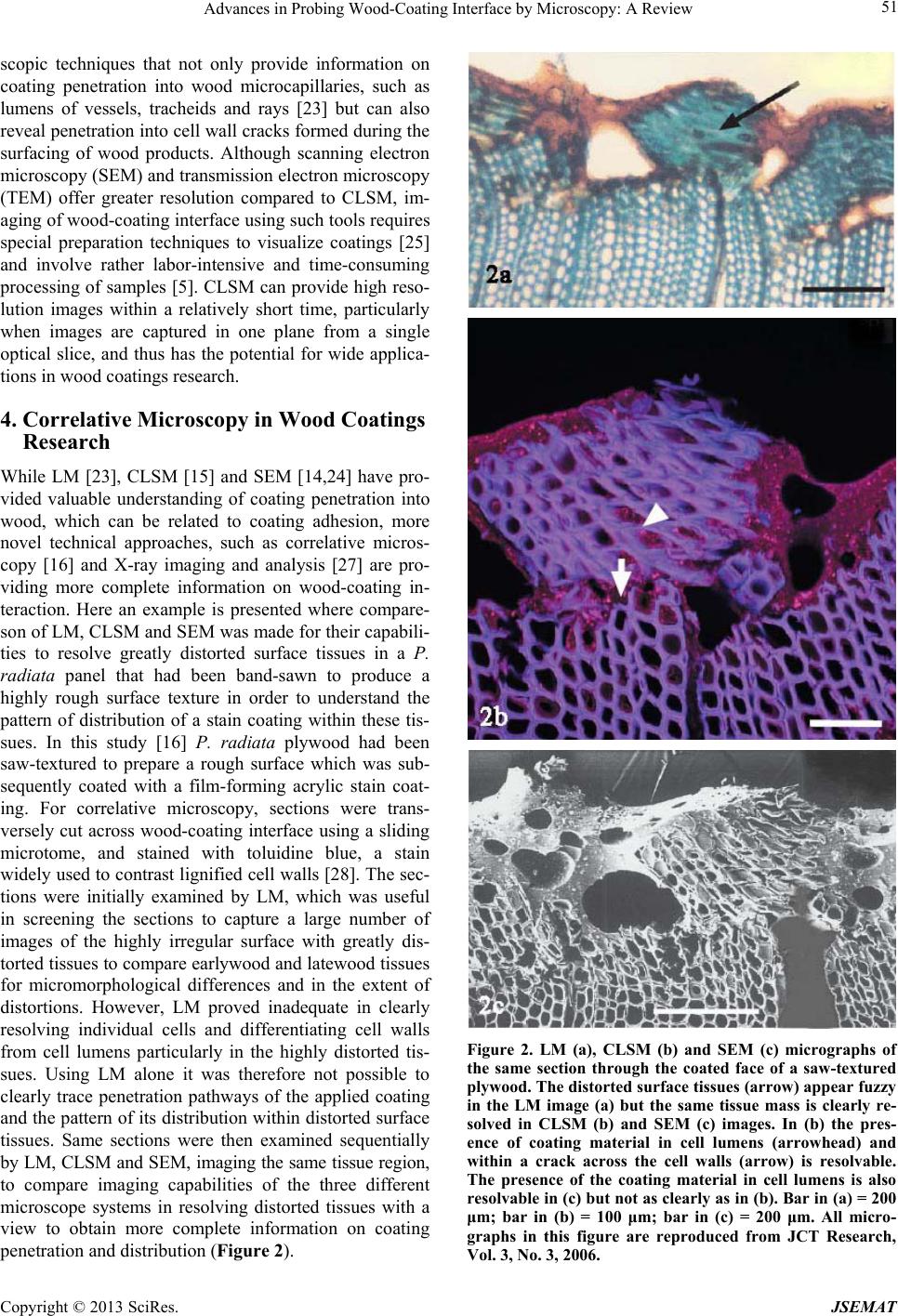 Advances in Probing Wood-Coating Interface by Microscopy: A Review 51 scopic techniques that not only provide information on coating penetration into wood microcapillaries, such as lumens of vessels, tracheids and rays [23] but can also reveal penetration into cell wall cracks formed during the surfacing of wood products. Although scanning electron microscopy (SEM) and transmission electron microscopy (TEM) offer greater resolution compared to CLSM, im- aging of wood-coating interface using such tools requires special preparation techniques to visualize coatings [25] and involve rather labor-intensive and time-consuming processing of samples [5]. CLSM can provide high reso- lution images within a relatively short time, particularly when images are captured in one plane from a single optical slice, and thus has the potential for wide applica- tions in wood coatings research. 4. Correlative Microscopy in Wood Coatings Research While LM [23], CLSM [15] and SEM [14,24] have pro- vided valuable understanding of coating penetration into wood, which can be related to coating adhesion, more novel technical approaches, such as correlative micros- copy [16] and X-ray imaging and analysis [27] are pro- viding more complete information on wood-coating in- teraction. Here an example is presented where compare- son of LM, CLSM and SEM was made for their capabili- ties to resolve greatly distorted surface tissues in a P. radiata panel that had been band-sawn to produce a highly rough surface texture in order to understand the pattern of distribution of a stain coating within these tis- sues. In this study [16] P. radiata plywood had been saw-textured to prepare a rough surface which was sub- sequently coated with a film-forming acrylic stain coat- ing. For correlative microscopy, sections were trans- versely cut across wood-coating interface using a sliding microtome, and stained with toluidine blue, a stain widely used to contrast lignified cell walls [28]. Th e sec- tions were initially examined by LM, which was useful in screening the sections to capture a large number of images of the highly irregular surface with greatly dis- torted tissues to compare earlywood and latewood tissues for micromorphological differences and in the extent of distortions. However, LM proved inadequate in clearly resolving individual cells and differentiating cell walls from cell lumens particularly in the highly distorted tis- sues. Using LM alone it was therefore not possible to clearly trace penetration pathways of the applied coating and the pattern of its distribution within distorted surface tissues. Same sections were then examined sequentially by LM, CLSM and SEM, imaging the same tissue region, to compare imaging capabilities of the three different microscope systems in resolving distorted tissues with a view to obtain more complete information on coating penetration and distribution (Figure 2). Figure 2. LM (a), CLSM (b) and SEM (c) micrographs of the same section through the coated face of a saw-textured plywood. The distorted surface tissues (arrow) appear fuzzy in the LM image (a) but the same tissue mass is clearly re- solved in CLSM (b) and SEM (c) images. In (b) the pres- ence of coating material in cell lumens (arrowhead) and within a crack across the cell walls (arrow) is resolvable. The presence of the coating material in cell lumens is also resolvable in (c) but not as clearly as in (b). Bar in (a) = 200 μm; bar in (b) = 100 μm; bar in (c) = 200 μm. All micro- graphs in this figure are reproduced from JCT Research, Vol. 3, No. 3, 2006. Copyright © 2013 SciRes. JSEMAT 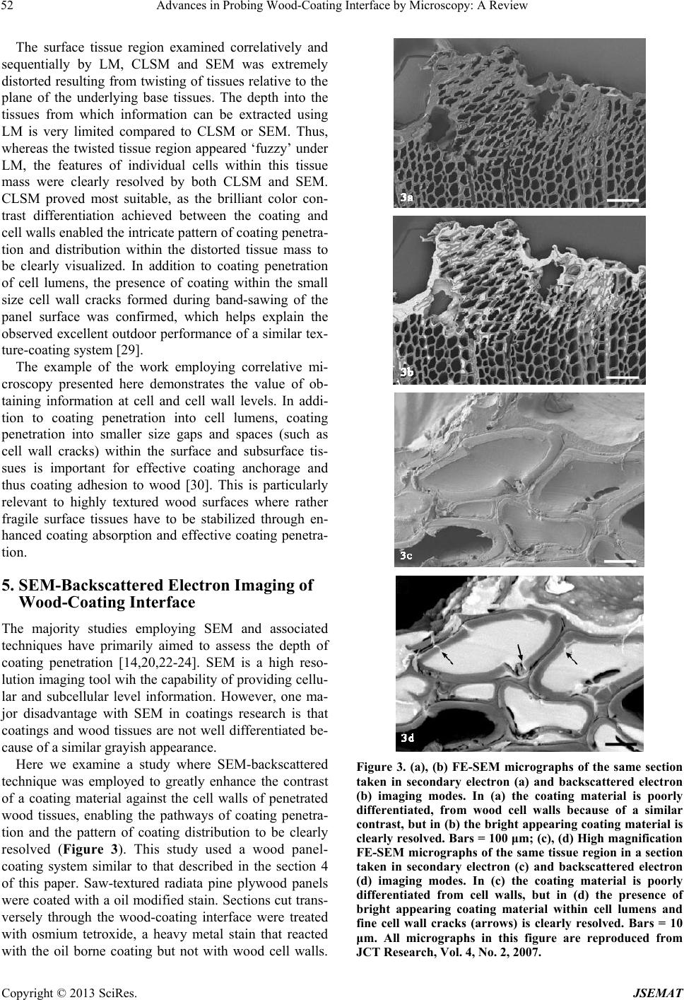 Advances in Probing Wood-Coating Interface by Microscopy: A Review 52 The surface tissue region examined correlatively and sequentially by LM, CLSM and SEM was extremely distorted resulting from twisting of tissues relative to the plane of the underlying base tissues. The depth into the tissues from which information can be extracted using LM is very limited compared to CLSM or SEM. Thus, whereas the twisted tissue region appeared ‘fuzzy’ under LM, the features of individual cells within this tissue mass were clearly resolved by both CLSM and SEM. CLSM proved most suitable, as the brilliant color con- trast differentiation achieved between the coating and cell walls enabled the intricate pattern of coatin g pene tra- tion and distribution within the distorted tissue mass to be clearly visualized. In addition to coating penetration of cell lumens, the presence of coating within the small size cell wall cracks formed during band-sawing of the panel surface was confirmed, which helps explain the observed excellent outdoor performance of a similar tex- ture-coating system [29]. The example of the work employing correlative mi- croscopy presented here demonstrates the value of ob- taining information at cell and cell wall levels. In addi- tion to coating penetration into cell lumens, coating penetration into smaller size gaps and spaces (such as cell wall cracks) within the surface and subsurface tis- sues is important for effective coating anchorage and thus coating adhesion to wood [30]. This is particularly relevant to highly textured wood surfaces where rather fragile surface tissues have to be stabilized through en- hanced coating absorption and effective coating penetra- tion. 5. SEM-Backscattered Electron Imaging of Wood-Coating Interface The majority studies employing SEM and associated techniques have primarily aimed to assess the depth of coating penetration [14,20,22-24]. SEM is a high reso- lution imaging tool wih the capability of pro viding cellu- lar and subcellular level information. However, one ma- jor disadvantage with SEM in coatings research is that coatings and wood tissues are not well differentiated be- cause of a similar grayish appearance. Here we examine a study where SEM-backscattered technique was employed to greatly enhance the contrast of a coating material against the cell walls of penetrated wood tissues, enabling the pathways of coating penetra- tion and the pattern of coating distribution to be clearly resolved (Figure 3). This study used a wood panel- coating system similar to that described in the section 4 of this paper. Saw-textured radiata pine plywood panels were coated with a oil modified stain. Sections cut trans- versely through the wood-coating interface were treated with osmium tetroxide, a heavy metal stain that reacted with the oil borne coating but not with wood cell walls. Figure 3. (a), (b) FE-SEM micrographs of the same section taken in secondary electron (a) and backscattered electron (b) imaging modes. In (a) the coating material is poorly differentiated, from wood cell walls because of a similar contrast, but in (b) the bright appearing coating material is clearly resolved. Bars = 100 μm; (c), (d) High magnification FE-SEM micrographs of the same tissue region in a section taken in secondary electron (c) and backscattered electron (d) imaging modes. In (c) the coating material is poorly differentiated from cell walls, but in (d) the presence of bright appearing coating material within cell lumens and fine cell wall cracks (arrows) is clearly resolved. Bars = 10 μm. All micrographs in this figure are reproduced from JCT Research, Vol. 4, No. 2, 2007. Copyright © 2013 SciRes. JSEMAT  Advances in Probing Wood-Coating Interface by Microscopy: A Review 53 The osmium treated sections were first imaged with SEI (secondary electron imaging) mode of a FE-SEM (field emission scanning electron microscope), and the same sections were then viewed under BEI (backscattered electron imaging) mode. Comparison of the two imaging modes using the same sections showed a distinct advan- tage of the BEI imaging over the SEI imaging. Whereas in the SEI mode the coating was poorly differentiated from wood tissues, in the BEI mode the coating material appeared bright and thus was readily differentiated from wood tissues, which enabled the intricate pathway of coating penetration and distribution within the highly distorted saw-textured surface tissues to be readily ex- amined, including penetration of th e coating material into very small size cell wall cracks present within these tis- sues. Osmium tetroxide staining of the coating material was a critical step in the process. Backscattered electron imaging provides information on the presence and location of high atomic number sub- stances within a specimen, which appear bright under this imaging mode due to high yields of backscattered electron signals, and this was the basis for coupling the heavy metal osmium tetroxide with the coating . Osmium tetroxide has been used previously as a stain or tracer in other systems involving backscattered electron imaging [31,32], but in the work described above it was the first time backscattered electron imaging was used to under- take high resolution imaging work on the penetration and distribution of a coating into wood. Similar approach can also work for examining other coating systems with wood tissues, provided suitable high atomic number sub- stances can be employed that can specifically react with the coatings of interest, and without affinity for wood cell walls. 6. Conclusion While light microscopy has been useful in determining the depth of coating penetration into wood, it is not suit- able for examining wood-coating interface because of its inability to clearly resolve particularly the interface be- tween the outermost surface layer of wood tissues and the applied coating. The novel and more specific mi- croscopy approaches employed in more recent years are proving valuable in wood-coating interaction studies, particularly involving rough-texture wood surfaces con- taining hig hl y di st ort ed tissues. 7. Future Prospects The technical advances discussed in imaging wood- coating interface are providing greater understanding of the physical aspects of wood-coating interaction. Com- bined use of microscopy techniques with complementary capabilities can provide more complete information, and in future wood-coating studies th e scope of this approach should expand . The majority microscopic studies undertaken have mainly focused on examining depth of coating penetra- tion. While understanding of this is impor tant in the con- text of evaluation of coating adhesion, higher resolution imaging should be combined to understand particularly the effect of surface preparation on wood tissues at finer levels. As documented by CLSM [15], even relatively smooth surfacing with planer knives can generate cracks of varying dimensions within cell walls, which collec- tively can significantly increase cell wall surface areas, thus enhancing the anchorage of penetrated coating ma- terial. While LM can still serve as the basic imaging tool for rapid assessment of coating penetration and distribu- tion, the scope in future studies should widen to include the assessment using also tools that can provide 3-D im- aging capability and high resolution, such as CLSM, FE-SEM and TEM, which in addition to revealing the finer aspects of wood-coating interaction can provide useful information for assessing coating performance, such as demonstrated in a study employing TEM [5]. 8. Acknowledgements This work was supported by the Korean Ministry of Education, Science and Technology and the Korean Federation of Science and Techno logy Societies. REFERENCES [1] R. M. Rowell, W. C. Feist and W. D. Ellis, “Weathering of Chemically Modified Southern Pine,” Wood Science, Vol. 13, No. 4, 1981, pp. 202-208. [2] D. N.-S. Hon, “Weathering Reactions and Protection of Wood Surfaces,” Journal of Applied Polymer Science, Vol. 37, No. 2, 1983, pp. 845-864. [3] W. C. Feist, R. M. Rowell and J. Youngquist, “Weather- ing and Finish Performance of Acetylated Aspen Fiber- board,” Wood an d Fiber Science, Vol. 23, No. 2, 1 991, pp. 260-272. [4] M. Kuo and H. N. Hu, “Ultrastructural Changes of Photo- degradation of Wood Surfaces Exposed to UV Light,” Holzforschung, Vol. 45, No. 5, 1991, pp. 347-353. doi:10.1515/hfsg.1991.45.5.347 [5] A. P. Singh, E. A. Dunningham and D. V. Plackett, “As- sessing the Performance of a Commercial Wood Stain by Transmission Electron Microscopy,” Holzforschung, Vol. 49, No. 3, 1995, pp. 255-258. [6] P. D. Evans, P. D. Thay and K. J. Sc hmalzl, “Degra dation of Wood Surfaces During Natural Weathering. Effect of Ligni n and Cellulose a nd on the Adhesion of Acrylic La- tex Primers,” Wood Science and Technology, Vol. 30, No. 6, 1996, pp. 411-422. doi:10.1007/BF00244437 [7] D. V. Plackett, E. A. Dunningham and A. P. Singh, “We a t h er i n g o f C h e mi c a l ly Modified Wood,” I n : D . N. - S . Copyright © 2013 SciRes. JSEMAT  Advances in Probing Wood-Coating Interface by Microscopy: A Review Copyright © 2013 SciRes. JSEMAT 54 Hon, Ed., Chemical Modification of Lignocellulosic Ma- terials, Marcel Dekker, Inc., New York, 1996, pp. 277- 294. [8] R. S. Williams, M. T. Knaebe and W. C. Feist, “Erosion Rates of Wood during Natural Weathering. Part II. Ear- lywood and Latewood Erosion Rates,” Wood and Fiber Science, Vol. 33, No. 1, 2001, pp. 43-49. [9] A. P. Singh and B. S. W. Dawson, “The Mechanism of Failure of Clear Coated Wooden Boards as Revealed by Microscopy,” IAWA Journal, Vol. 24, No. 1, 2003, pp. 1- 11. [10] P. D. Evans, “Weathering of Wood and Wood Compos- ites,” In: R. M. Rowell, Ed., Handbook of Wood Chemis- try and Wood Composites, 2nd Edition, CRC Press, Tay- lor and Francis Group, Boca Raton, 2013, pp. 151-216. [11] R. A. Bl anc hette, T . N ilsson, G. F. Daniel and A. R. Ab ad, “Biological Degradation of Wood,” In: R. M. Rowell and J. Barbour, Eds., Archaeological Wood: Properties, Che- mistry and Preservation, American Chemical Society, Washington DC, 1990, pp. 147-174. [12] A. P. Singh and J. A. Butcher, “Bacterial Degradation of Wood Cell Walls: A Review of Degradation Patterns,” Journal of the Institute of Wood Science, Vol. 12, 1991, pp. 143-157. [13] R. S. Williams and W. C. Feist, “Durability of Paint or Solid-Color Stain Applied to Preweathered Wood,” For- est Products Journal, Vol. 43, No. 1, 1993, pp. 8-14. [14] M. de Meijer and H. Militz, “Wet Adhesion of Low-VOC Coatings on Wood: A Quantitative Analysis,” Progress in Organic Coatings, Vol. 38, No. 3-4, 2000, pp. 223-240. doi:10.1016/S0300-9440(00)00108-9 [15] A. P. Singh and B. S. W. Dawson, “Confocal Micro- scope—A Valuable Tool For Examining Wood-Coating Interface,” Journal of Coatings Technology and Research, Vol. 1, No. 3, 2004, pp. 235-237. doi:10.1007/s11998-004-0017-z [16] A. P. Singh and B. S. W. Dawson, “Microscopic Assess- ment of the Effect of Saw-Textured Pinus radiata Ply- wood Surface on the Distribution of a Film-Forming Acrylic Stain,” Journal of Coatings Technology and Re- search, Vol. 3, No. 3, 2006, pp. 193-201. doi:10.1007/BF02774508 [17] M. H. Schneider and W. A. Côté, “Studies of Wood and Coating Interactions Using Fluorescence Microscopy and Pyrolysis Gas-Liquid Chromatography,” Journal of Paint Technology, Vol. 39, No. 511, 1967, pp. 465-471. [18] W. A. Côté and R. G. Robinson, “A Comparative Study of Wood-Coating Interaction Using Incident Fluorescence and Transmitted Fluorescence Microscopy,” Journal of Paint Technology, Vol. 40, No. 525, 1968, pp. 427-432. [19] M. H. Schneider, “Coating Penetration into Wood Sub- stance Studied with Electron Microscopy Using Replica Techniques,” Journal of Paint Technology, Vol. 42, No. 547, 1970, pp. 457-460. [20] M. H. Schneider, “Scanning Electron Microscope Study of a Coating Component Deposited from Solution into Wood,” Journal of the Oil and Color Chemists Associa- tion, Vol. 62, No. 11, 1979, pp. 441-444. [21] M. H. Schneider, “Microscopic Distribution of Linseed Oil after Application to Wood Surface,” Journal of Coat- ings Technology, Vol. 52, No. 665, 1980, pp. 64-67. [22] R. M. Nussbaum, “Penetration of Water-Borne Alkyd Emulsions and Solvent Borne Alkyds into Wood,” Holz als Roh-und Werkstoff, Vol. 52, No. 6, 1994, pp. 389-393. doi:10.1007/BF02615391 [23] M. de Meijer, K. Thurich and H. Militz, “Comparative Study on Penetration Characteristics of Modern Wood Coatings,” Wood Science and Technology, Vol. 32, No. 5, 1998, pp. 347-365. doi:10.1007/BF00702791 [24] R. E. Hernández and J. Cool, “Evaluation of Three Sur- facing Methods on Paper Birch Wood in Relation to Wa- ter- and Solvent-Borne Coating Performance,” Wood and Fibe r Science, Vol. 40, No. 3, 2008, pp. 459-469. [25] A. P. Singh, A. Ratz and B. S. W. Dawson, “A Novel Method for High Resolution Imaging of Coating Distri- bution within a Rough Textured Plywood Surface,” Jour- nal of Coatings Technology and Research, Vol. 4, No. 2, 2007, pp. 207-210. doi:10.1007/s11998-007-9019-y [26] L. F. de Moura and R. E. Hernández, “Evaluation of Var- nish Coating Performance for Two Surfacing Methods on Sugar Maple Wood,” Wood and Fiber Science, Vol. 37, No. 2, 2005, pp. 355-366. [27] J. Van den Bulcke, M. Boone, J. Van Acker and L. Van Hoorebeke, “High-Resolution X-Ray Imaging and Analy- sis of Coatings on and in Wood,” Journal of Coatings Technology and Research, Vol. 7, No. 2, 2010, pp. 271- 277. doi:10.1007/s11998-009-9182-4 [28] T. P. O’Brien and M. E. McCully, “Plant Structure and Development: A Pictorial and Physiological Approach,” The Macmillan Company/Collier-Macmillan Ltd., London, 1969. [29] R. S. Williams and W. C. Feist, “Effect of Preweathering, Surface Roughness and Wood Species on the Perform- ance of Paints and Stains,” Journal of Coatings Tech- nology, Vol. 66, No. 828, 1994, p. 109. [30] B. S. W. Dawson, S. Gallagher and A. P. Singh, “Micro- scopic View of Wood and Coating Interaction and Coat- ing Performance on Wood,” Forest Research Bullet in, No. 228, New Zealand Forest Research Institute, Rotorua, 2003. [31] P. B. DeNee and R. L. Carpenter, “Application of Heavy Metal Staining (OsO4)/Backscattered Electron Imaging Technique to the Study of Organic Aerosols,” Proceed- ings of the 14th Annual Conference of the Microbeam Analysis Society, San Antonio, 1979, pp. 8-10. [32] D. E. Schraufnagel and D. P. Ganesan, “Tracers in Vas- cular Casting Resins Enhance Backscattering Brightness,” Scanning Microscopy, Vol. 12, No. 4, 1998, pp. 631-639. |