

Paper Menu >>
Journal Menu >>
 International Journal of Medical Physics, Clinical Engineering and Radiation Oncology, 2013, 2, 39-43 Published Online February 2013 (http://www.scirp.org/journal/ijmpcero) http://dx.doi.org/10.4236/ijmpcero.2013.21006 Copyright © 2013 SciRes. IJMPCERO Endovascular Treatment Options of Acute Limb Ischemia Di Zhang, Wensheng Lou, Guoping Chen, Xindao Yin, Jianping Gu Department of Radiation Physics, Nanjing Hospital, Nanjing Medical University, Nanjing, China Email: [email protected], [email protected] Received October 14, 2012; revised November 15, 2012; accepted November 22, 2012 ABSTRACT Acute limb ischemia is an urgent condition which occurs when there is an abrupt interruption of blood flow into an ex- tremity usually because of either embolic or thrombotic vascular occlusion. Restoration of perfusion through early in- tervention can decrease amputation and mortality. Contemporary treatment includes both surgery and endovascular te- chniques. There is a rapid progress in endovascular intervention therapy. This article aims to make a comprehensive review of the endovascular intervention options of acute limb ischemia. Keywords: Acute Limb Ischemia; Arterial Embolus; Arterial Thrombosis; Endovascular Therapy 1. Introduction Acute limb ischemia (ALI) of the lower extremities re- mains a challenging clinical dilemma, which occurs when there is an abrupt interruption of blood flow into an ex- tremity usually because of either embolic or thrombotic vascular occlusion [1,2]. The major clinical sign of ALI is “6P”, i.e., pain, parasthesia, paralysis, pulselessness, pallor and poikilothermia [3]. The overall prognosis is poor, the mortality rate was 5.3% - 12%, amputation rate was 5.3% - 14% [1,4-6]. When profound ischemia ensues, this represents an emergency in which restoration of per- fusion through early intervention can lead to limb sal- vage, whereas delay may result in significant morbidity, including limb loss and, potentially, death. Therefore, timely and correctly treatment of this disease is impor- tant. There are three management options in acute lower limb ischemia: 1) clot removal by catheter-directed thro- mbolysis with or without percutaneous mechanical thro- mbectomy; 2) surgical thromboembolectomy followed by correction of underlying arterial lesions; and 3) anti- coagulation with continued observation. We can catego- rize the level of the patient’s limb ischemia utilizing cli- nical assessment of motor and sensory function and in- terrogating ankle arterial flow velocity signals into seve- ral levels [7]. As shown in Table 1, those with level I ischemia, particularly if they have significant comorbi- dities, can and should be treated with heparin and ob- servation, being watched closely while attention is given to treating associated comorbidities. Conversely, in pa- tients with the same level of ischemia (I), i.e., active pa- tients without significant comorbidities, it is practical to proceed directly with endovascular revascularization (CDT and, possibly, percutaneous mechanical thrombec- tomy). This same management choice is equally appro- priate for those at level IIA, and at both of these levels of ischemia there should be sufficient time for restoration of patency using either endovascular or open surgical tech- niques [8]. In the past, the decisional breakpoint in choosing between endovascular and surgical revascu- larization came between class IIA and class IIB, Treat- ment of ALI has shifted toward endovascular therapies because of rapidly improving technology and delivery systems unless there is a contraindication, such as pro- found critical limb ischemia, renal dysfunction, or con- trast allergy. Regardless of which kind of therapy to be chosen, the patient should be immediately anticoagu- lated to prevent further clot extension. This article aims to make a comprehensive review of the endovascular op- tions with intra-arterial thrombolysis and/or adjuvant en- dovascular techniques. 2. Catheter Directed Thrombolysis (CDT) The development of effective medications to dissolve oc- cluding thrombus led to the increasing use of throm- bolytic therapy in patients with ALI. Randomized control trials have provided a rationale for thrombolysis as a first step in patients with ALI vs immediate operative revas- cularization. Catheter directed thrombolysis offers seve- ral potential advantages to lessen the pitfalls of open techniques. By utilizing an endovascular approach and local anesthesia, the risks of general anesthesia are mini- mized within a subgroup of patients that are at their physiologic limits [9]. In addition, enzymatic dissolution of thrombus allows for more effective clot resolution particularly within distal arterial beds that are often re- 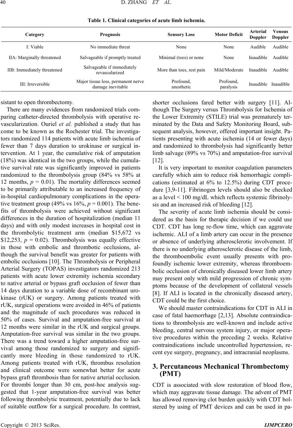 D. ZHANG ET AL. 40 Table 1. Clinical categories of acute limb ischemia. Category Prognosis Sensory Loss Motor Deficit Arterial Doppler Venous Doppler I: Viable No immediate threat None None AudibleAudible IIA: Marginally threatened Salvageable if promptly treated Minimal (toes) or none None InaudibleAudible IIB: Immediately threatened Salvageable if immediately revascularized More than toes, rest pain Mild/Moderate InaudibleAudible III: Irreversible Major tissue loss, permanent nerve damage inevitable Profound, anesthetic Profound, paralysis Inaudible Inaudible sistant to open thrombectomy. There are many evidences from randomized trials com- paring catheter-directed thrombolysis with operative re- vascularization. Ouriel et al. published a study that has come to be known as the Rochester trial. The investiga- tors randomized 114 patients with acute limb ischemia of fewer than 7 days duration to urokinase or surgical in- tervention. At 1 year, the cumulative risk of amputation (18%) was identical in the two groups, while the cumula- tive survival rate was significantly improved in patients randomized to the thrombolysis group (84% vs 58% at 12 months, p = 0.01). The mortality differences seemed to be primarily attributable to an increased frequency of in-hospital cardiopulmonary complications in the opera- tive treatment group (49% vs 16%, p = 0.001). The bene- fits of thrombolysis were achieved without significant differences in the duration of hospitalization (median 11 days) and with only modest increases in hospital cost in the thrombolytic treatment arm (median $15,672 vs $12,253, p = 0.02). Thrombolysis was equally effective in those with embolic and thrombotic occlusions, al- though the survival benefit was greater for patients with embolic occlusions [10]. The Thrombolysis or Peripheral Arterial Surgery (TOPAS) investigators randomized 213 patients with acute lower extremity ischemia secondary to native arterial or bypass graft occlusion of fewer than 14 days duration to a variable dose of recombinant uro- kinase (rUK) or surgery. Among patients treated with rUK, surgical operations were avoided in 46% of patients and the magnitude of such procedures was reduced in 50% of cases. Survival and amputation-free survival at 12 months were similar in the rUK and surgical groups. Amputation-free survival was similar in the two groups. There was a trend toward a higher amputation-free sur- vival among those randomized to surgery and signifi- cantly more bleeding in those randomized to rUK. Among patients treated with rUK, thrombus resolution and clinical outcome were somewhat better for acute bypass graft thrombosis than for native arterial occlusion. For thrombi longer than 30 cm, post-hoc analysis sug- gested that 1-year amputation-free survival was better following thrombolytic treatment, potentially due to lack of suitable outflow for a surgical procedure. In contrast, shorter occlusions fared better with surgery [11]. Al- though The Surgery versus Thrombolysis for Ischemia of the Lower Extremity (STILE) trial was prematurely ter- minated by the Data and Safety Monitoring Board, sub- sequent analysis, however, offered important insight. Pa- tients presenting with acute ischemia (14 or fewer days) and randomized to thrombolysis had significantly better limb salvage (89% vs 70%) and amputation-free survival [12]. It is very important to monitor coagulation parameters carefully which aim to reduce risk hemorrhagic compli- cations (estimated at 6% to 12.5%) during CDT proce- dure [3,9-11]. Fibrinogen levels should also be checked as a level < 100 mg/dL which reflects systemic fibrinoly- sis and an increased risk of bleeding [12]. The severity of acute limb ischemia should be consi- dered as the basis for therapic decision if we could use CDT. CDT has long re-flow time, which can aggravate ischemic. ALI of a limb artery can occur in the presence or absence of underlying atherosclerotic involvement. If there is no underlying atherosclerotic disease of the limb, the thromboembolic event usually presents with pro- foundly ischemic lower extremity, whereas thromboem- bolic occlusion of chronically diseased lower limb artery may present only with mild progression of chronic sym- ptoms because of the development of collateral vessels [8]. If ALI is located in the chronically diseased artery, CDT could be the first choice. We should master contraindications for CDT in ALI in case of fatal haemorrhage [2,13]. Absolute contraindica- tions to thrombolysis are well-known and include active bleeding, central nervous system injury, or major opera- tive procedures within the preceding 2 weeks. Relative contraindications include uncontrolled hypertension, re- cent eye surgery, pregnancy, and intracranial neoplasms. 3. Percutaneous Mechanical Thrombectomy (PMT) CDT is associated with slow restoration of blood flow, which may aggravate tissue damage. The advent of PMT has allowed removing clot burden quickly with CDT bol- stered by using of PMT devices and can be used in pa- Copyright © 2013 SciRes. IJMPCERO  D. ZHANG ET AL. 41 tients with acute profoundly limb ischemia. The earlier clot-removing methods are applied, the better the out- comes. Although no randomized prospective study showed that safety and efficacy of percutaneous mechanical thro- mbectomy or combined with thrombolysis in the treat- ment of ALI, some data has shown its advantage in fast- reflow and improvement of patients’ symptoms, and PMT may be the only available treatment option in pa- tients at high risk for open surgery or with contraindica- tions to pharmacologic thrombolysis. K. Kasirajan et al. published that thrombus removal rate was 85%, using a PMT catheter as an initial treatment for acute (<2 weeks) and subacute (2 weeks to 4 months) arterial occlusion of the limbs. Angiographic outcome was not dependent on the duration of occlusion or the conduit type [14]. Ansel GM and his colleagues showed results of treatment of acute limb ischemia with a percutaneous mechanical thro- mbectomy-based endovascular approach. Angiography following PMT showed thrombus removal complete/ substantial 63.6%, partial 28.0%, and minimal 8.8%, res- pectively, and amputation free survival of acute limb is- chemia treated with PMT alone or in combination with thrombolysis was 94.7% at five-year follow-up [15]. Oguzkurt L et al. reported that complete thrombus re- moval with PMT was achieved in 90% with acute occlu- sions. Amputation-free survival rate was 100% at one month, 93% at one year, and 93% at two years [16]. There is a new effective method to dissolve occluding thrombus reported recently which using isolated phar- maco-mechanical thrombolysis-thrombectomy (IPMT) to isolate the thrombus between two balloons and utilize wire oscillation to increase the thombus-lytic exposure surface area followed by aspiration. IPMT decreases sys- temic lytic exposure, procedure time, and distal embo- lism [17]. PMT has generally been used as an adjunctive method to CDT, but reverse was also true, and CDT could be an adjunctive to PMT. Total dose of thrombolytic drug used was low because PMT removed most of the thrombi in most cases [14,15]. Main disadvantages of PMT were the need for larger vascular sheath for large guiding catheter than usual and the dissection of the artery. It should be noted that a patient’s symptoms may get transiently wor- se as the thrombus fragments with distal emboli. Distal embolisation has been reported in 10% of cases with the PMT procedure [16]. So during PMT, the operator should perform gently to reduce risk of distal embolisation and vascular injury. 4. Angioplasty Successful clot dissolution will unveil a “culprit” lesion responsible for initiating thrombosis. Angioplasty is not the preferred initial method of treatment for ALI, only in patients considered unfit for thrombolysis or in patients who had undergone unsuccessful surgical recanalisation or treatment with residual stenosis, as well as dealing with intraoperative complications remedies [18]. Arte- riography following removal of the thrombus by throm- bolysis, and/or PMT usually delineates the responsible lesion and, a decision must then be made in regard to how best to manage this lesion and eliminate the threat of recurrence it poses. Discrete atherosclerotic lesions are well managed by either balloon angioplasty or, if acces- sible in the surgical field, by surgical revision with patch angioplasty, both yielding durable results. However, longer, more extensive lesions or multiple stenoses-in- series are still best treated by bypass. Kashyap et al. re- ported in an overwhelming majority of cases (91%), an adjuvant procedure was performed to treat the “culprit lesion” leading to thrombosis. This was a purely percu- taneous endovascular procedure in 56%, an open surgical procedure in 15% and a combined approach in 28% [4]. Plate et al. [19] reported that angioplasty ratio after CDT and/or PMT was 68%; PTA was 60%, 5% stent implan- tation, aneurysm exclusion 2%, and 1% endarterectomy. Kim et al. reported 15 cases of ALI underwent stent im- plantation, technical success rate was 100%. Duration of the intervention ranged between 20 min and 50 min. During the average follow-up of 14.4 months, no re-oc- clusion or restenosis was observed by CT angiography or duplex USG [18]. Oguzkurt et al. [16] reported using long-duration balloon inflation with/ without stent place- ment to fix artery dissection. The articles about stenting in ALI gradual increase in recent years, with good clini- cal efficacy and no occurrence of distal embolization [20, 21]. Stent-assisted recanalisation may be an effective treatment for patients with acute limb ischaemia who are considered unfit for thrombolysis or surgical recanalisa- tion or who have severe ischaemia requiring immediate recanalisation or occlusion in the iliac arteries. Berczi et al. [22] treated seven acute thrombotic occlusions in the iliac arteries by primary stent implantation without distal embolisation. 5. Embolic Protection Devices (EPDs) Distal embolization following percutaneous revasculari- zation procedures is a universal phenomenon that has been reported in various vascular beds, including the ca- rotid and renal arteries and the lower extremities. Distal embolization may make it necessitate the use of addi- tional interventions, including thrombectomy or throm- bolysis, resulting in longer procedure time, greater amount of contrast used, and larger radiation exposure. EPDs are considered as a ‘‘standard of care’’ during per- cutaneous carotid interventions, coronary artery bypass graft PCI, and even renal angioplasty based on recent da- ta. Although the FDA has not ratified any EPDs for pe- ripheral arterial, several works have shown the safety and Copyright © 2013 SciRes. IJMPCERO  D. ZHANG ET AL. 42 feasibility of EPDs use in this vascular territory. There was a report, the first time in 2003, about using EPDs in peripheral vascular angioplasty, and filter membranes were evaluated with scanning electron microscopy. Sys- tem delivery was successful in all patients [23]. D. Siab- lis et al. [24] investigated the feasibility, safety and distal emboli protection capability during recanalization of lower extremities’ acute and subacute occlusions. Tech- nical success rate of deployment and utilization of the filtration devices was 100%. Macroscopic particulate de- bris was extracted from all the filters containing fresh thrombus, calcification minerals, cholesterol and fibrin. Shammas et al. [25] reported that there were no compli- cations related to the release and recovery process of the device. Although many researchers believe patients with high risk for distal embolic, poor outflow tract and direct plaque excision will derive the greatest benefit from the use of this technology, large multicenter registry is need- ed to define the exact effects for these devices, to im- prove the technical design for this unique vascular bed, and clearly to identify best indications [23-25]. 6. Summary The treatment methods chosen should be based on the patient’s conditions and angiographic results during cli- nical practice. Now more and more interventional thera- pies for ALI which derive from combinations of several procedures above are used to restore blood flow quickly and reduce complications. CDT combined with PMT plus PTA/stent for ALI can be effective and minimally invasive. In the future, one can expect to improve endo- vascular techniques to the point where it will more sig- nificantly impact on the management of acute as well as chronic lower extremity occlusive diseases. REFERENCES [1] J. L. ELiason, R. M. Wainess, M. C. Proctor, J. B. Dimi- ck, J. A. Cowan Jr., et al., “A National and Single Institu- tional Experience in the Contemporary Treatment of Acu- te Lower Extremity Ischemia,” Annals of Surgery, Vol. 238, No. 3, 2003, pp. 382-390. [2] C. Kyriakides, W. G. Austen Jr., Y. Wang, J. Favuzza, F. D. Moore Jr., et al., “Neutrophil Mediated Remote Or- gan Injury after Lower Torso Ischemia and Reperfusion Is Selectin and Complement Dependent,” The Journal of Trauma, Vol. 48, No. 1, 2000, pp. 32-38. doi:10.1097/00005373-200001000-00006 [3] M. Dvorak, M. Vlasin, M. Dvorakova, P. Rauser and L. Lexmaulova, “Heparin and Its Derivatives in the Treat- ment of Arterial Thrombosis: A Review,” Veterinarni Medicina, Vol. 55, No. 11, 2010, pp. 523-546. [4] V. S. Kashyap, R. Gilani, J. F. Bena, M. Bannazadeh and T. P. Sarac, “Endovascular Therapy for Acute Limb Is- chemia,” Journal of Vascular Surgery, Vol. 53, No. 2, 2011, pp. 340-346. doi:10.1016/j.jvs.2010.08.064 [5] Z. Mitrev, F. Beyersdorf, R. Hallmann, Y. Poloczek, K. Ihnken, et al., “Reperfusion Injury in Skeletal Muscle: Controlled Limb Reperfusion Reduces Local and Sys- temic Complications after Prolonged Ischaemia,” Car- diovascular Surgery, Vol. 2, No. 6, 1994, pp. 737-748. [6] K. Kasirajan, F. P. Beavers, D. G. Clair, D. G. Clair, R. Greenberg, et al., “Rheolytic Thrombectomy in the Mana- gement of Acute and Subacute Limb Threatening Ische- mia,” Journal of Vascular and Interventional Radiology, Vol. 12, No. 4, 2001, pp. 413-420. doi:10.1016/S1051-0443(07)61878-8 [7] R. B. Rutherford, J. D. Bake, C. Ernst, et al., Recom- mended Standards for Reports Dealing with Lower Ex- tremity Ischemia: Revised Version,” Journal of Vascular Surgery, Vol. 26, No. 3, 1997, pp. 517-538. doi:10.1016/S0741-5214(97)70045-4 [8] R. B. Rutherford, “Clinical Staging of Acute Limb Ische- mia as the Basis for Choice of Revascularization Method: When and How to Intervene,” Seminars in Vascular Sur- gery, Vol. 22, No. 1, 2009, pp. 5-9. doi:10.1053/j.semvascsurg.2008.12.003 [9] K. Ouriel and F. J. Veith, “Acute Lower Limb Isehemia: Determinants of Outcome,” Surgery, Vol. 124, No. 4, 1998, pp. 336-342. doi:10.1016/S0039-6060(98)70139-4 [10] K. Ouriel, C. K. Shortell, J. A. DeWeese, et al., “A com- parison of Thrombolytic Therapy with Operative Revas- cularization in the Initial Treatment of Acute Peripheral Arterial Ischemia,” Journal of Vascular Surgery, Vol. 19, No. 6, 1994, pp. 1021-1030. doi:10.1016/S0741-5214(94)70214-4 [11] K. Ouriel, F. J. Veith and A. A. Sasahara, “A Comparison of Recombinant Urokinase with Vascular Surgery as Ini- tial Treatment for Acute Arterial Occlusion of the Legs. Thrombolysis or Peripheral Arterial Surgery (TOPAS) Investigators,” The New England Journal of Medicine Vol. 338, No. 16, 1998, pp. 1105-1111. doi:10.1056/NEJM199804163381603 [12] The STILE Investigators, “Results of a Prospective Ran- domized Trial Evaluating Surgery versus Thrombolysis for Ischemia of the Lower Extremity. The STILE Trial: The STILE Investigators (Appendix A),” Annals of Sur- gery, Vol. 220, No. 3, 1994, pp. 251-268. doi:10.1097/00000658-199409000-00003 [13] A. S. Ward, S. K. Andaz and S. Bygrave, “Thrombolysis with Tissue—Plasminogen Activator: Results with a High- Dose Transthrombus Technique,” Journal of Vascular S ur- gery, Vol. 19, No. 3, 1994, pp. 503-508. doi:10.1016/S0741-5214(94)70078-8 [14] K. Kasirajan, F. P. Beavers, D. G. Clair, R. Greenberg, et al., “Rheolytic Thrombectomy in the Management of Acute and Subacute Limb Threatening Ischemia,” Jour- nal of Vascular and Interventional Radiology, Vol. 12, No. 4, 2001, pp. 413-420. doi:10.1016/S1051-0443(07)61878-8 [15] G. M. Ansel, C. F. Botti and M. J. Silver, “Treatment of Acute Limb Ischemia with a Percutaneous Mechanical Thrombectomy-Based Endovascular Approach: 5-Year Limb Salvage and Survival Results from a Single Center Copyright © 2013 SciRes. IJMPCERO  D. ZHANG ET AL. Copyright © 2013 SciRes. IJMPCERO 43 Series,” Catheterization and Cardiovascular Interven- tions, Vol. 72, No. 3, 2008, pp. 325-330. doi:10.1002/ccd.21641 [16] L. Oguzkurt, U. Ozkan, B. Gumus, I. Coşkun, N. Koca, et al., “Percutaneous Aspiration Thrombectomy in the Treat- ment of Lower Extremity Thromboembolic Occlusions,” Diagnostic and Interventional Radiology Diagnostic, Vol. 16, No. 1, 2010, pp. 79-83. [17] R. Gupta and T. A. Hennebry, “Percutaneous Isolated Pharmaco-Mechanical Thrombolysis-Thrombectomy Sys- tem for the Management of Acute Arterial Limb Ischemia: 30-Day Results from a Single-Center Experience,” Cathe- terization and Cardiovascular Interventions, Vol. 80, No. 4, 2012, pp. 636-643. doi:10.1002/ccd.24283 [18] C. Kim, W. Jeon, T. Shin, D. Choi, J. Kim, et al., “Stent- Assisted Recanalisation of Acute Occlusive Arteries in Patients with Acute Limb Ischaemia,” European Journal of Vascular and Endovascular Surgery, Vol. 39, No. 1, 2010, pp. 89-96. doi:10.1016/j.ejvs.2009.09.023 [19] G. Plate, I. Jansson, C. Forssell, P. Weber and S. Oreds- son, “Thrombolysis for Acute Lower Limb Ischaemia—A Prospective, Randomised, Multicentre Study Comparing Two Strategies,” European Journal of Vascular and En- dovascular Surgery, Vol. 31, No. 6, 2006, pp. 651-660. doi:10.1016/j.ejvs.2005.11.017 [20] J. Raja, G. Munneke, R. Morgan and A. M. Belli, “Stent- ing in Acute Lower Limb Arterial Occlusions,” Cardio- vascular and Interventional Radiology, Vol. 31, No. S2, 2008, pp. S41-S44. doi:10.1007/s00270-007-9052-3 [21] S. Yilmaz, T. Sindel and E. Lüleci, “Primary Stenting of Embolic Occlusions in Iliac Arteries,” Journal of Endo- vascular Therapy, Vol. 10, No. 3, 2003, pp. 629-635. doi:10.1583/1545-1550(2003)010<0629:PSOEOI>2.0.C O;2 [22] V. Berczi, S. M. Thomas, D. R. Turner, J. R. Bottomley, T. J. Cleveland, et al., “Stent Implantation for Acute Iliac Artery Occlusions: Initial Experience,” Journal of Vas- cular and Interventional Radiology , Vol. 17, No. 4, 2006, pp. 645-649. doi:10.1097/01.RVI.0000203918.91835.73 [23] C. W. König, B. Pusich, G. Tepe, H. P. Wendel, U. Hahn, et al., “Frequent Embolization in Peripheral Angioplasty: Detection with an Embolism Protection Device (Angio- guard) and Electron Microscopy,” Cardiovascular and Interventional Radiology, Vol. 26, No. 4, 2003, pp. 334- 349. doi:10.1007/s00270-003-2656-3 [24] D. Siablis, D. Karnabatidis, K. Katsanos, P. Ravazoula, P. Kraniotis, et al., “Outflow Protection Filters during Per- cutaneous Recanalization of Lower Extremities’ Arteri- alocclusions: A Pilot Study,” European Journal of Radi- ology, Vol. 55, No. 2, 2005, pp. 243-249. doi:10.1016/j.ejrad.2004.07.010 [25] N. W. Shammas, E. J. Dippel, D. Coiner, G. A. Shammas, M. Jerin, et al., “Preventing Lower Extremity Distal Em- bolization Using Embolic Filter Protection: Results of the PROTECT Registry,” Journal of Endovascular Therapy, Vol. 15, No. 3, 2008, pp. 270-276. doi:10.1583/08-2397.1 |