

Paper Menu >>
Journal Menu >>
 Open Journal of Respiratory Diseases, 2013, 3, 4-12 http://dx.doi.org/10.4236/ojrd.2013.31002 Published Online February 2013 (http://www.scirp.org/journal/ojrd) Cigarette Smoke Induces Apoptosis by Activation of Caspase-3 in Isolated Fetal Rat Lung Type II Alveolar Epithelial Cells in Vitro Asra Ahmed1, James A. Thliveris2, Anthony Shaw1,3, Michael Sowa1,3, James Gilchrist1, J. Elliott Scott1,2,4* 1Departments of Oral Biology, Manitoba Institute for Child Health, University of Manitoba, Winnipeg, Canada 2Human Anatomy and Cell Science, Manitoba Institute for Child Health, University of Manitoba, Winnipeg, Canada 3Manitoba Institute for Child Health, The National Research Council Biodiagnostics Institute, University of Manitoba, Winnipeg, Canada 4The Biology of Breathing Group, Manitoba Institute for Child Health, University of Manitoba, Winnipeg, Canada Email: *mailto:[email protected] Received December 6, 2012; revised January 8, 2013; accepted January 20, 2013 ABSTRACT Smoking during pregnancy is a major source of fetal exposure to numerous harmful agents present in tobacco smoke. Lung development involves complex biochemical processes resulting in dramatic changes which continue even after birth. In addition to type I cells which form the blood-air barrier, type II alveolar epithelial (AE) cells have important and diverse functions related to immunological protection and stabilization of the alveolus through synthesis and secre- tion of the pulmonary surfactant. Apoptosis or programmed cells death is an important physiological process during lung embryogenesis and for the proper maintenance of homeostasis. Caspases are proteases that play important roles in regulating apoptosis. Caspase-3 is the key executioner caspase in the cascade of events leading to cell death by apop- tosis. We explored the hypothesis that cigarette smoke extract (CSE) induces apoptosis in fetal rat lung type II AE cells by activation of caspase-3. To analyze these factors, isolated fetal rat lung type II AE cells were used. The cells were exposed to different concentrations of CSE (5%, 10% or 15%) (v/v) for 60 min. The results of the present study showed that CSE induced apoptosis in fetal rat lung type II AE cells with a significant increase (p < 0.05) in caspase-3 activity and decrease in cell proliferation at CSE concentrations of 10% and 15% (v/v). These observations indicate that ciga- rette smoke extract induces apoptosis by activation of caspase-3 in fetal rat lung type II AE cells in a dose-dependent manner and may potentially alter the regulated development of the lung and the appearance of the surfactant-producing type II alveolar cells which are critical for the establishment of adequate gas exchange at birth. Keywords: Cigarette Smoke Toxicity; Fetal Rat Lung Type II Alveolar Cells; Apoptosis; Protease; Caspase-3; Lung Development; Developmental Toxicity; Maternal Smoking 1. Introduction Cigarette smoke contains more than 4000 chemicals in- cluding addictive and carcinogenic agents which signifi- cantly contribute to the progression of pulmonary disease. Cigarette smoking during pregnancy is a major source of prenatal exposure to harmful agents in tobacco smoke to the developing fetus. Despite the consequences, 30% - 40% of women smoke during pregnancy worldwide [1]. Exposure of tobacco smoke and second-hand smoke in utero has been associated with neonatal mortality, growth retardation [2], low birth weight, sudden infant death syndrome, preterm delivery and higher incidence of still- birth [3]. The respiratory system is one of the most structurally complex and critical systems of the body and its deve- lopment begins during the fifth week of gestation [4]. The adult lungs are comprised of forty different types of cells. Among these the major cell types include fibro- blasts, type I alveolar epithelial (AE) cells, type II alveo- lar epithelial (AE) cells and macrophages. The alveolar epithelium is formed of type I AE cells and type II AE cells. The type II AE cells cover only ~5% of the total alveolar epithelial surface comprising 16% of the total lung cell population [5]. They are interspersed between type I AE cells and are thought to be progenitor cells of type I AE cells during lung development [6] and injury [7]. Furthermore, these cells release growth factors which help regulate cell growth following injury. Type II AE *Corresponding author. C opyright © 2013 SciRes. OJRD  A. AHMED ET AL. 5 cells are characterized morphologically by the presence of large intracellular concentric membrane bounded sto- rage units of surfactant, known as lamellar bodies (LB). The major functions of type II AE cells include synthesis, secretion and regulation of surfactant which essential for normal biophysical and immuno-modulatory functions of the lung. It is the first site of defense against inhaled com- ponents of air such as cigarette smoke (CS). There is pre- liminary evidence that intrauterine fetal smoke exposure alters lung development [3] and the infants born to mo- thers who smoke during pregnancy are small for their gestational age. Typically, in normal lung development apoptosis or programmed cell death plays an important role to main- tain equilibrium between cell death and cell proliferation. A defect in apoptotic processes during embryogenesis may lead to developmental abnormalities [8]. Apoptosis is an irreversible, timely regulated form of cell death es- sential for proper maintenance of homeostasis, during em- bryogenesis and for functional regulation of the immune system [9]. Cells undergoing apoptosis show characteris- tic well defined morphologic changes differentiating it from necrosis. Apoptotic stimuli can be initiated extra- cellularly or intracellularly. The process of apoptosis is not associated with any inflammatory response as the cellular contents of dying cells are not released in the surround- ing interstitial tissues. Furthermore, apoptosis is a com- plex, multi-step process which involves biochemical events including activation of an intracellular proteolytic cas- pase cascade, which is important in the regulation and execution of apoptotic cell death. Knowledge of the im- portance of caspases in apoptosis has been made possible through studies on knockout animals deficient of parti- cular caspases which confirmed profound defects in apo- ptosis [10]. Moreover, studies involving use of inhibitors of caspases which effectively inhibit apoptosis also help understand the important role of caspase activation in apoptosis [11]. Caspases, are proteases which belong to the family of cysteine-aspartic acid endopeptidases. Caspases are pri- marily localized in the cytoplasm and are synthesized as inactive enzyme precursors or zymogens [12]. Recent stu- dies report the existence of caspases in the mitochondrial intra-membrane space (pro-caspase-2, -3, -8 and -9), en- doplasmic reticulum (pro-caspase-12) nucleus and Golgi apparatus (pro-caspase-2) [13]. Caspases are broadly classified into two groups; one which is thought to play a central role in apoptosis (cas- pases-2, -3, -6, -7, -8, -9, -10, and -12) and a second group which is primarily involved in cytokine processing during inflammation (caspases-1, -4, and -5) [14]. Of these enzymes caspase-3 also known as CPP32, YAMA or apopain is considered as the major execution- ers of apoptosis. It is the first of all the effector caspases to be activated for amplifying downstream apoptotic processes. Caspase-3 can be activated through caspase-8 and caspase-9 by extrinsic or intrinsic signaling, respec- tively [15], suggesting, that the apoptotic signals from ei- ther extrinsic or intrinsic pathways converge for active- tion of caspase-3. Activated caspase-3 has been reported to contribute mainly to the morphologic changes in apo- ptotic cells including membrane blebbing, chromatin con- densation and DNA fragmentation [10] and has been reported in developing lung in the pseudoglandular bran- ching morphogenesis phase [16]. The aim of the present study was to examine the effect of cigarette smoke extract on fetal lung type II AE cells. We hypothesized that cigarette smoke extract induces apoptosis in type II AE cells through activation of cas- pase-3. 2. Materials and Methods 2.1. Preparation of Cigarette Smoke Extract Cigarette smoke extract (CSE) was prepared according to method designed by Janoff and Carp [17]. Unfiltered research cigarettes from University of Kentucky, each containing 2.45 mg nicotine/cigarette [18] were used. Cigarette smoke was drawn from each cigarette into a 50 ml syringe for two seconds maintaining a gap of 20 sec- onds between each draw with the syringe and bubbled through 50 ml of minimum essential medium (MEM) at room temperature. This cycle was repeated until the end of the cigarette. 50 ml of fresh MEM was used for the next cigarette. The resulting smoke extracted MEM was considered to be 100% CSE. It was filtered using 0.22 µm pore filters (Millipore) to sterile and stored at −80˚C. Further dilutions (5%, 10% and 15%) were made in se- rum-free media containing antibiotics and fungizone. Before treating cells with conditioned media, the pH was adjusted to 7.2. 2.2. Isolation and Culture of Type II AE Cells Use of animals was approved according to the Canadian Council on Animal Care guidelines. Timed pregnant Sprague-Dawley rats purchased from Central Animal Services, University of Manitoba were used to isolate fe- tal lung type II alveolar cells. Rats were euthanized with an intra-peritoneal injection of 1 ml Euthanyl (240 mg/ml sodium pentobarbital) on gestational day 21 (day 22.5 is term gestation). Fetuses were removed by hysterotomy, decapitated and placed in cold, sterile Hanks Balanced Salt Solution (HBSS, Gibco, ON Canada). Lungs were dissected from fetuses by making an incision in the mid- sternal region. Lungs were minced using a Sorval tissue chopper (Sorval Instruments, Newton, CT) in a laminar flow hood. The minced lung tissue was dissociated by incubating with trypsin-EDTA (0.05%/0.02%) in HBSS at 37˚C for 45 minutes in a water-jacketed trypsinization flask which was placed on a magnetic stirrer. Minimal Copyright © 2013 SciRes. OJRD 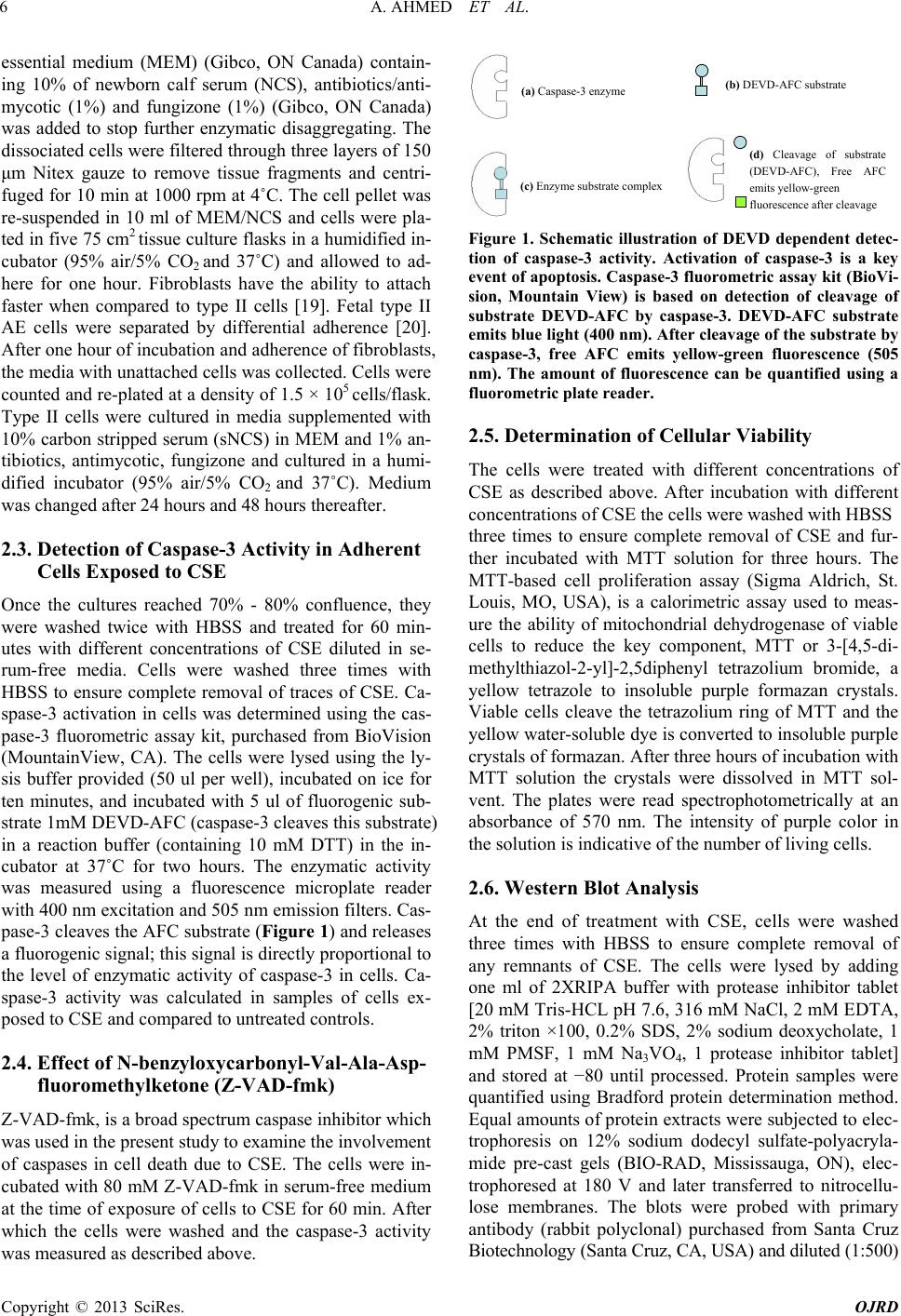 A. AHMED ET AL. 6 essential medium (MEM) (Gibco, ON Canada) contain- ing 10% of newborn calf serum (NCS), antibiotics/anti- mycotic (1%) and fungizone (1%) (Gibco, ON Canada) was added to stop further enzymatic disaggregating. The dissociated cells were filtered through three layers of 150 μm Nitex gauze to remove tissue fragments and centri- fuged for 10 min at 1000 rpm at 4˚C. The cell pellet was re-suspended in 10 ml of MEM/NCS and cells were pla- ted in five 75 cm2 tissue culture flasks in a humidified in- cubator (95% air/5% CO2 and 37˚C) and allowed to ad- here for one hour. Fibroblasts have the ability to attach faster when compared to type II cells [19]. Fetal type II AE cells were separated by differential adherence [20]. After one hour of incubation and adherence of fibroblasts, the media with unattached cells was collected. Cells were counted and re-plated at a density of 1.5 × 10⁵ cells/flask. Type II cells were cultured in media supplemented with 10% carbon stripped serum (sNCS) in MEM and 1% an- tibiotics, antimycotic, fungizone and cultured in a humi- dified incubator (95% air/5% CO2 and 37˚C). Medium was changed after 24 hours and 48 hours thereafter. 2.3. Detection of Caspase-3 Activity in Adherent Cells Exposed to CSE Once the cultures reached 70% - 80% confluence, they were washed twice with HBSS and treated for 60 min- utes with different concentrations of CSE diluted in se- rum-free media. Cells were washed three times with HBSS to ensure complete removal of traces of CSE. Ca- spase-3 activation in cells was determined using the cas- pase-3 fluorometric assay kit, purchased from BioVision (MountainView, CA). The cells were lysed using the ly- sis buffer provided (50 ul per well), incubated on ice for ten minutes, and incubated with 5 ul of fluorogenic sub- strate 1mM DEVD-AFC (caspase-3 cleaves this substrate) in a reaction buffer (containing 10 mM DTT) in the in- cubator at 37˚C for two hours. The enzymatic activity was measured using a fluorescence microplate reader with 400 nm excitation and 505 nm emission filters. Cas- pase-3 cleaves the AFC substrate (Figure 1) and releases a fluorogenic signal; this signal is directly proportional to the level of enzymatic activity of caspase-3 in cells. Ca- spase-3 activity was calculated in samples of cells ex- posed to CSE and compared to untreated controls. 2.4. Effect of N-benzyloxycarbonyl-Val-Ala-Asp- fluoromethylketone (Z-VAD-fmk) Z-VAD-fmk, is a broad spectrum caspase inhibitor which was used in the present study to examine the involvement of caspases in cell death due to CSE. The cells were in- cubated with 80 mM Z-VAD-fmk in serum-free medium at the time of exposure of cells to CSE for 60 min. After which the cells were washed and the caspase-3 activity was measured as described above. (a) Caspase-3 enzyme (b) DEVD-AFC substrate (c) Enzyme substrate complex (d) Cleavage of substrate (DEVD-AFC), Free AFC emits yellow-green fluorescence after cleavage Figure 1. Schematic illustration of DEVD dependent detec- tion of caspase-3 activity. Activation of caspase-3 is a key event of apoptosis. Caspase-3 fluorometric assay kit (BioVi- sion, Mountain View) is based on detection of cleavage of substrate DEVD-AFC by caspase-3. DEVD-AFC substrate emits blue light (400 nm). After cleavage of the substrate by caspase-3, free AFC emits yellow-green fluorescence (505 nm). The amount of fluorescence can be quantified using a fluorometric plate reader. 2.5. Determination of Cellular Viability The cells were treated with different concentrations of CSE as described above. After incubation with different concentrations of CSE the cells were washed with HBSS three times to ensure complete removal of CSE and fur- ther incubated with MTT solution for three hours. The MTT-based cell proliferation assay (Sigma Aldrich, St. Louis, MO, USA), is a calorimetric assay used to meas- ure the ability of mitochondrial dehydrogenase of viable cells to reduce the key component, MTT or 3-[4,5-di- methylthiazol-2-yl]-2,5d iphenyl tetrazolium bromide, a yellow tetrazole to insoluble purple formazan crystals. Viable cells cleave the tetrazolium ring of MTT and the yellow water-soluble dye is converted to insoluble purple crystals of formazan. After three hours of incubation with MTT solution the crystals were dissolved in MTT sol- vent. The plates were read spectrophotometrically at an absorbance of 570 nm. The intensity of purple color in the solution is indicative of the number of living cells. 2.6. Western Blot Analysis At the end of treatment with CSE, cells were washed three times with HBSS to ensure complete removal of any remnants of CSE. The cells were lysed by adding one ml of 2XRIPA buffer with protease inhibitor tablet [20 mM Tris-HCL pH 7.6, 316 mM NaCl, 2 mM EDTA, 2% triton ×100, 0.2% SDS, 2% sodium deoxycholate, 1 mM PMSF, 1 mM Na3VO4, 1 protease inhibitor tablet] and stored at −80 until processed. Protein samples were quantified using Bradford protein determination method. Equal amounts of protein extracts were subjected to elec- trophoresis on 12% sodium dodecyl sulfate-polyacryla- mide pre-cast gels (BIO-RAD, Mississauga, ON), elec- trophoresed at 180 V and later transferred to nitrocellu- lose membranes. The blots were probed with primary antibody (rabbit polyclonal) purchased from Santa Cruz Biotechnology (Santa Cruz, CA, USA) and diluted (1:500) Copyright © 2013 SciRes. OJRD 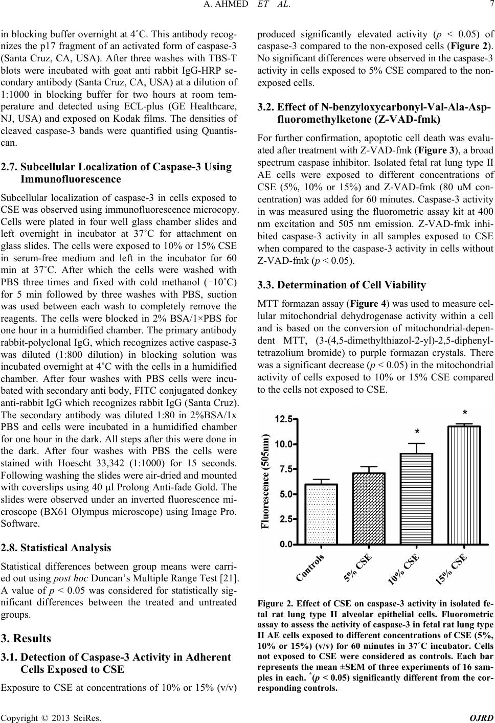 A. AHMED ET AL. 7 in blocking buffer overnight at 4˚C. This antibody recog- nizes the p17 fragment of an activated form of caspase-3 (Santa Cruz, CA, USA). After three washes with TBS-T blots were incubated with goat anti rabbit IgG-HRP se- condary antibody (Santa Cruz, CA, USA) at a dilution of 1:1000 in blocking buffer for two hours at room tem- perature and detected using ECL-plus (GE Healthcare, NJ, USA) and exposed on Kodak films. The densities of cleaved caspase-3 bands were quantified using Quantis- can. 2.7. Subcellular Localization of Caspase-3 Using Immunofluorescence Subcellular localization of caspase-3 in cells exposed to CSE was observed using immunofluorescence microcopy. Cells were plated in four well glass chamber slides and left overnight in incubator at 37˚C for attachment on glass slides. The cells were exposed to 10% or 15% CSE in serum-free medium and left in the incubator for 60 min at 37˚C. After which the cells were washed with PBS three times and fixed with cold methanol (−10˚C) for 5 min followed by three washes with PBS, suction was used between each wash to completely remove the reagents. The cells were blocked in 2% BSA/1×PBS for one hour in a humidified chamber. The primary antibody rabbit-polyclonal IgG, which recognizes active caspase-3 was diluted (1:800 dilution) in blocking solution was incubated overnight at 4˚C with the cells in a humidified chamber. After four washes with PBS cells were incu- bated with secondary anti body, FITC conjugated donkey anti-rabbit IgG which recognizes rabbit IgG (Santa Cruz). The secondary antibody was diluted 1:80 in 2%BSA/1x PBS and cells were incubated in a humidified chamber for one hour in the dark. All steps after this were done in the dark. After four washes with PBS the cells were stained with Hoescht 33,342 (1:1000) for 15 seconds. Following washing the slides were air-dried and mounted with coverslips using 40 μl Prolong Anti-fade Gold. The slides were observed under an inverted fluorescence mi- croscope (BX61 Olympus microscope) using Image Pro. Software. 2.8. Statistical Analysis Statistical differences between group means were carri- ed out using post ho c Duncan’s Multiple Range Test [21]. A value of p < 0.05 was considered for statistically sig- nificant differences between the treated and untreated groups. 3. Results 3.1. Detection of Caspase-3 Activity in Adherent Cells Exposed to CSE Exposure to CSE at concentrations of 10% or 15% (v/v) produced significantly elevated activity (p < 0.05) of caspase-3 compared to the non-exposed cells (Figure 2). No significant differences were observed in the caspase-3 activity in cells exposed to 5% CSE compared to the non- exposed cells. 3.2. Effect of N-benzyloxycarbonyl-Val-Ala-Asp- fluoromethylketone (Z-VAD-fmk) For further confirmation, apoptotic cell death was evalu- ated after treatment with Z-VAD-fmk (Figure 3), a broad spectrum caspase inhibitor. Isolated fetal rat lung type II AE cells were exposed to different concentrations of CSE (5%, 10% or 15%) and Z-VAD-fmk (80 uM con- centration) was added for 60 minutes. Caspase-3 activity in was measured using the fluorometric assay kit at 400 nm excitation and 505 nm emission. Z-VAD-fmk inhi- bited caspase-3 activity in all samples exposed to CSE when compared to the caspase-3 activity in cells without Z-VAD-fmk (p < 0.05). 3.3. Determination of Cell Viability MTT formazan assay (F igure 4) was used to measure cel- lular mitochondrial dehydrogenase activity within a cell and is based on the conversion of mitochondrial-depen- dent MTT, (3-(4,5-dimethylthiazol-2-yl)-2,5-diphenyl- tetrazolium bromide) to purple formazan crystals. There was a significant decrease (p < 0.05) in the mitochondrial activity of cells exposed to 10% or 15% CSE compared to the cells not exposed to CSE. Figure 2. Effect of CSE on caspase-3 activity in isolated fe- tal rat lung type II alveolar epithelial cells. Fluorometric assay to assess the activity of caspase-3 in fetal rat lung type II AE cells exposed to different concentrations of CSE (5%, 10% or 15%) (v/v) for 60 minutes in 37˚C incubator. Cells not exposed to CSE were considered as controls. Each bar represents the mean ±SEM of three experiments of 16 sam- ples in each. *(p < 0.05) significantly different from the cor- responding controls. Copyright © 2013 SciRes. OJRD 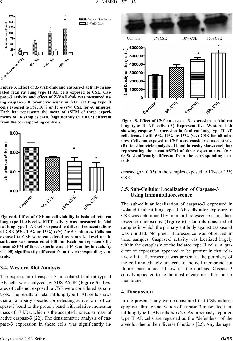 A. AHMED ET AL. 8 Figure 3. Effect of Z-VAD-fmk and caspase-3 activity in iso- lated fetal rat lung type II AE cells exposed to CSE. Cas- pase-3 activity and effect of Z-VAD-fmk was measured us- ing caspase-3 fluorometric assay in fetal rat lung type II cells exposed to 5%, 10% or 15% (v/v) CSE for 60 minutes. Each bar represents the mean of ±SEM of three experi- ments of 16 samples each. *significantly (p < 0.05) different from the corresponding controls. Figure 4. Effect of CSE on cell viability in isolated fetal rat lung type II AE cells. MTT activity was measured in fetal rat lung type II AE cells expose d to different concentrations of CSE (5%, 10% or 15%) (v/v) for 60 minutes. Cells not exposed to CSE were considered as controls. Level of ab- sorbance was measured at 540 nm. Each bar represents the mean ±SEM of three experiments of 16 samples in each. *(p < 0.05) significantly different from the corresponding con- trols. 3.4. Western Blot Analysis The expression of caspase-3 in isolated fetal rat type II AE cells was analyzed by SDS-PAGE (Figure 5). Lys- ates of cells not exposed to CSE were considered as con- trols. The results of fetal rat lung type II AE cells shows that an antibody specific for detecting active form of ca- spase-3 bond to the protein band with relative molecular mass of 17 kDa, which is the accepted molecular mass of active caspase-3 [22]. The densitometric analysis of cas- pase-3 expression in these cells was significantly in- Controls 5% CSE 10% CSE 15% CSE Figure 5. Effect of CSE on caspase-3 expression in fetal ra reased (p < 0.05) in the samples exposed to 10% or 15% 3.5. Sub-Cellular Localization of Caspase-3 The sub-3 expressed in we demonstrated that CSE induces t lung type II AE cells. (A) Representative Western bolt showing caspase-3 expression in fetal rat lung type II AE cells treated with 5%, 10% or 15% (v/v) CSE for 60 min- utes. Cells not exposed to CSE were considered as controls. (B) Densitometric analysis of band intensity shows each bar representing the mean ±SEM of three experiments. *(p < 0.05) significantly different from the corresponding con- trols. c CSE. Using Immunofluorescence -cellular localization of caspase isolated fetal rat lung type II AE cells after exposure to CSE was determined by immunofluorescence using fluo- rescence microscopy (Figure 6). Controls consisted of samples in which the primary antibody against caspase -3 was omitted. No green fluorescence was observed in these samples. Caspase-3 activity was localized largely within the cytoplasm of the isolated type II cells. A gra- dient of expression appeared to be present in that rela- tively little fluorescence was present at the periphery of the cell immediately adjacent to the cell membrane but fluorescence increased towards the nucleus. Caspase-3 activity appeared to be the most intense near the nuclear membrane. 4. Discussion In the present study apoptosis through activation of caspase-3 in isolated fetal rat lung type II AE cells in vitro. As previously reported type II AE cells are regarded as the “defenders” of the alveolus due to their diverse functions [22]. Any damage Copyright © 2013 SciRes. OJRD 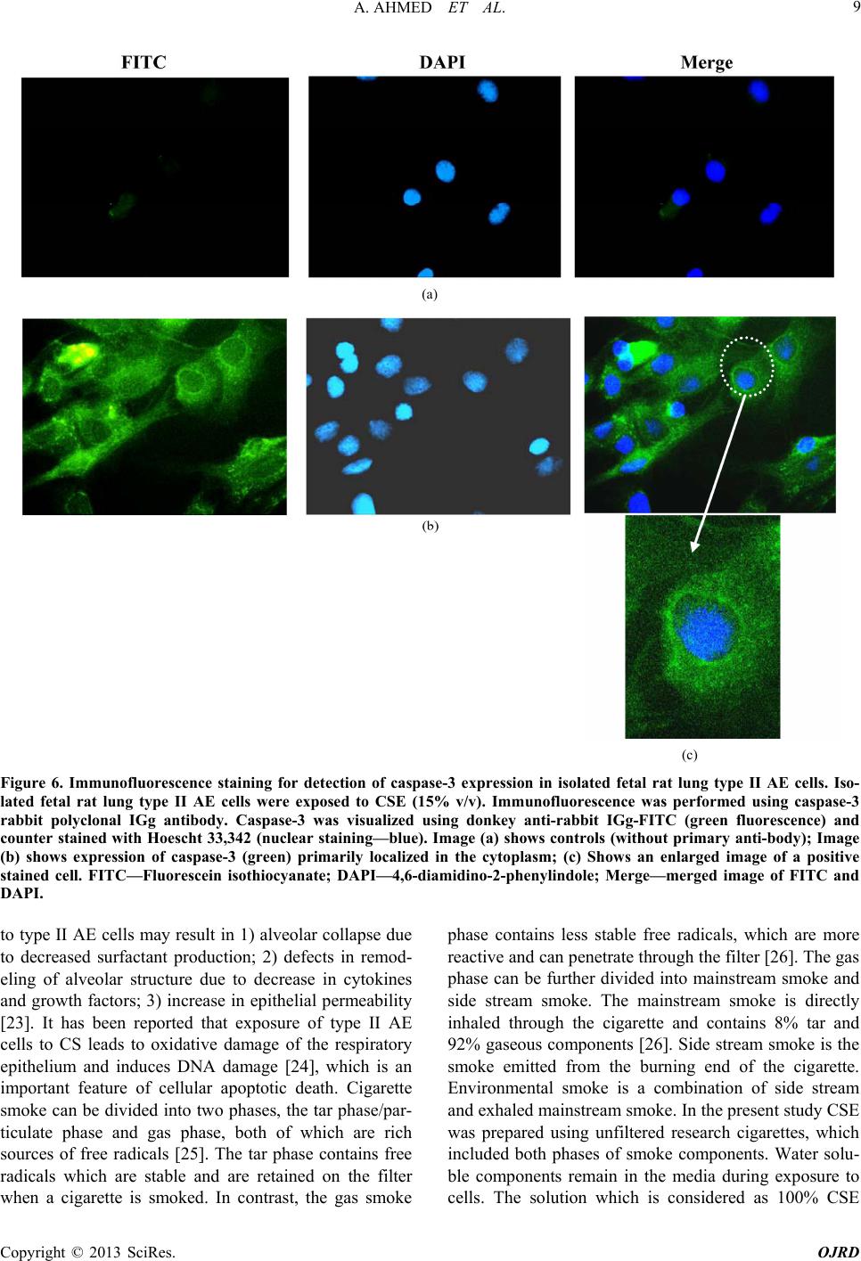 A. AHMED ET AL. Copyright © 2013 SciRes. OJRD 9 (a) (c) Figure 6. Immunofluorype II AE cells. Iso- type II AE cells may result in 1) alveolar collapse due phase contains less stable free radicals, which are more escence staining for detection of caspase-3 expression in isolated fetal rat lung t lated fetal rat lung type II AE cells were exposed to CSE (15% v/v). Immunofluorescence was performed using caspase-3 rabbit polyclonal IGg antibody. Caspase-3 was visualized using donkey anti-rabbit IGg-FITC (green fluorescence) and counter stained with Hoescht 33,342 (nuclear staining—blue). Image (a) shows controls (without primary anti-body); Image (b) shows expression of caspase-3 (green) primarily localized in the cytoplasm; (c) Shows an enlarged image of a positive stained cell. FITC—Fluorescein isothiocyanate; DAPI—4,6-diamidino-2-phenylindole; Merge—merged image of FITC and DAPI. to to decreased surfactant production; 2) defects in remod- eling of alveolar structure due to decrease in cytokines and growth factors; 3) increase in epithelial permeability [23]. It has been reported that exposure of type II AE cells to CS leads to oxidative damage of the respiratory epithelium and induces DNA damage [24], which is an important feature of cellular apoptotic death. Cigarette smoke can be divided into two phases, the tar phase/par- ticulate phase and gas phase, both of which are rich sources of free radicals [25]. The tar phase contains free radicals which are stable and are retained on the filter when a cigarette is smoked. In contrast, the gas smoke reactive and can penetrate through the filter [26]. The gas phase can be further divided into mainstream smoke and side stream smoke. The mainstream smoke is directly inhaled through the cigarette and contains 8% tar and 92% gaseous components [26]. Side stream smoke is the smoke emitted from the burning end of the cigarette. Environmental smoke is a combination of side stream and exhaled mainstream smoke. In the present study CSE was prepared using unfiltered research cigarettes, which included both phases of smoke components. Water solu- ble components remain in the media during exposure to cells. The solution which is considered as 100% CSE 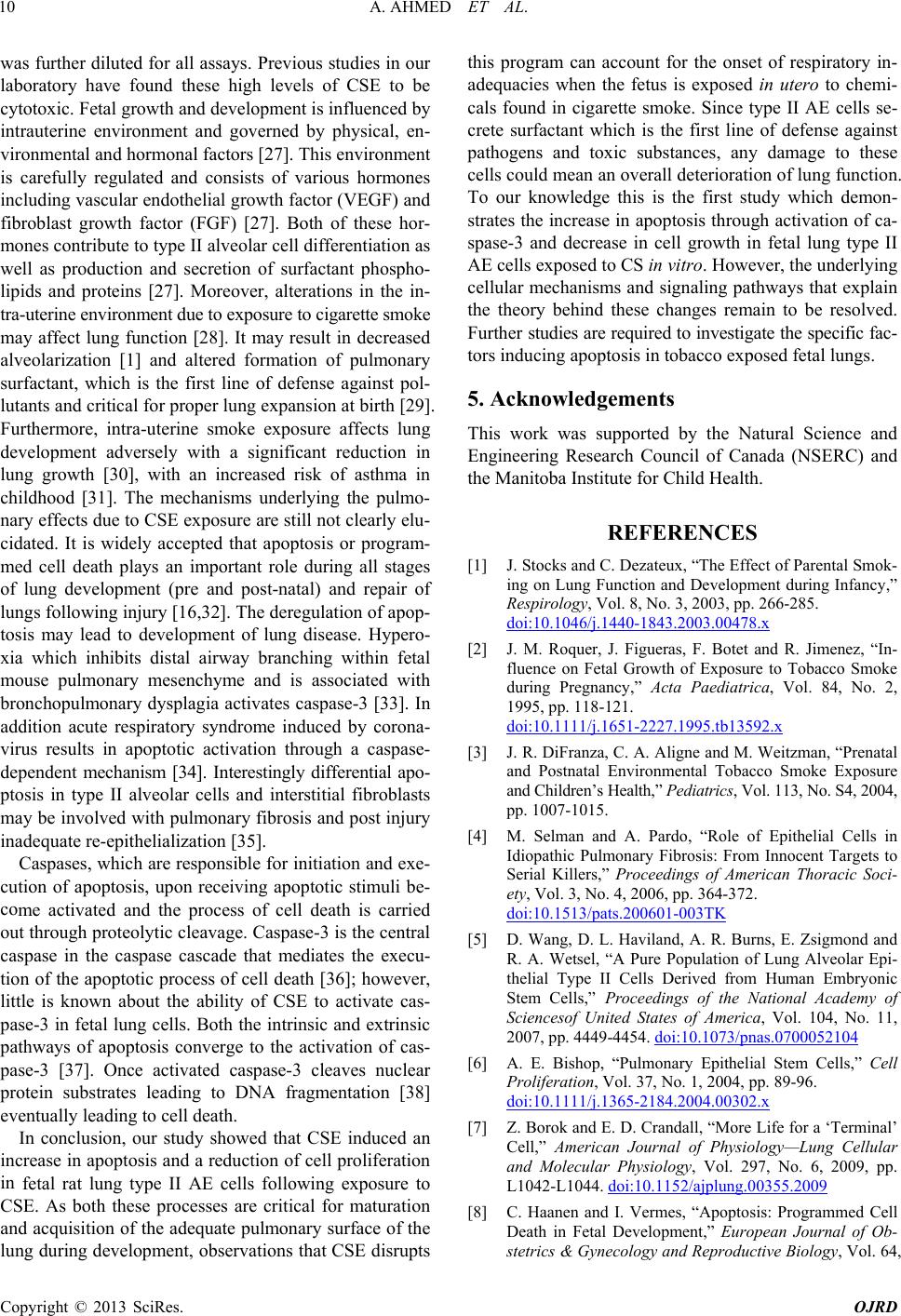 A. AHMED ET AL. 10 was further diluted for all assays. Previous studies in our laboratory have found these high levels of CSE to be cytotoxic. Fetal growth and development is influenced by intrauterine environment and governed by physical, en- vironmental and hormonal factors [27]. This environment is carefully regulated and consists of various hormones including vascular endothelial growth factor (VEGF) and fibroblast growth factor (FGF) [27]. Both of these hor- mones contribute to type II alveolar cell differentiation as well as production and secretion of surfactant phospho- lipids and proteins [27]. Moreover, alterations in the in- tra-uterine environment due to exposure to cigarette smoke may affect lung function [28]. It may result in decreased alveolarization [1] and altered formation of pulmonary surfactant, which is the first line of defense against pol- lutants and critical for proper lung expansion at birth [29]. Furthermore, intra-uterine smoke exposure affects lung development adversely with a significant reduction in lung growth [30], with an increased risk of asthma in childhood [31]. The mechanisms underlying the pulmo- nary effects due to CSE exposure are still not clearly elu- cidated. It is widely accepted that apoptosis or program- med cell death plays an important role during all stages of lung development (pre and post-natal) and repair of lungs following injury [16,32]. The deregulation of apop- tosis may lead to development of lung disease. Hypero- xia which inhibits distal airway branching within fetal mouse pulmonary mesenchyme and is associated with bronchopulmonary dysplagia activates caspase-3 [33]. In addition acute respiratory syndrome induced by corona- virus results in apoptotic activation through a caspase- dependent mechanism [34]. Interestingly differential apo- ptosis in type II alveolar cells and interstitial fibroblasts may be involved with pulmonary fibrosis and post injury inadequate re-epithelialization [35]. Caspases, which are responsible for initiation and exe- cution of apoptosis, upon receiving apoptotic stimu co proliferation in y the Natural Science and il of Canada (NSERC) and [1] J. Stocks and C of Parental Smok- ing on Lung Fnt during Infancy,” li be- me activated and the process of cell death is carried out through proteolytic cleavage. Caspase-3 is the central caspase in the caspase cascade that mediates the execu- tion of the apoptotic process of cell death [36]; however, little is known about the ability of CSE to activate cas- pase-3 in fetal lung cells. Both the intrinsic and extrinsic pathways of apoptosis converge to the activation of cas- pase-3 [37]. Once activated caspase-3 cleaves nuclear protein substrates leading to DNA fragmentation [38] eventually leading to cell death. In conclusion, our study showed that CSE induced an increase in apoptosis and a reduction of cell fetal rat lung type II AE cells following exposure to CSE. As both these processes are critical for maturation and acquisition of the adequate pulmonary surface of the lung during development, observations that CSE disrupts this program can account for the onset of respiratory in- adequacies when the fetus is exposed in utero to chemi- cals found in cigarette smoke. Since type II AE cells se- crete surfactant which is the first line of defense against pathogens and toxic substances, any damage to these cells could mean an overall deterioration of lung function. To our knowledge this is the first study which demon- strates the increase in apoptosis through activation of ca- spase-3 and decrease in cell growth in fetal lung type II AE cells exposed to CS in vitro. However, the underlying cellular mechanisms and signaling pathways that explain the theory behind these changes remain to be resolved. Further studies are required to investigate the specific fac- tors inducing apoptosis in tobacco exposed fetal lungs. 5. Acknowledgements This work was supported b Engineering Research Counc the Manitoba Institute for Child Health. REFERENCES . Dezateux, “The Effect unction and Developme Respirology, Vol. 8, No. 3, 2003, pp. 266-285. doi:10.1046/j.1440-1843.2003.00478.x [2] J. M. Roquer, J. Figueras, F. Botet and R. Jime fluence on Fetal Growth of Exposure to nez, “In- Tobacco Smoke during Pregnancy,” Acta Paediatrica, Vol. 84, No. 2, 1995, pp. 118-121. doi:10.1111/j.1651-2227.1995.tb13592.x [3] J. R. DiFranza, C. A. and Postnatal Environmental Tobacco Sm Aligne and M. Weitzman, “Prenatal oke Exposure onary Fibrosis: From Innocent Targets to and Children’s Health,” Pediatrics , Vol. 113, No. S4, 2004, pp. 1007-1015. [4] M. Selman and A. Pardo, “Role of Epithelial Cells in Idiopathic Pulm Serial Killers,” Proceedings of American Thoracic Soci- ety, Vol. 3, No. 4, 2006, pp. 364-372. doi:10.1513/pats.200601-003TK [5] D. Wang, D. L. Haviland, A. R. Burns, R. A. Wetsel, “A Pure Population E. Zsigmond and of Lung Alveolar Epi- thelial Type II Cells Derived from Human Embryonic Stem Cells,” Proceedings of the National Academy of Sciencesof United States of America, Vol. 104, No. 11, 2007, pp. 4449-4454. doi:10.1073/pnas.0700052104 [6] A. E. Bishop, “Pulmonary Epithelial Stem Cells,” Cell Proliferation, Vol. 37, No. 1, 2004, pp. 89-96. doi:10.1111/j.1365-2184.2004.00302.x [7] Z. Borok and E. D. Crandall, “More Life for a ‘T Cell,” American Journal of Physiologyerminal’ —Lung Cellular and Molecular Physiology, Vol. 297, No. 6, 2009, pp. L1042-L1044. doi:10.1152/ajplung.00355.2009 [8] C. Haanen and I. Vermes, “Apoptosis: Programmed Cell Death in Fetal Development,” European Journal of Ob- stetrics & Gynecology and Reproductive Biology, Vol. 64, Copyright © 2013 SciRes. OJRD  A. AHMED ET AL. 11 No. 1, 1996, pp. 129-133. doi:10.1016/0301-2115(95)02261-9 [9] S. Elmore, “Apoptosis: A Review of Programmed Cell Death,” Toxicologic Pathology, Vol. 35, No 495-516. . 4, 2007, pp. 20337doi:10.1080/019262307013 002, pp. 97- [10] C. Kohler, S. Orrenius and B. Zhivotovsky, “Evaluation of Caspase Activity in Apoptotic Cells,” Journal of Im- munological Methods, Vol. 265, No. 1-2, 2 110. doi:10.1016/S0022-1759(02)00073-X [11] P. G. Ekert, J. Silke and D. L. Vaux, “Caspase Inhibi- tors,” Cell Death & Differentiation, Vol. 6, No. 11, 1999, pp. 1081-1086. doi:10.1038/sj.cdd.4400594 [12] D. W. Nicholson and N. A. Thornberry, “Caspases: Killer Proteases,” Trends in Biochemical Sciences, Vol. 22, No. 8, 1997, pp. 299-306. doi:10.1016/S0968-0004(97)01085-2 [13] D. Chandra and D. G. Tang, “Mitochondrially Localized Active Caspase-9 and Caspase-3 Re Translocation from the Cytosol and Pa sult Mostly from rtly from Caspase- Mediated Activation in the Organelle. Lack of Evidence for Apaf-1-Mediated Procaspase-9 Activation in the Mi- tochondria,” The Journal of Biological Chemistry, Vol. 278, No. 19, 2003, pp. 17408-17420. doi:10.1074/jbc.M300750200 [14] B. Fadeel, S. Orrenius and B. Zhivotovsky, “The Most Unkindest Cut of All: On the Multip malian Caspases,” Leukemia, le Roles of Mam Vol. 14, No. 8, 2000, pp - . 1514-1525. doi:10.1038/sj.leu.2401871 [15] N. A. Thornberry and Y. Lazebnik, “Caspases: Enemies within,” Science, Vol. 281, No. 5381, 1998, pp. 1312- 1316. doi:10.1126/science.281.5381.1312 ogenesis, [16] C. Wongtrakool and J. Roman, “Apoptosis of Mesen- chymal Cells during the Pseudoglandular Stage of Lung Development Affects Branching Morph” Ex- perimental Lung Research, Vol. 34, No. 8, 2008, pp. 481- 499. doi:10.1080/01902140802271842 [17] A. Janoff and H. Carp, “Possible Mechanisms of Emphy- sema in Smokers: Cigarette Smoke Condensate Supp- resses Protease Inhibition in Vitro,” American Review Re- spiratory Disease, Vol. 116, No. 1, 1977, pp. 65-72. [18] A. R. Tabassian, E. S. Nylen, A. E. Giron, R. H. Snider, M. M. Cassidy and K. L. Becker, “Evidence for Cigarette Smoke-Induced Calcitonin Secretion from Lungs of Man and Hamster,” Life Sciences, Vol. 42, No. 23, 1988, pp. 2323-2329. doi:10.1016/0024-3205(88)90185-3 [19] J. E. Scott, S. Y. Yang, E. Stanik and J. E. Anderson, “In- fluence of Strain on [3H]Thymidine Incorporation, Sur- factant-Related Phospholipid Synthesis, and Camp Levels t Lung by Differential Adherence in Fetal Type II Alveolar Cells,” American Journal of Re- spiratory Cell and Molecular Biology, Vol. 8, No. 3, 1993, pp. 258-265. [20] J. J. Batenburg, C. J. Otto-Verberne, A. A. Ten Have-Op- broek and W. Klazinga, “Isolation of Alveolar Type II Cells from Fetal Ra in Monolayer Culture,” Biochimica et Biophysica Acta, Vol. 960, No. 3, 1988, pp. 441-453. doi:10.1016/0005-2760(88)90053-7 [21] L. Ott, “An Introduction to Statistical Methods and Data Analysis,” Duxbury Press, Pacific [22] H. Fehrenbach, “Alveolar Epithelial Grove, 1977. Type II Cell: Defen- der of the Alveolus Revisited,” Respiratory Research, Vol. 2, No. 1, 2001, pp. 33-46. doi:10.1186/rr36 [23] Y. Hoshino, T. Mio, S. Nagai, H. Miki, I. Ito and T. Izu- mi, “Cytotoxic Effects of Cigarette Smoke Extract on an Alveolar Type II Cell-Derived Cell Line,” American Jour- nal of Physiology—Lung Cellular and Molecular Physi- ology, Vol. 281, No. 2, 2001, pp. L509-L516. [24] K. Aoshiba, M. Koinuma, N. Yokohori and A. Nagai, “Immunohistochemical Evaluation of Oxidative Stress in Murine Lungs after Cigarette Smoke Exposure,” Inhala- tion Toxicology, Vol. 15, No. 10, 2003, pp. 1029-1038. [25] W. A. Pryor, “Cigarette Smoke Radicals and the Role of Free Radicals in Chemical Carcinogenicity,” Environ- mental Health Perspectives, Vol. 105, No. S4, 1997, pp. 875-882. [26] J. A. Ambrose and R. S. Barua, “The Pathophysiology of Cigarette Smoking and Cardiovascular Disease: An Up- date,” Journal of the American College of Cardiology, Vol. 43, No. 10, 2004, pp. 1731-1737. doi:10.1016/j.jacc.2003.12.047 [27] S. Joshi and S. Kotecha, “Lung Growth and Develop- ment,” Early Human Development, Vol. 83, No. 12, 2007, pp. 789-794. doi:10.1016/j.earlhumdev.2007.09.007 [28] R. J. Rona, M. C. Gulliford and S. Chinn, “Effects of Pre- maturity and Intrauterine Growth on Respiratory Health and Lung Function in Childhood,” British Medical Jour- nal, Vol. 306, No. 6881, 1993, pp. 817-820. doi:10.1136/bmj.306.6881.817 [29] L. A. Creuwels, L. M. van Golde and H. P. Haagsman, “The Pulmonary Surfactant System: Biochemical and Cli- nical Aspects,” Lung, Vol. 175, No. 1, 1997, pp. 1-39. doi:10.1007/PL00007554 [30] M. Sexton and J. R. Hebel, “A Clinical Trial of Change in Maternal Smoking and Its Effect on Birth Weight,” Jour- nal of the American Medical Association, Vol. 251, No. 7, 1984, pp. 911-915. doi:10.1001/jama.1984.03340310025013 [31] E. von Mutius, “Environmental Factors Influencing the Development and Progression of Pediatric Asthma,” Jour- nal of Allergy and Clinical Immunology, Vol. 10 2002, pp. S525-S532. 9, No. 6, 2.124565doi:10.1067/mai.200 [32] X. Li, R. Shu, G. Filippatos and B. D. Uhal, “Apoptosis in Lung Injury and Remodeling,” Journal of Applied Phy- siology, Vol. 97, No. 4, 2004, pp. 1535-1542. doi:10.1152/japplphysiol.00519.2004 [33] H. I. Dieperink, T. S. Blackwell and L. S. Prince, “Hy- peroxia and Apoptosis in Developing Mouse Lung Me- senchyme,” Pediatric Researc h, Vol. 59, No. 2, 185-190. doi:10.1203/01.pdr.00001963 2006, pp. 71.85945.3a [34] Y. J. Tan, B. C. Fielding, P. Y. Goh, S. Shen, T. H. Tan, S. G. Lim and W. Hong, “Overexpression of 7a, a Protein Specifically Encoded by the Severe Acute Respiratory Syndrome Coronavirus, Induces Apoptosis via a Caspase- Dependent Pathway,” Journal of Virology, Vol. 78, No. 24, 2004, pp. 14043-14047. doi:10.1128/JVI.78.24.14043-14047.2004 [35] F. Drakopanagiotakis, A. Xifteri, V. Polychronopoulos and D. Bouros, “Apoptosis in Lung Injury and Fibrosis,” Copyright © 2013 SciRes. OJRD  A. AHMED ET AL. Copyright © 2013 SciRes. OJRD 12 l, Vol. 32, No. 6, 2008, pp 7 European Respiratory Journa 1631-1638. . doi:10.1183/09031936.0017680 [36] S. Kumar, “Caspase Function in Programmed Cell Death,” Cell Death & Differentiation, Vol. 14, No. 1, 2007, pp. 32-43. doi:10.1038/sj.cdd.4402060 [37] R. Ganesan, P. R. Mittl, S. Jelakovic and M. G. Grutter . 5, 2006, pp. 1378- , “Extended Substrate Recognition in Caspase-3 Revealed by High Resolution X-Ray Structure Analysis,” Journal of Molecular Biology, Vol. 359, No 1388. doi:10.1016/j.jmb.2006.04.051 [38] B. B. Wolf, M. Schuler, F. Echeverri and D. R. Green, “Caspase-3 Is the Primary Activator of Apoptotic DNA Fragmentation via DNA Fragmentation Factor-45/Inhi- bitor of Caspase-Activated DNase Inactivation,” The Jour- nal of Biological Chemistry, Vol. 274, No. 43, 1999, pp. 30651-30656. doi:10.1074/jbc.274.43.30651 |