

Paper Menu >>
Journal Menu >>
 Advances in Nanoparticles, 2013, 2, 6-10 http://dx.doi.org/10.4236/anp.2013.21002 Published Online February 2013 (http://www.scirp.org/journal/anp) Synthesis of La/N Co-Doped SrTiO3 Using Polymerized Complex Method for Visible Light Photocatalysis Uyi Sulaeman1, Shu Yin2, Tsugio Sato2 1Department of Chemistry, Jenderal Soedirman University, Purwokerto, Indonesia 2Institute of Multidisciplinary Research for Advanced Materials, Tohoku University, Sendai, Japan Email: [email protected] Received December 16, 2012; revised January 15, 2013; accepted January 25, 2013 ABSTRACT Lanthanum and nitrogen co-doped SrTiO3 was synthesized using polymerized complex method with Ti(OC3H7)4, SrCl2·6H2O and La(NO3)3·6H2O as starting materials followed by calcinations in NH3. Ethylene glycol and anhydrous citric acid were used as the precursors of synthesis. The samples were characterized using XRD, TEM, DRS, BET, EDX and XPS. The cubic-perovskite type of La/N co-doped SrTiO3 nanoparticle could be successfully synthesized. The photocatalytic activity of SrTiO3 for DeNOx ability in visible light region (λ > 510 nm) could be improved by co-doping of La3+ and N3−. The high visible light photocatalytic activity of this substance was caused by a narrow band gap energy that enables to absorb visible light. Keywords: Photocatalysis; Visible Light; SrTiO3; Polymerized Complex; La-Doping; N-Doping 1. Introduction Recently, synthesis of strontium titanate based photo- catalyst for converting visible light energy to photoreac- tion has been a great attention [1-6]. Among the modify- ing of SrTiO3, doping with nitrogen is the most effective to enhance photoreaction in visible light. The mixing of N 2p with O 2p states narrows the band gap energy and enhances the photocatalytic ability in visible light. How- ever, substituting O2− by N3− will generate residual ani- onic vacancies which suppress the photocatalytic activity of SrTiO3. To solve this problem, co-doping of lantha- num and nitrogen into SrTiO3, can decrease the band gap energy without forming lattice defect and lattice strain, and consequently lead to generate high visible light photocatalytic activity. The La3+ can substitute Sr2+ without large lattice strain because of the similar ionic radius. Many researchers had developed the synthesis of La/N co-doped SrTiO3 catalysts. Miyauchi et al. [7] reported that the synthesis of La/N co-doped SrTiO3 catalysts us- ing sol-gel method decreased the ionic vacancy and then increased the photocatalytic activity in a visible light. Wang et al. [8] found that the synthesis of La/N co- doped SrTiO3 catalysts using mechanochemical reaction enhanced the photocatalytic activity in a visible light. However, they had a large particle and low specific sur- face area which limited the catalytic ability. To improve the potocatalytic ability, synthesis of fine particle which has a large specific surface area should be realized. Polymerized complex method using citric acid and ethylene glycol as polymeric precursors has been widely used for metal oxide synthesis [9,10]. The polymerized complex process has great advantages over other synthe- sis techniques due to mixing of several components in atomic scale, good stoichiometry control, high purity, low cost and relatively low processing temperature [11]. The metallic ions are dispersed in the polymeric network at the atomic scale without precipitation and phase seg- regation [12]. Based on this consideration, the fine parti- cles of La-doped SrTiO3 could be synthesized by the polymerized complex process and then followed by cal- cinations in ammonia to obtain the La/N co-doped SrTiO3 nanoparticles. In the present paper, we report the synthesis of La/N co-doped SrTiO3 using polymerized complex method. The lanthanum and nitrogen co-doping effectively nar- rowed the band-gap energy of SrTiO3. The photocatalytic activity of SrTiO3 for NO decomposition in visible light region (λ > 510 nm) could be enhanced. The high visible light photocatalytic activity of this substance might be caused by the low band gap energy and high specific surface area. 2. Experiment 2.1. Preparation of Catalyst The La/N co-doped SrTiO3 with variation of lanthanum doping was prepared by the polymerized complex method C opyright © 2013 SciRes. ANP 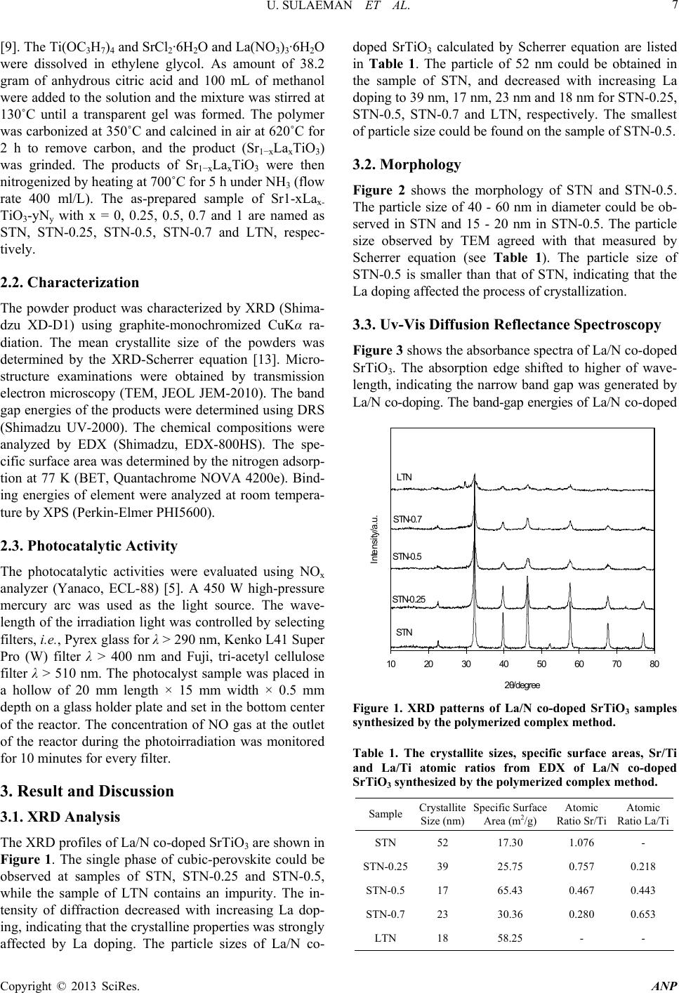 U. SULAEMAN ET AL. 7 [9]. The Ti(OC3H7)4 and SrCl2·6H2O and La(NO3)3·6H2O were dissolved in ethylene glycol. As amount of 38.2 gram of anhydrous citric acid and 100 mL of methanol were added to the solution and the mixture was stirred at 130˚C until a transparent gel was formed. The polymer was carbonized at 350˚C and calcined in air at 620˚C for 2 h to remove carbon, and the product (Sr1−xLaxTiO3) was grinded. The products of Sr1−xLaxTiO3 were then nitrogenized by heating at 700˚C for 5 h under NH3 (flow rate 400 ml/L). The as-prepared sample of Sr1-xLax- TiO3-yNy with x = 0, 0.25, 0.5, 0.7 and 1 are named as STN, STN-0.25, STN-0.5, STN-0.7 and LTN, respec- tively. 2.2. Characterization The powder product was characterized by XRD (Shima- dzu XD-D1) using graphite-monochromized CuKα ra- diation. The mean crystallite size of the powders was determined by the XRD-Scherrer equation [13]. Micro- structure examinations were obtained by transmission electron microscopy (TEM, JEOL JEM-2010). The band gap energies of the products were determined using DRS (Shimadzu UV-2000). The chemical compositions were analyzed by EDX (Shimadzu, EDX-800HS). The spe- cific surface area was determined by the nitrogen adsorp- tion at 77 K (BET, Quantachrome NOVA 4200e). Bind- ing energies of element were analyzed at room tempera- ture by XPS (Perkin-Elmer PHI5600). 2.3. Photocatalytic Activity The photocatalytic activities were evaluated using NOx analyzer (Yanaco, ECL-88) [5]. A 450 W high-pressure mercury arc was used as the light source. The wave- length of the irradiation light was controlled by selecting filters, i.e., Pyrex glass for λ > 290 nm, Kenko L41 Super Pro (W) filter λ > 400 nm and Fuji, tri-acetyl cellulose filter λ > 510 nm. The photocalyst sample was placed in a hollow of 20 mm length × 15 mm width × 0.5 mm depth on a glass holder plate and set in the bottom center of the reactor. The concentration of NO gas at the outlet of the reactor during the photoirradiation was monitored for 10 minutes for every filter. 3. Result and Discussion 3.1. XRD Analysis The XRD profiles of La/N co-doped SrTiO3 are shown in Figure 1. The single phase of cubic-perovskite could be observed at samples of STN, STN-0.25 and STN-0.5, while the sample of LTN contains an impurity. The in- tensity of diffraction decreased with increasing La dop- ing, indicating that the crystalline properties was strongly affected by La doping. The particle sizes of La/N co- doped SrTiO3 calculated by Scherrer equation are listed in Table 1. The particle of 52 nm could be obtained in the sample of STN, and decreased with increasing La doping to 39 nm, 17 nm, 23 nm and 18 nm for STN-0.25, STN-0.5, STN-0.7 and LTN, respectively. The smallest of particle size could be found on the sample of STN-0.5. 3.2. Morphology Figure 2 shows the morphology of STN and STN-0.5. The particle size of 40 - 60 nm in diameter could be ob- served in STN and 15 - 20 nm in STN-0.5. The particle size observed by TEM agreed with that measured by Scherrer equation (see Table 1). The particle size of STN-0.5 is smaller than that of STN, indicating that the La doping affected the process of crystallization. 3.3. Uv-Vis Diffusion Reflectance Spectroscopy Figure 3 shows the absorbance spectra of La/N co-doped SrTiO3. The absorption edge shifted to higher of wave- length, indicating the narrow band gap was generated by La/N co-doping. The band-gap energies of La/N co-doped 10 20 3040 5060 70 80 2θ/degree Intensity/a.u. STN-0.25 STN-0.5 STN-0.7 LTN STN Figure 1. XRD patterns of La/N co-doped SrTiO3 samples synthesized by the polyme rized co mple x me thod. Table 1. The crystallite sizes, specific surface areas, Sr/Ti and La/Ti atomic ratios from EDX of La/N co-doped SrTiO3 synthesized by the polymer i zed complex me thod. Sample Crystallite Size (nm) Specific Surface Area (m2/g) Atomic Ratio Sr/Ti Atomic Ratio La/Ti STN 52 17.30 1.076 - STN-0.2539 25.75 0.757 0.218 STN-0.517 65.43 0.467 0.443 STN-0.723 30.36 0.280 0.653 LTN 18 58.25 - - Copyright © 2013 SciRes. ANP 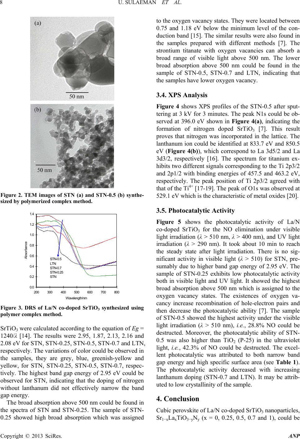 U. SULAEMAN ET AL. 8 Figure 2. TEM images of STN (a) and STN-0.5 (b) synthe- sized by polymeri zed comple x method. 0.0 0.2 0.4 0.6 0.8 1.0 1.2 1.4 200 300 400 500600 700 800 Wavelength/nm Absorbance STN-0.5 LTN STN-0.7 STN-0.25 STN Figure 3. DRS of La/N co-doped SrTiO3 synthesized using polymer complex method. SrTiO3 were calculated according to the equation of Eg = 1240/λ [14]. The results were 2.95, 1.87, 2.13, 2.16 and 2.08 eV for STN, STN-0.25, STN-0.5, STN-0.7 and LTN, respectively. The variations of color could be observed in the samples, they are grey, blue, greenish-yellow and yellow, for STN, STN-0.25, STN-0.5, STN-0.7, respec- tively. The highest band gap energy of 2.95 eV could be observed for STN, indicating that the doping of nitrogen without lanthanum did not effectively narrow the band gap energy. The broad absorption above 500 nm could be found in the spectra of STN and STN-0.25. The sample of STN- 0.25 showed high broad absorption which was assigned to the oxygen vacancy states. They were located between 0.75 and 1.18 eV below the minimum level of the con- duction band [15]. The similar results were also found in the samples prepared with different methods [7]. The strontium titanate with oxygen vacancies can absorb a broad range of visible light above 500 nm. The lower broad absorption above 500 nm could be found in the sample of STN-0.5, STN-0.7 and LTN, indicating that the samples have lower oxygen vacancy. 3.4. XPS Analys is Figure 4 shows XPS profiles of the STN-0.5 after sput- tering at 3 kV for 3 minutes. The peak N1s could be ob- served at 396.0 eV shown in Figure 4(a), indicating the formation of nitrogen doped SrTiO3 [7]. This result proves that nitrogen was incorporated in the lattice. The lanthanum ion could be identified at 833.7 eV and 850.5 eV (Figure 4(b)), which correspond to La 3d5/2 and La 3d3/2, respectively [16]. The spectrum for titanium ex- hibits two different signals corresponding to the Ti 2p3/2 and 2p1/2 with binding energies of 457.5 and 463.2 eV, respectively. The peak position of Ti 2p3/2 agreed with that of the Ti4+ [17-19]. The peak of O1s was observed at 529.1 eV which is the characteristic of metal oxides [20]. 3.5. Photocatalytic Activity Figure 5 shows the photocatalytic activity of La/N co-doped SrTiO3 for the NO elimination under visible light irradiation (λ > 510 nm, λ > 400 nm), and UV light irradiation (λ > 290 nm). It took about 10 min to reach the steady state after light irradiation. There is no sig- nificant activity in visible light (λ > 510) for STN, pre- sumably due to higher band gap energy of 2.95 eV. The sample of STN-0.25 exhibits low photocatalytic activity both in visible light and UV light. It showed the highest broad absorption above 500 nm which is assigned to the oxygen vacancy states. The existences of oxygen va- cancy increase recombination of hole-electron pairs and then decrease the photocatalytic ability [7]. The sample of STN-0.5 showed the highest activity under the visible light irradiation (λ > 510 nm), i.e. , 28.8% NO could be destructed. Moreover, the photocatalytic ability of STN- 0.5 was also higher than TiO2 (P-25) in the ultraviolet light, i.e., 42.3% of NO could be destructed. The excel- lent photocatalytic was attributed to both narrow band gap energy and high specific surface area (see Table 1). The photocatalytic activity decreased with increasing lanthanum doping (STN-0.7 and LTN). It may be attrib- uted to low crystallinity of the sample. 4. Conclusion Cubic perovskite of La/N co-doped SrTiO3 nanoparticles, Sr1−xLaxTiO3−yNy (x = 0, 0.25, 0.5, 0.7 and 1), could be Copyright © 2013 SciRes. ANP 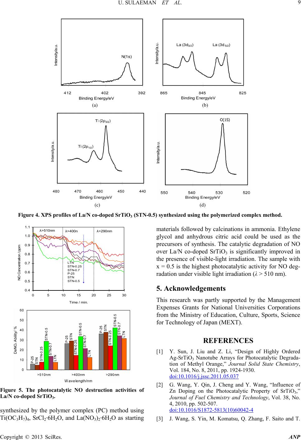 U. SULAEMAN ET AL. Copyright © 2013 SciRes. ANP 9 (a) (b) (c) (d) Figure 4. XPS profiles of La/N co-doped SrTiO3 (STN-0.5) synthesized using the polymerized complex method. 0.4 0.5 0.6 0.7 0.8 0.9 1.0 1.1 0510 15 20 2530 Time / min. NO Concentration / ppm LTN STN-0.25 STN-0.7 P-25 STN STN-0.5 λ>510nm λ>400n λ>290nm Figure 5. The photocatalytic NO destruction activities of La/N co-doped SrTiO3. synthesized by the polymer complex (PC) method using Ti(OC3H7)4, SrCl2·6H2O, and La(NO3)2·6H2O as starting materials followed by calcinations in ammonia. Ethylene glycol and anhydrous citric acid could be used as the precursors of synthesis. The catalytic degradation of NO over La/N co-doped SrTiO3 is significantly improved in the presence of visible-light irradiation. The sample with x = 0.5 is the highest photocatalytic activity for NO deg- radation under visible light irradiation (λ > 510 nm). 5. Acknowledgements This research was partly supported by the Management Expenses Grants for National Universities Corporations from the Ministry of Education, Culture, Sports, Science for Technology of Japan (MEXT). REFERENCES [1] Y. Sun, J. Liu and Z. Li, “Design of Highly Ordered Ag-SrTiO3 Nanotube Arrays for Photocatalytic Degrada- tion of Methyl Orange,” Journal Solid State Chemistry, Vol. 184, No. 8, 2011, pp. 1924-1930. doi:10.1016/j.jssc.2011.05.037 [2] G. Wang, Y. Qin, J. Cheng and Y. Wang, “Influence of Zn Doping on the Photocatalytic Property of SrTiO3,” Journal of Fuel Chemistry and Technology, Vol. 38, No. 4, 2010, pp. 502-507. doi:10.1016/S1872-5813(10)60042-4 [3] J. Wang, S. Yin, M. Komatsu, Q. Zhang, F. Saito and T.  U. SULAEMAN ET AL. 10 Sato, “Preparation and Characterization of Nitrogen Doped SrTiO3 Photocatalyst,” Journal of Photochemistry and Photobiology A: Chemistry, Vol. 165, No. 1-3, 2004, pp. 149-156. doi:10.1016/j.jphotochem.2004.02.022 [4] A. Jia, Z. Su, L. Lou and S. Liu, “Synthesis and Charac- terization of Highly-Active Nickel and Lanthanum Co- Doped SrTiO3,” Solid State Science, Vol. 12, No. 7, 2010, pp. 1140-1145. doi:10.1016/j.solidstatesciences.2010.04.005 [5] J. Wang, H. Li, H. Li, S. Yin and T. Sato, “Preparation and Photocatalytic Activity of Visible Light-Active Sul- fur and Nitrogen Co-Doped SrTiO3,” Solid State Science, Vol. 11, No. 1, 2009, pp. 182-188. doi:10.1016/j.solidstatesciences.2008.04.010 [6] T. Ohno, T. Tsubota, Y. Nakamura and K. Sayama, “Pre- paration of S, C Cation-Codoped SrTiO3 and Its Photo- catalytic Activity under Visible Light,” Applied Catalysis A: General, Vol. 288, No. 1-2, 2005, pp. 74-79. doi:10.1016/j.apcata.2005.04.035 [7] M. Miyauchi, M. Takashio and H. Tobimatsu, “Photo- catalytic Activity of SrTiO3 Codoped with Nitrogen and Lanthanum under Visible Light Illumination,” Langmuir, Vol. 20, No. 1, 2004, pp. 232-236. doi:10.1021/la0353125 [8] J. Wang, S. Yin, M. Komatsu and T. Sato, “Lanthanum and Nitrogen Co-Doped SrTiO3 Powders as Visible Light Sensitive Photocatalyst,” Journal of the European Ce- ramic Society, Vol. 25, No. 13, 2005, pp. 3207-3212. doi:10.1016/j.jeurceramsoc.2004.07.027 [9] A. Kasahara, K. Nukumizu, G. Hitoki, T. Takata, J.N. Kondo, M. Hara, H. Kobayashi and K. Domen, “Photo- reactions on LaTiO2N under Visible Light Irradiation,” The Journal of Physical Chemistry A, Vol. 106, No. 29, 2002, pp. 6750-6753. doi:10.1021/jp025961+ [10] S. Farhikhteh, A. Maghsoudipour and B. Raissi, “Synthe- sis of Nanocrystalline YSZ (ZrO2-8Y2O3) Powder by Po- lymerized Complex Method,” Journal of Alloys and Com- pounds, Vol. 491, No. 1-2, 2010, pp. 402-405. doi:10.1016/j.jallcom.2009.10.196 [11] J. P. Zhao, M. H. Quan and L. Zhang, “Preparation of Potassium Lithium Niobate Powders with Low Li Content via the Pechini Method,” Ceramics International, Vol. 32, No. 7, 2006, pp. 843-846. doi:10.1016/j.ceramint.2005.05.010 [12] S. Qiu, H. Fan and X. Zheng, “Pb(Zr0.95Ti0.05)O3 Powders Synthesized by Pechini Method: Effect of Molecular Weight of Polyester on the Phase and Morphology,” Journal of Sol-Gel Science and Technology, Vol. 42, No.1, 2007, pp. 21-26. doi:10.1007/s10971-006-1509-3 [13] B. Dcullity, “Elements of X-Ray Diffraction,” 3rd Edition, Addison-Wesley Publishing Co., Boston, 1976. [14] S. X. Liu, X. Y. Chen and X. Chen, “Preparation of N-Doped Visible Light Response Nanosize TiO2 Photo- catalyst Using the Acid-Catalyzed Hydrolysis Method,” Chinese Journal of Catalysis, Vol. 27, No. 8, 2006, pp. 697-702. doi:10.1016/S1872-2067(06)60037-5 [15] D. C. Cronemeyer, “Infrared Absorption of Reduced Rutile TiO2 Single Crystals,” Physical Review, Vol. 113, No. 5, 1959, pp. 1222-1226. doi:10.1103/PhysRev.113.1222 [16] A. J. Nelson, T. Buuren, T. M. Willey, C. Bostedt, J. J. Adams, K. I. Schaffers, L. Terminello and T. A. Callcott, “Electronic Structure of Lanthanum Calcium Oxoborate LaCa4O(BO3)3,” Journal of Electron Spectroscopy and Re- lated Phenomena, Vol. 137-140, No. 2004, pp. 541-546. [17] S. Martınez-Mendez, Y. Henrıquez, O. Domınguez, L. D’Ornelas and H. Krentzien, “Catalytic Properties of Sil- ica Supported Titanium, Vanadium and Niobium Oxide Nanoparticles towards the Oxidation of Saturated and Unsaturated Hydrocarbons,” Journal of Molecular Ca- talysis A: Chemical, Vol. 252, No. 1-2, 2006, pp. 226- 234. doi:10.1016/j.molcata.2006.02.041 [18] M. Ziolek and I. Nowak, “Characterization Techniques Employed in the Study of Niobium and Tantalum-Con- taining Materials,” Catalysis Today, Vol. 78, No. 1-4, 2003, pp. 543-553. doi:10.1016/S0920-5861(02)00353-X [19] R. E. Tanner, Y. Liang and E. I. Altman, “Structure and Chemical Reactivity of Adsorbed Carboxylic Acids on Anatase TiO2(0 0 1),” Surface Science, Vol. 506, No. 3, 2002, pp. 251-271. doi:10.1016/S0039-6028(02)01388-2 [20] M. Z. Atashbar, H. T. Sun, B. Gong, W. Wlodarski and R. Lam, “XPS Study of Nb-Doped Oxygen Sensing TiO2 Thin Films Prepared by Sol-Gel Method,” Thin Solid Films, Vol. 326, No. 1-2, 1998, pp. 238-244. doi:10.1016/S0040-6090(98)00534-3 Copyright © 2013 SciRes. ANP |