

Paper Menu >>
Journal Menu >>
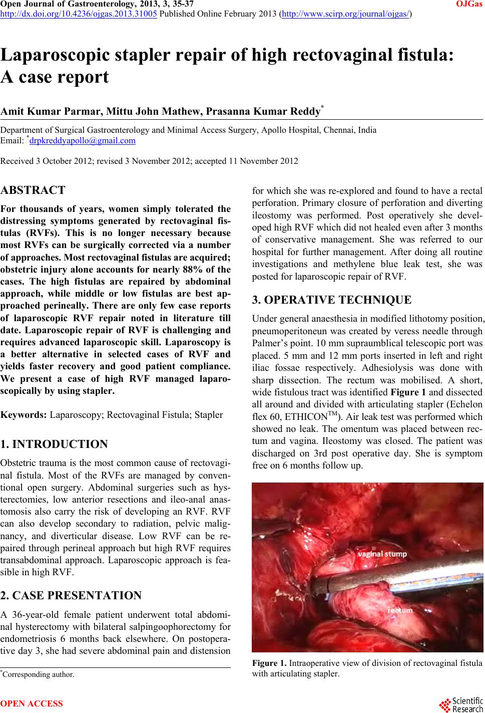 Open Journal of Gastroenterology, 2013, 3, 35-37 OJGas http://dx.doi.org/10.4236/ojgas.2013.31005 Published Online February 2013 (http://www.scirp.org/journal/ojgas/) Laparoscopic stapler repair of high rectovaginal fistula: A case report Amit Kumar Parmar, Mittu John Mathew, Prasanna Kumar Reddy* Department of Surgical Gastroenterology and Minimal Access Surgery, Apollo Hospital, Chennai, India Email: *[email protected] Received 3 October 2012; revised 3 November 2012; accepted 11 November 2012 ABSTRACT For thousands of years, women simply tolerated the distressing symptoms generated by rectovaginal fis- tulas (RVFs). This is no longer necessary because most RVFs can be surgically corrected via a number of approaches. Most rectovaginal fistulas are acquired; obstetric injury alone accounts for nearly 88% of the cases. The high fistulas are repaired by abdominal approach, while middle or low fistulas are best ap- proached perineally. There are only few case reports of laparoscopic RVF repair noted in literature till date. Laparoscopic repair of RVF is challenging and requires advanced laparoscopic skill. Laparoscopy is a better alternative in selected cases of RVF and yields faster recovery and good patient compliance. We present a case of high RVF managed laparo- scopically by using stapler. Keywords: Laparoscopy; Rectovaginal Fistula; Stapler 1. INTRODUCTION Obstetric trauma is the most common cause of rectovagi- nal fistula. Most of the RVFs are managed by conven- tional open surgery. Abdominal surgeries such as hys- terectomies, low anterior resections and ileo-anal anas- tomosis also carry the risk of developing an RVF. RVF can also develop secondary to radiation, pelvic malig- nancy, and diverticular disease. Low RVF can be re- paired through perineal approach but high RVF requires transabdominal approach. Laparoscopic approach is fea- sible in high RVF. 2. CASE PRESENTATION A 36-year-old female patient underwent total abdomi- nal hysterectomy with bilateral salpingoophorectomy for endometriosis 6 months back elsewhere. On postopera- tive day 3, she had severe abdominal pain and distension for which she was re-explored and found to have a rectal perforation. Primary closure of perforation and diverting ileostomy was performed. Post operatively she devel- oped high RVF which did not healed even after 3 months of conservative management. She was referred to our hospital for further management. After doing all routine investigations and methylene blue leak test, she was posted for laparoscopic repair of RVF. 3. OPERATIVE TECHNIQUE Under general anaesthesia in modified lithotomy position, pneumoperitoneun was created by veress needle through Palmer’s point. 10 mm supraumblical telescopic port was placed. 5 mm and 12 mm ports inserted in left and right iliac fossae respectively. Adhesiolysis was done with sharp dissection. The rectum was mobilised. A short, wide fistulous tract was identified Figure 1 and dissected all around and divided with articulating stapler (Echelon flex 60, ETHICONTM). Air leak test was performed w h i ch showed no leak. The omentum was placed between rec- tum and vagina. Ileostomy was closed. The patient was discharged on 3rd post operative day. She is symptom free on 6 mon t hs fo l low up. Figure 1. Intraoperative view of division of rectovaginal fistula with articulating stapler. *Corresponding a uthor. OPEN ACCESS  A. K. Parmar et al. / Open Journal of Gastroenterology 3 (2013) 35-37 36 4. DISCUSSION Rectovaginal fistula is epithelial lined communication between rectum and vagina. Most common cause of RVF is obstetric trauma. It can occur as a complication after variety of rectal, vaginal and pelvic operations including hysterectomy, low anterior resection, ileal-pouch anal anastomosis and stapled hemorroidectomy [1]. RVF can be associated with pelvic malignancy or radiation ther- apy for malignancy, and inflammatory bowel disease. RVFs can be classified into low and high varieties. Low RVF is between the lower third of the rectum and the lower half of the vagina. A high RVF is between the middle third of the rectum and the posterior vaginal fornix. Small-sized fistulas are less than 0.5 cm in di- ameter, medium-sized fistulas are 0.5 - 2.5 cm, and large-sized fistulas exceed 2.5 cm [2]. Clinical features of the rectovaginal fistula are stool and air passage from the vagina. Other symptoms include recurrent urinary tract infection and perineal skin inflammation or infec- tion. Symptoms of chronic inflammat ion and irritation in these patients have an effect on their social life and psy- chology and lead to sexual dysfunction. Most fistulas at the lower rectum are palpable by digital rectal examina- tion. Contrast radiography is the most dependable meth od for diagnosing small and high rectovaginal fistulas. High fistulas may not be readily apparent on physical exami- nation or vaginal inspection and may even be missed by endoscopy. Methylene blue enema with a vaginal tam- pon in place, looking for staining on the tampoon is used to confirm the diagnosis. Vaginography with a water soluble contrast medium has a reported sensitivity of 79% to 100% [3-5]. CT and MRI also play a role in the diagnosis and evaluation of the RVF as they may give insight into the un derlying cause of the fistula. Spontaneous healing may occur with adequate medical treatment such as total parenteral nutrition, antibiotics, and long-time fasting. However, surgical therapy re- mains the mainstay for managing complex fistula was not suitable for conservative management or underwent prolonged conservative management (without resolution). Operative access to this type of lesion includes fecal di- version, and/or transperineal approach of resection, or rectal anastomosis or repair. Operation of the middle and lower rectum is associated with complications, including urinary and sexual dysfunction. The management of RVF depends on size, location, cause, anal sphincter function and overall health status of the patient. Low fistulas can be repaired through perineal approach. Transabdominal approach is standard for high fistula. Total laparoscopic repair of RVF is still rare. Nezhat CH et al. [6] reports correction of two cases of RVF by laparoscopy. Pelosi et al. [1] reported laparoscopic upper rectovaginal mobili- sation with transvaginal repair of recurrent RVF. Pala- nivelu et al. [2] reported 2 cases of high RVF managed laparoscopically. Schwenk et al. [7] reported a case of intracorporeal colorectal anastomosis for which they had performed a laparoscopic resection of the sigmoid colon with the fistulous tract and. They all concluded that laparoscopic repair of RVF is feasible but it demands adequate experience in advanced laparoscopic proce- dures and proper identification of tissue planes. Good preparation of the bowel is essential to avoid any faecal contamination of the operative area. Fistulous tract is generally divided and closed by using suture. But, in our case we used linear stapler for this purpose. The idea behind using it was to make procedure simpler and faster and avoidance of faecal contamination as well. And thus, we were able to close the ileostomy at the same time and avoided one more surgical burden on the patient. This is probably first case of laparoscopic stapler repair of RVF in literature. 5. CONCLUSION Laparoscopic repair of RVF is challenging and requires advanced laparoscopic skill. Laparoscopy is a promising alternative in selected cases of RVF and yields faster recovery and good patient compliance. We found that stapler repair as compared to primary intracorporeal clo- sure makes the procedure simpler, faster, and easy. Safety and long term outcomes of laparoscopic repair is yet to be proved by long term follow up and further studies. REFERENCES [1] Pelosi III, M.A. and Pelosi, M.A. (1997) Transvaginal repair of recurrent rectovaginal fistula with laparoscopic- assisted rectovaginal mobilization. Journal of Laparoen- doscopic & Advanced Surgical Techniques, 7, 379-383. doi:10.1089/lap.1997.7.379 [2] Palanivelu, C., Rangarajan, M., Senthilkumar, R., Ma- dankumar, M.V. and Kalyanakumari, V. (2007) Laparo- scopic management of iatrogenic high rectovaginal fistu- las (type VI). Singapore Medical Journal, 48, e96- e 9 8. [3] Arnold, M.W., Aguilar, P.S. and Stewart, W.R.C. (1990) Vaginography: An easy and safe technique for diagnosis of colovaginal fistulas. Diseases of the Colon & Rectum, 33, 344-345. doi:10.1007/BF02055482 [4] Bird, D., Taylor, D. and Lee, P. (1993) Vaginography: The investigation of choice for vaginal fistulae? Austra- lian and New Zealand Journal of Surgery, 63, 894-896. doi:10.1111/j.1445-2197.1993.tb00366.x [5] Giordano, P., Drew, P.J. and Taylor, D. (1996) Vagino- graphy—Investigation of choice for clinically suspected vaginal fistulas. Diseases of the Colon & Rectum, 39, 568-572. doi:10.1007/BF02058713 [6] Nezhat, C.H., Bastidas, J.A., Pennington, E., Nezhat, F.R., Raga, F. and Nezhat, C.R. (1998) Laparoscopic treatment Copyright © 2013 SciRes. OPEN ACCESS  A. K. Parmar et al. / Open Journal of Gastroenterology 3 (2013) 35-37 Copyright © 2013 SciRes. OPEN ACCESS 37 of type IV rectovaginal fistula. Journal of the American Association of Gynecologic Laparoscopists, 5, 297-299. doi:10.1016/S1074-3804(98)80036-4 [7] Schwenk, W., Bohm, B., Grundel, K. and Muller, J. (1997) Laparoscopic resection of high rectovaginal fistula with intracorporeal colorectal anastomosis and omento- plasty. Surgical Endoscopy, 11, 147-149. doi:10.1007/s004649900318 |