

Paper Menu >>
Journal Menu >>
 Advances in Bioscience and Biotechnology, 2013, 4, 113-117 ABB http://dx.doi.org/10.4236/abb.2013.41A016 Published Online January 2013 (http://www.scirp.org/journal/abb/) Complications of airway self-expandable metallic stent in benign airway diseases Fu-Tsai Chung1,2,3*#, Shu-Min Lin1#, Chun-Liang Chou1,3, Hao-Cheng Chen3,4, Chih-Hsi Kuo1, Horng-Chyuan Lin1, Chien-Ying Liu1, Chun-Hua Wang1, Han-Pin Kuo1, Chih-Teng Yu1* 1Department of Thoracic Medicine, Chang Gung Memorial Hospital, College of Medicine, Chang Gung University, Taipei, Taiwan 2Graduate Institute of Clinical Medical Sciences, College of Medicine, Chang Gung University, Taoyuan, Taiwan 3Department of Thoracic Medicine, Saint Paul’s Hospital, Taoyuan, Taiwan 4Department of Thoracic Medicine, Shuang Ho Hospital, Taipei Medical University, Taipei, Taiwan Email: *[email protected] Received 5 November 2012; revised 17 December 2012; accepted 14 January 2013 ABSTRACT Objectives: To present the complications of Ultraflex Self-Expandable Metallic Stents (SEMSs) applied in patients with benign tracheobronchial diseases. Meth- ods: Eighty patients received 124 SEMSs were retro- spectively reviewed in a tertiary hospital. Results: The baseline characteristics of patients rec eived S EM S implantation including age (mean + standard devia- tion: 62.5 + 14.8 years), gender (male, 60.5%), smok- ing (46.8%), forced expiratory volume in first one second (FEV1) (mean + standard deviation, 0.92 + 0.41 L/s), follow-up days after SEMS implantation (median (interquartile range); 457 (131 - 897)) and covered SEMS (33.9%) were listed. Symptoms im- proved after SEMS implantation among 95 patients (76.6%). Time to complications developed was 236 (median; interquartile range, 59 - 672) days. The overall complication rate was 41.9% after SEMS im- plantation. The complications included SEMS migra- tion (6.5%), granuloma (19.3%) and SEMS fracture (16.1%). Successful management rates of SEMS mi- gration, granuloma and SEMS fracture were up to 100%, 83.3%, and 85% respectively. Conclusions: Patients received SEMS implantation due to benign conditions had poor lung function and were old. The complication rate in patients with benign conditions was high after longer follow-up period, however, suc- cessful management achieved in most patients with complications. Keywords: Ultraflex Self-Expandable Metallic Stent; Benign Airway Disease; Complication; Management 1. INTRODUCTION Patients with symptomatic central airway obstruction could be managed in the past ten years [1-3]. Interven- tional bronchoscopic techniques have been widely ap- plied in patients with central airway lesions because of the advances in endobronchial stents and insertion skills [4-7]. Bronchoscopy with rigid or flexible bronchoscope is the common procedure of stent placement in those patients. However, some patients are not suitable to re- ceive surgical intervention with rigid bronchoscope with a general anesthetic, due to illness severity and comor- bidities. Self-Expandable Metallic Stents (SEMSs) can be successfully inserted using a flexible bronchoscope while the patient receives conscious sedation and a local anesthetic [8-10]. For some patients who are not eligible for surgical procedures or rigid bronchoscopy, self-ex- pandable metallic stents (SEMSs) insertion offers a ch an ce to relieve their symptoms [10,11]. Despite SEMSs could relieve central airway obstruc- tion in large tracheobronchial diseases, some complica- tions such as migration, granuloma formation, stent frac- ture, recurrent lumen obstruction of the stent and in- creased bacterial colonization and impaired mucocilliary clearance were reported [10,12-14]. The United States Food and Drug Administration (FDA) has warned that SEMS implantation should be considered only if the pa- tient is not eligible for surgery, rigid bronchoscopy, or silicone stent implantation due to these potentially haz- ardous complications. Despite the fact, studies [10,15] also concluded that the SEMS are useful management options for central airway obstruction symptoms in pa- tients with benign and malignant diseases. The complica- tions of this management modality in patients with be- nign obstructing lesions after placement of SEMSs re- main the important issues to be concerned. *Corresponding authors. #Drs. F.-T. Chung and S.-M. Lin equally contribute to this article as f irs t authors. None of the authors have any conflicts of interest to disclose.The goal of this study was to describe the baseline OPEN ACCESS 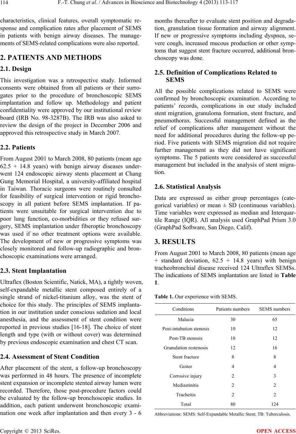 F.-T. Chung et al. / Advances in Bioscience and Biotechnology 4 (2013) 113-117 114 characteristics, clinical features, overall symptomatic re- sponse and complication rates after placement of SEMS in patients with benign airway diseases. The manage- ments of SEMS-related complications were also reported. 2. PATIENTS AND METHODS 2.1. Design This investigation was a retrospective study. Informed consents were obtained from all patients or their surro- gates prior to the procedure of bronchoscopic SEMS implantation and follow up. Methodology and patient confidentiality were approved b y our institutional review board (IRB No. 98-3287B). The IRB was also asked to review the design of the project in December 2006 and approved this retrospective study in March 2007. 2.2. Patients From August 2001 to March 2008, 80 p atients (mean age 62.5 + 14.8 years) with benign airway diseases under- went 124 endoscopic airway stents placement at Chang Gung Memorial Hospital, a university-affiliated hospital in Taiwan. Thoracic surgeons were routinely consulted for feasibility of surgical intervention or rigid broncho- scopy in all patient before SEMS implantation. If pa- tients were unsuitable for surgical intervention due to poor lung function, co-morbidities or they refused sur- gery, SEMS implantation under fiberoptic bronchoscopy was used if no other treatment options were available. The development of new or progressive symptoms was closely monitored and follow-up radiographic and bron- choscopic examinations were arranged. 2.3. Stent Implantation Ultraflex (Bosto n Scien tif ic, Natick , MA), a tigh tly w ove n, self-expandable metallic stent composed entirely of a single strand of nickel-titanium alloy, was the stent of choice for this study. The principles of SEMS implanta- tion in our institution under conscious sedation and local anesthesia, and the assessment of stent condition were reported in previous studies [16-18]. The choice of stent length and type (with or without cover) was determined by previous endoscopic examination and chest CT scan. 2.4. Assessment of Stent Condition After placement of the stent, a follow-up bronchoscopy was performed in 48 hours. The presence of incomplete stent expansion or incomplete stented airway lumen were recorded. Therefore, those post-procedure factors could be evaluated by the follow-up bronchoscopic studies. In addition, each patient underwent bronchoscopic exami- nation one week after implantation and then every 3 - 6 months thereafter to evaluate stent position and degrada- tion, granulation tissue formation and airway alignment. If new or progressive symptoms including dyspnea, se- vere cough, increased mucous production or other symp- toms that suggest stent fracture occurred, additional br on- choscopy was done. 2.5. Definition of Complications Related to SEMS All the possible complications related to SEMS were confirmed by bronchoscopic examination. According to patients’ records, complications in our study included stent migration, granuloma formation, stent fracture, and pneumothorax. Successful management defined as the relief of complications after management without the need for additional procedures during the follow-up pe- riod. Five patients with SEMS migration did not require further management as they did not have significant symptoms. The 5 patients were considered as successful management but included in the analysis of stent migra- tion. 2.6. Statistical Analysis Data are expressed as either group percentages (cate- gorical variables) or mean ± SD (continuous variables). Time variables were expressed as median and Interquar- tile Range (IQR). All analysis used GraphPad Prism 3.0 (GraphPad Sof t ware, San Diego, Calif). 3. RESULTS From August 2001 to March 2008, 80 p atients (mean age + standard deviation, 62.5 + 14.8 years) with benign tracheobronchial disease received 124 Ultraflex SEMSs. The indications of SEMS implantation are listed in Table 1. Table 1. Our experience with SEMS. Conditions Patients numbers SEMS numbers Malacia 30 65 Post-intubation st enosis10 12 Post-TB stenosis 10 12 Granulation restenosis12 16 Stent fracture 8 8 Goiter 4 4 Corrosive injury 2 3 Mediastinitis 2 2 Tracheitis 2 2 Total 80 124 Abbreviations: SEMS: Self-Expandable Metallic Stent; TB: Tuberculosis. Copyright © 2013 SciRes. OPEN ACCESS 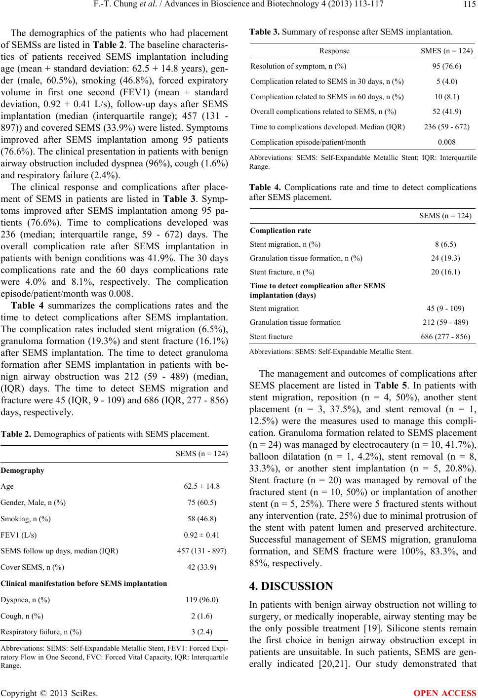 F.-T. Chung et al. / Advances in Bioscience and Biotechnology 4 (2013) 113-117 11 5 The demographics of the patients who had placement of SEMSs are listed in Table 2. The baseline characteris- tics of patients received SEMS implantation including age (mean + standard deviation: 62.5 + 14.8 years), gen- der (male, 60.5%), smoking (46.8%), forced expiratory volume in first one second (FEV1) (mean + standard deviation, 0.92 + 0.41 L/s), follow-up days after SEMS implantation (median (interquartile range); 457 (131 - 897)) and covered SEMS (33.9%) were listed. Symptoms improved after SEMS implantation among 95 patients (76.6%). The clinical presentation in patients with benign airway obstruction included dyspnea (96%), cough (1.6 %) and respiratory failure (2.4%). The clinical response and complications after place- ment of SEMS in patients are listed in Table 3. Symp- toms improved after SEMS implantation among 95 pa- tients (76.6%). Time to complications developed was 236 (median; interquartile range, 59 - 672) days. The overall complication rate after SEMS implantation in patients with benign conditions was 41.9%. The 30 days complications rate and the 60 days complications rate were 4.0% and 8.1%, respectively. The complication episode/patient/month was 0. 008. Table 4 summarizes the complications rates and the time to detect complications after SEMS implantation. The complication rates included stent migration (6.5%), granuloma formation (19.3%) and stent fracture (16.1%) after SEMS implantation. The time to detect granuloma formation after SEMS implantation in patients with be- nign airway obstruction was 212 (59 - 489) (median, (IQR) days. The time to detect SEMS migration and fracture were 45 (IQ R, 9 - 109) and 686 (IQR, 2 77 - 856 ) days, respectively. Table 2. Demographics of patients with SEMS placement. SEMS (n = 124) Demography Age 62.5 ± 14.8 Gender, Male, n (%) 75 (60.5) Smoking, n (%) 58 (46.8) FEV1 (L/s) 0.92 ± 0.41 SEMS follow up days, median (IQR) 457 (131 - 897) Cover SEMS, n (%) 42 (33.9) Clinical manifestation before SEMS implantation Dyspnea, n (%) 119 (96.0) Cough, n (%) 2 (1.6) Respiratory failure, n (%) 3 (2.4) Abbreviations: SEMS: Self-Expandable Metallic Stent, FEV1: Forced Expi- ratory Flow in One Second, FVC: Forced Vital Capacity, IQR: Interquartile Range. Table 3. Summary of response after SEMS implantation. Response SMES (n = 124) Resolution of symptom, n (%) 95 (76.6) Complication related to SEMS in 30 days, n (%) 5 (4.0) Complication related to SEMS in 60 days, n (%) 10 (8.1) Overall complications related to SEMS, n (%) 52 (41.9) Time to complicat ions developed. Median (IQR) 236 (59 - 672) Complication ep isode/patient/month 0.008 Abbreviations: SEMS: Self-Expandable Metallic Stent; IQR: Interquartile Range. Table 4. Complications rate and time to detect complications after SEMS placement. SEMS (n = 124) Complication rate Stent migration, n (%) 8 (6.5) Granulation tissue formation, n (%) 24 (19.3) Stent fracture, n (%) 20 (16.1) Time to detect complication after SEMS implantation (days) Stent migration 45 (9 - 109) Granulation tissue formation 212 (59 - 489) Stent fracture 686 (277 - 856) Abbreviations: SEMS: Self-Expandable Metallic Stent. The management and outcomes of complications after SEMS placement are listed in Table 5. In patients with stent migration, reposition (n = 4, 50%), another stent placement (n = 3, 37.5%), and stent removal (n = 1, 12.5%) were the measures used to manage this compli- cation. Granuloma formation related to SEMS placement (n = 24) was managed by electrocautery (n = 10, 41.7%), balloon dilatation (n = 1, 4.2%), stent removal (n = 8, 33.3%), or another stent implantation (n = 5, 20.8%). Stent fracture (n = 20) was managed by removal of the fractured stent (n = 10, 50%) or implantation of another stent (n = 5, 25%). There were 5 fractured stents without any intervention (rate, 25%) due to minimal protrusion of the stent with patent lumen and preserved architecture. Successful management of SEMS migration, granuloma formation, and SEMS fracture were 100%, 83.3%, and 85%, respectively. 4. DISCUSSION In patients with benign airway obstruction not willing to surgery, or medically inoperable, airway stenting may be the only possible treatment [19]. Silicone stents remain the first choice in benign airway obstruction except in patients are unsuitable. In such patients, SEMS are gen- erally indicated [20,21]. Our study demonstrated that Copyright © 2013 SciRes. OPEN ACCESS 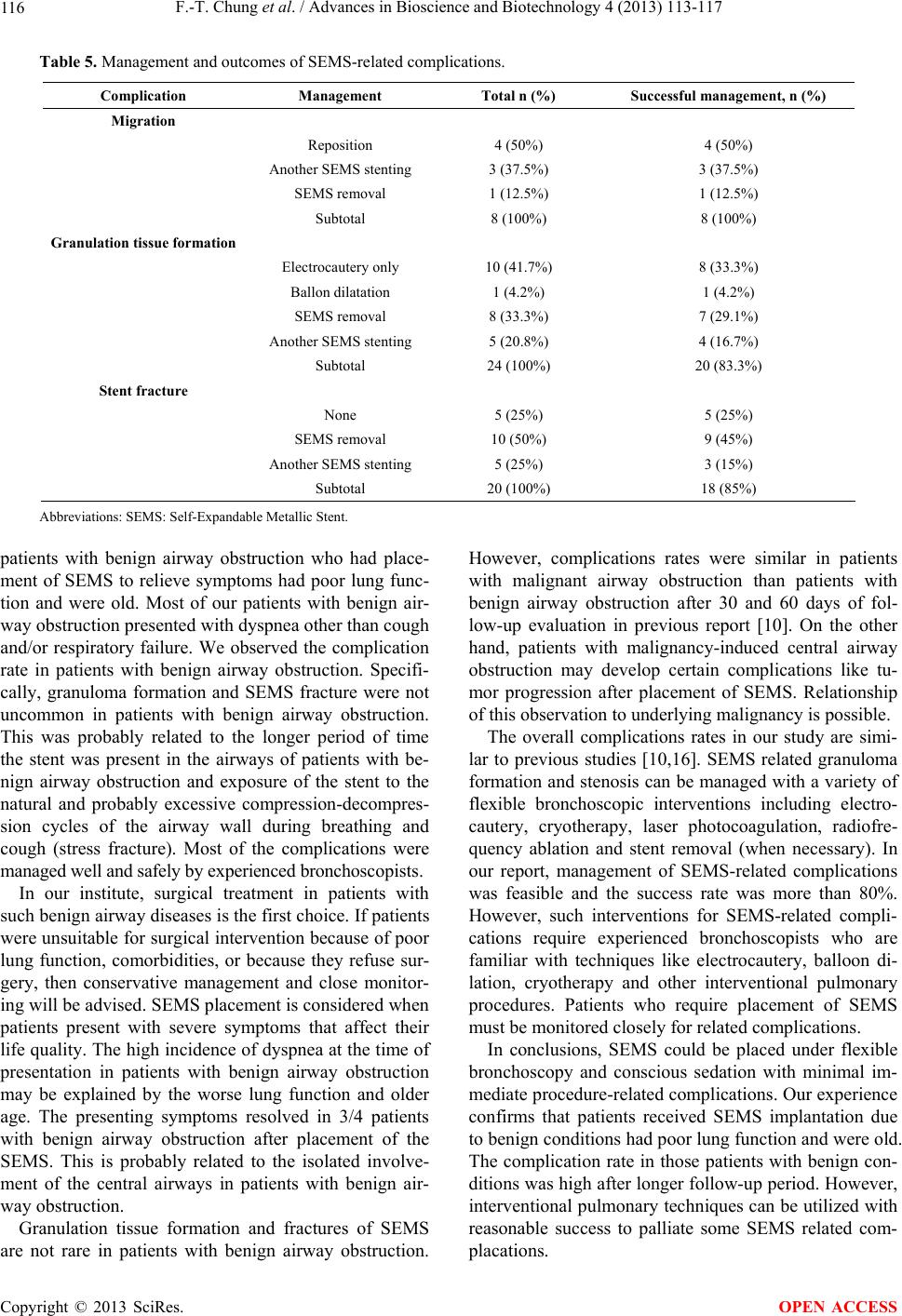 F.-T. Chung et al. / Advances in Bioscience and Biotechnology 4 (2013) 113-117 Copyright © 2013 SciRes. 116 OPEN ACCESS Table 5. Management and outcomes of SEMS-related complications. Complication Management Total n (%) Successful management, n (%) Migration Reposition 4 (50%) 4 (50%) Another SEMS stenting 3 (37.5%) 3 (37.5%) SEMS removal 1 (12.5%) 1 (12.5%) Subtotal 8 (100%) 8 (1 0 0%) Granulation tissue formation Electrocautery only 10 (41.7%) 8 (33.3%) Ballon dilatation 1 (4.2%) 1 (4.2%) SEMS removal 8 (33.3%) 7 (29.1%) Another SEMS stenting 5 (20.8%) 4 (16.7%) Subtotal 24 (100%) 20 (83.3%) Stent fracture None 5 (25%) 5 (25%) SEMS removal 10 (50%) 9 (45%) Another SEMS stenting 5 (25%) 3 (15%) Subtotal 20 (100%) 18 (85%) Abbreviations: SEMS: Self-Expandable Metallic Stent. patients with benign airway obstruction who had place- ment of SEMS to relieve symptoms had poor lung func- tion and were old. Most of our patients with benign air- way obstruction presented with dyspnea other than cough and/or respiratory failure. We observed the complication rate in patients with benign airway obstruction. Specifi- cally, granuloma formation and SEMS fracture were not uncommon in patients with benign airway obstruction. This was probably related to the longer period of time the stent was present in the airways of patients with be- nign airway obstruction and exposure of the stent to the natural and probably excessive compression-decompres- sion cycles of the airway wall during breathing and cough (stress fracture). Most of the complications were managed well and safely by experienced bronchoscopists. In our institute, surgical treatment in patients with such benign airway diseases is the first choice. If patients were unsuitable for surgical intervention because of poor lung function, comorbidities, or because they refuse sur- gery, then conservative management and close monitor- ing will be advised. SEMS placement is considered when patients present with severe symptoms that affect their life quality. The high incidence of dyspnea at the time of presentation in patients with benign airway obstruction may be explained by the worse lung function and older age. The presenting symptoms resolved in 3/4 patients with benign airway obstruction after placement of the SEMS. This is probably related to the isolated involve- ment of the central airways in patients with benign air- way obstruction. Granulation tissue formation and fractures of SEMS are not rare in patients with benign airway obstruction. However, complications rates were similar in patients with malignant airway obstruction than patients with benign airway obstruction after 30 and 60 days of fol- low-up evaluation in previous report [10]. On the other hand, patients with malignancy-induced central airway obstruction may develop certain complications like tu- mor progression after placement of SEMS. Relationship of this observation to underlying malignancy is possible. The overall complications rates in our study are simi- lar to previous studies [10,16]. SEMS related granuloma formation and stenosis can be managed with a variety of flexible bronchoscopic interventions including electro- cautery, cryotherapy, laser photocoagulation, radiofre- quency ablation and stent removal (when necessary). In our report, management of SEMS-related complications was feasible and the success rate was more than 80%. However, such interventions for SEMS-related compli- cations require experienced bronchoscopists who are familiar with techniques like electrocautery, balloon di- lation, cryotherapy and other interventional pulmonary procedures. Patients who require placement of SEMS must be monitored closely for related complications. In conclusions, SEMS could be placed under flexible bronchoscopy and conscious sedation with minimal im- mediate procedure-related complications. Our experience confirms that patients received SEMS implantation due to benign conditions had poor lung function and were old. The complication rate in those patients with benign con- ditions was high after longer follow-up period. However, interventional p ulmonary techniques can be utilized with reasonable success to palliate some SEMS related com- placations.  F.-T. Chung et al. / Advances in Bioscience and Biotechnology 4 (2013) 113-117 11 7 5. ACKNOWLEDGEMENTS This study was partial supported by grants from Change Gung Memo- rial Hospital (CMRPG391211 by Drs. F.T.C., C.T.Y., and C.S.K.; CMRPG3B0981-3 by Dr. F.T.C. and CMRPG3B0831-3 by Dr. F.T.C.). REFERENCES [1] Seijo, L.M. and Sterman, D.H. (2001) Interventional pul- monology. The New England Journal of Medicine, 344, 740-749. doi:10.1056/NEJM200103083441007 [2] Bolliger, C.T., Mathur, P.N., Beamis, J.F., et al. (2002) ERS/ATS statement on interventional pulmonology. Euro- pean Respiratory Society/American Thoracic Society. European Respiratory Journal, 19, 356-373. [3] Brichet, A., Verkindre, C., Dupont, J., et al. (1999) Mul- tidisciplinary approach to management of postintubation tracheal stenoses. European Respiratory Journal, 13, 888- 893. doi:10.1034/j.1399-3003.1999.13d32.x [4] Al-Kattan, K., Simonds, A., Chung, K.F. and Kaplan, D.K. (1997) Kyphoscoliosis and bronchial torsion. Chest, 111, 1134-1137. doi:10.1378/chest.111.4.1134 [5] Colt, H.G. and Harrell, J.H. (1997) Therapeutic rigid bronchoscopy allows level of care changes in patients with acute respiratory failure from central airways ob- struction. Chest, 112, 202-206. doi:10.1378/chest.112.1.202 [6] Shaffer, J.P. and Allen, J.N. (1998) The use of expand- able metal stents to facilitate extubation in patients with large airway obstruction. Chest, 114, 1378-1382. doi:10.1378/chest.114.5.1378 [7] Noppen, M., Stratakos, G., Amjadi, K., et al. (2007) Stent - ing allows weaning and extubation in ventilator- or tra- cheostomy dependency secondary to benign airway dis- ease. Respiratory Medicine, 101, 139-145. doi:10.1016/j.rmed.2006.03.037 [8] Hautmann, H., Bauer, M., Pfeifer, K.J. and Huber, R.M. (2000) Flexible bronchoscopy: A safe method for metal stent implantation in bronchial disease. The Annals of Tho- racic Surgery, 69, 398-401. doi:10.1016/S0003-4975(99)01398-3 [9] Madden, B.P., Datta, S. and Charokopos, N. (2002) Ex- perience with Ultraflex expandable metallic stents in the management of endobronchial pathology. The Annals of Thoracic Surgery, 73, 938-944. doi:10.1016/S0003-4975(01)03460-9 [10] Chung, F.T., Chen, H.C., Chou, C.L., et al. (2011) An outcome analysis of self-expandable metallic stents in central airway obstruction: A cohort study. Journal of Cardiothoracic Surgery, 6, 46. doi:10.1186/1749-8090-6-46 [11] Madden, B.P., Loke, T.K. and Sheth, A.C. (2006) Do expandable metallic airway stents have a role in the man- agement of patients with benign tracheobronchial disease? The Annals of Thoracic Surgery, 82, 274-278. doi:10.1016/j.athoracsur.2006.02.028 [12] Zakaluzny, S.A., Lane, J.D. and Mair, E.A. (2003) Com- plications of tracheobronchial airway stents. Otolaryn- gology—Head and Neck Surgery, 128, 478-488. doi:10.1016/S0194-5998(03)00002-0 [13] Gaissert, H.A., Grillo, H.C., Wright, C.D., et al. (2003) Complication of benign tracheobronchial strictures by self-expanding metal stents. The Journal of Thoracic and Cardiovascular Surgery, 126, 744-747. doi:10.1016/S0022-5223(03)00361-1 [14] Noppen, M., Meysman, M., Claes, I., D’Haese, J., et al. (1999) Screw-thread vs Dumon endoprosthesis in the management of tracheal stenosis. Chest, 115, 532-535. doi:10.1378/chest.115.2.532 [15] Husain, S.A., Finch, D., Ahmed, M., et al. (2007) Long- term follow-up of ultraflex metallic stents in benign and malignant central airway obstruction. The Annals of Tho- racic Surgery, 83, 1251-1256. doi:10.1016/j.athoracsur.2006.11.066 [16] Chung, F.T., Lin, S.M., Chen, H.C., et al. (2010) Factors to obstructive grangulation tissue formation after ultraflex stent implantation in benign tracheal stenosis. The Jour- nal of Thoracic and Cardiovascular Surgery, 58, 102- 107. doi:10.1055/s-0029-1186266 [17] Chung, F.T., Chen, G.Y., Chou, C.L., et al. (2012) Re- move airway ultraflex stents by flexible bronchoscope study. The American Jou rnal of the Medical Sciences, 343, 267-272. doi:10.1097/MAJ.0b013e31822a6bc3 [18] Chung, F.T., Lin, H.C., Chou, C.L., et al. (2012) Airway ultraflex stenting in esophagorespiratory fistula caused by esophageal cancer. The American Journal of the Medical Sciences, 344, 105-109. doi:10.1097/MAJ.0b013e3182367b6a [19] Wood, D.E., Liu, Y.H., Vallieres, E., et al. (2003) Airway stenting for malignant and benign tracheobronchial steno- sis. The Annals of Thoracic Surgery, 76, 167-172. doi:10.1016/S0003-4975(03)00033-X [20] Noppen, M., Stratakos, G., D’Haese, J., et al. (2005) Removal of covered self-expanda ble metallic airway st e n t s in benign disorders: Indications, technique, and outcomes. Chest, 127, 482-487. doi:10.1378/chest.127.2.482 [21] Nesbitt, J.C. and Carrasco, H. (1996) Expandable stents. Chest Surgery Clinics of North America, 6, 305-328. Copyright © 2013 SciRes. OPEN ACCESS |