

Paper Menu >>
Journal Menu >>
 Advances in Computed Tomography, 2012, 1, 17-20 http://dx.doi.org/10.4236/act.2012.13004 Published Online December 2012 (http://www.SciRP.org/journal/act) Left Pulmonary Artery Sling Associated with Patent Ductus Arteriosus and Atrial Septal Defect: Evaluation with Multidetector CT Rukeshman Shakya, Jianhua Liu, Xiaolin Xu, Mohit Godar, Qinghai Yuan Department of Radiology, The Second Hospital of Jilin University, Changchun, China Email: [email protected] Received October 16, 2012; revised November 18, 2012; accepted November 26, 2012 ABSTRACT We report a case of left pulmonary artery sling associated with patent ductus arteriosus and atrial septal defect in a 21-month-old child. 256-slice MDCT provides valuable information, such as abnormal origin of the left pulmonary ar- tery, the relationship between pulmonary artery and airway, the diameter of the patent ductus artery and atrial septal defect. The information is helpful in d iagnosis, pre-operative evaluation and post-operative follow-up of LPS. Keywords: Left Pulmonary Artery Sling; Patent Ductus Arteriosus; Atrial Septal Defect; Multidetector CT; CT Angiography 1. Introduction The left pulmonary artery sling (LPS) is a rare congenital abnormality, and is defined as that the LPA arising from the posterior aspect of RPA, crossing over the right mainstem bronchus near its origin, and coursing leftward between the trachea and esophagus to enter the left hemithorax. Atrial Septal Defect (ASD) is a congenital heart disease in which abnormal blood flows between left atrium and right atrium through defect in interatrial sep- tum. Patent Ductus Arteriosus (PDA) is heart problem that occurs due to abnormal blood flow between aorta and pulmonary artery caused by persistent ductus arteri- osus (tube) which didn’t close soon after child birth or after few days. The clinical manifestations of left pulmonary artery sling are mild or occasional stridor, breathless, inspira- tory and expiratory rhonchi, productive cough, cyanosis, difficulty in feeding. It is due to external compression of trachea or severe compression from complete tracheal rings. Hypoplasia of entire tracheobronchial tree or res- piratory distress with pneumonia, atelectasis, intercostal recession or air trapping may occur. When it is associ- ated with ASD and PDA, patient can present with short- ness of breath, swelling of limbs, systolic ejection mur- mur, wide splitting of P2, accentuation of right precordial thrust, hoarse cry, cough, lower respiratory tract infec- tions, failure to thrive, tachypnea, atelectasis or pneumo- nia. We report a rare case of LPS associated with patent ductus arteriosus and atrial septal defect. 2. Case Report A 21-month-old child presented to the pediatric depart- ment with recurrent cough for half a year and aggrava- tion of the symptoms with wheezing for 3 days. She was admitted after being diagnosed with lobar pneumonia on chest radiography and CT (Figures 1(a)-(c)). The medi- cal history revealed recurrent episodes of productive cough with fever, and on auscultation, continuous systolic mur- mur was heard along the upper left sternal border. Echo- cardiography showed a patent ductus artery (PDA) and atrial septal defect (ASD) (Figures 1(d) and (e)). Two weeks later, after recovery from pneumonia (Figure 1(f)), the patient was transferred to the depart- ment of cardiac surgery. CT angiography (CTA) was done using a 256-slice MDCT scanner both for diagnos- tic and pre-operative evaluation (Figures 2(a)-(e)). CTA showed a left pulmonary artery sling (PAS) associated with PDA and ASD. During surgery, the pulmonary ar- tery and aorta were separated and the patent ductus arte- riosus was doubly ligated and divided. The left pulmo- nary artery was dissected circumferentially to the hilar branches, and then anastomosed to the origin of PDA (Figure 2(f)). The ASD was also repaired. Two weeks later, the patient was discharged without complication. The case report was approved by the patient’s parents. 3. Discussion Aberrant left pulmonary artery (LPA) from the right pulmonary artery (RPA) is arare congen ital abnormality. C opyright © 2012 SciRes. ACT 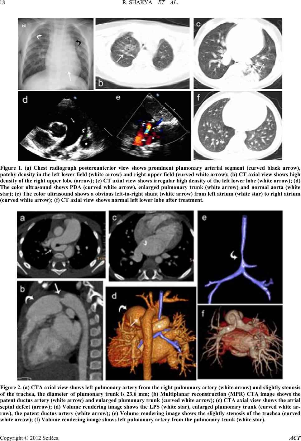 R. SHAKYA ET AL. 18 Figure 1. (a) Chest radiograph posteroanterior view shows prominent plumonary arterial segment (curved black arrow), patchy density in the le ft lower field (white arrow ) and right upper field (c urved white arrow); (b) CT axial view shows high density of the right upper lobe (arrow); (c) CT axial view shows irregular high density of the left lower lobe (w hite arr ow ); (d) The color ultrasound shows PDA (curved white arrow), enlarged pulmonary trunk (white arrow) and normal aorta (white star); (e) The color ultrasound shows a obvious left-to-right shunt (white arrow) from left atrium (white star) to right atrium (curved white arrow); (f) CT axial view shows normal left lowe r lo be aft er treatmen t. Figure 2. (a) CTA axial view shows left pulmonary artery from the right pulmonary artery (white arrow) and slightly stenosis of the trachea, the diameter of plumonary trunk is 23.6 mm; (b) Multiplanar reconstruction (MPR) CTA image shows the patent ductus arte ry (white arrow ) and enlarged plumonar y trunk (curve d white arrow); (c ) CTA axial view shows the atrial septal defect (arrow ); (d) Volume rendering image shows the LPS (white star), enlarged plumonary trunk (cur ved white ar- row), the patent ductus artery (white arrow); (e) Volume rendering image shows the slightly stenosis of the trachea (curved hite arrow); (f) Volume rendering image shows left pulmonary artery from the pulmonar y trunk (white star). w Copyright © 2012 SciRes. ACT 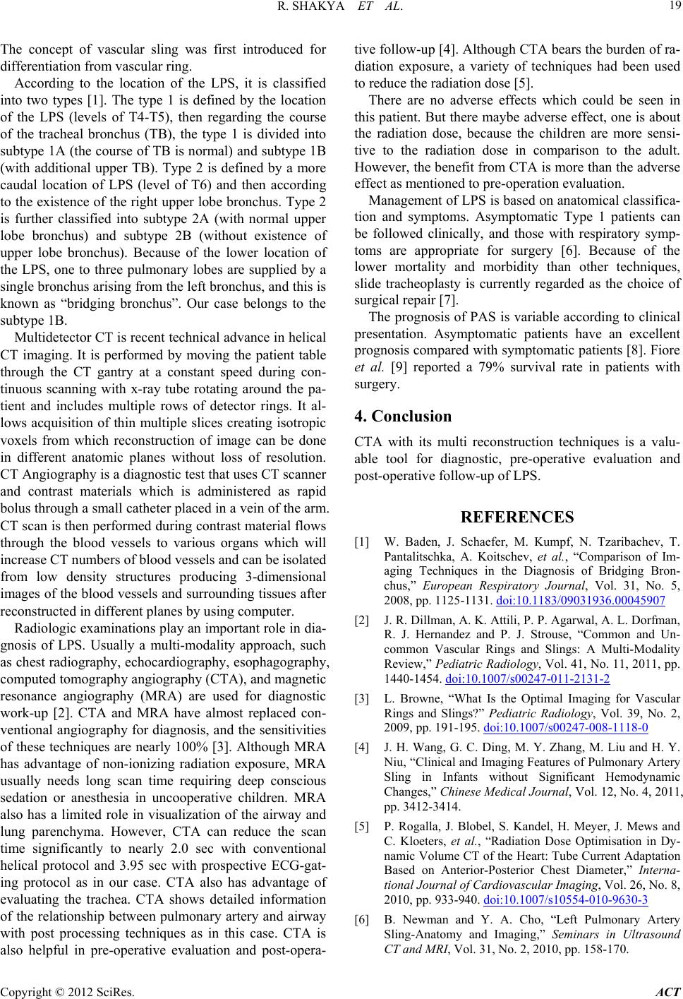 R. SHAKYA ET AL. 19 The concept of vascular sling was first introduced for differe nt iation fr om vascular ri ng. According to the location of the LPS, it is classified into two types [1]. The type 1 is defined by the location of the LPS (levels of T4-T5), then regarding the course of the tracheal bronchus (TB), the type 1 is divided into subtype 1A (the course of TB is normal) and subtype 1B (with additional upper TB). Type 2 is defined by a more caudal location of LPS (level of T6) and then according to the existence of the right upper lobe bronchus. Type 2 is further classified into subtype 2A (with normal upper lobe bronchus) and subtype 2B (without existence of upper lobe bronchus). Because of the lower location of the LPS, one to three pulmonary lobes are supplied by a single bronchus arising fr om the left bron chus, and this is known as “bridging bronchus”. Our case belongs to the subtype 1B . Multidetector CT is recent technical advance in helical CT imaging. It is performed by moving the patient table through the CT gantry at a constant speed during con- tinuous scanning with x-ray tube rotating around the pa- tient and includes multiple rows of detector rings. It al- lows acquisition of thin multiple slices creating isotropic voxels from which reconstruction of image can be done in different anatomic planes without loss of resolution. CT Angiography is a diagnostic test that uses CT scanner and contrast materials which is administered as rapid bolus through a small catheter placed in a vein of the arm. CT scan is then performed during contrast material flows through the blood vessels to various organs which will increase CT numbers of blood vessels and can be isolated from low density structures producing 3-dimensional images of the blood vessels and su rrounding tissues after reconstructed in different planes by usin g c omputer. Radiologic examinations play an important role in dia- gnosis of LPS. Usually a multi-modality approach, such as chest radiograph y, echocardiography, esophagogr aphy, computed tomography angiography (CTA), and magnetic resonance angiography (MRA) are used for diagnostic work-up [2]. CTA and MRA have almost replaced con- ventional ang iography for diagnosis, and the sens itivities of these techniques are nearly 100% [3]. Although MRA has advantage of non-ionizing radiation exposure, MRA usually needs long scan time requiring deep conscious sedation or anesthesia in uncooperative children. MRA also has a limited role in visualization of the airway and lung parenchyma. However, CTA can reduce the scan time significantly to nearly 2.0 sec with conventional helical protocol and 3.95 sec with prospective ECG-gat- ing protocol as in our case. CTA also has advantage of evaluating the trachea. CTA shows detailed information of the relationship between pulmonary artery and airway with post processing techniques as in this case. CTA is also helpful in pre-operative evaluation and post-opera- tive follow-u p [4]. Although CTA bears th e burden of ra- diation exposure, a variety of techniques had been used to reduce the radiatio n dose [5]. There are no adverse effects which could be seen in this patient. But there maybe adverse effect, one is about the radiation dose, because the children are more sensi- tive to the radiation dose in comparison to the adult. However, the benefit from CTA is more than the adverse effect as mentioned to pre-operation evaluation. Management of LPS is based on anatomical classifica- tion and symptoms. Asymptomatic Type 1 patients can be followed clinically, and those with respiratory symp- toms are appropriate for surgery [6]. Because of the lower mortality and morbidity than other techniques, slide tracheoplasty is currently regarded as the choice of surgical repair [7]. The prognosis of PAS is variable according to clinical presentation. Asymptomatic patients have an excellent prognosis compared with symptomatic patients [8]. Fiore et al. [9] reported a 79% survival rate in patients with surgery. 4. Conclusion CTA with its multi reconstruction techniques is a valu- able tool for diagnostic, pre-operative evaluation and post-operative follow-up of LPS. REFERENCES [1] W. Baden, J. Schaefer, M. Kumpf, N. Tzaribachev, T. Pantalitschka, A. Koitschev, et al., “Comparison of Im- aging Techniques in the Diagnosis of Bridging Bron- chus,” European Respiratory Journal, Vol. 31, No. 5, 2008, pp. 1125-1131. doi:10.1183/09031936.00045907 [2] J. R. Dillman, A. K. Attili, P. P. Agarwal, A. L. Dorfman, R. J. Hernandez and P. J. Strouse, “Common and Un- common Vascular Rings and Slings: A Multi-Modality Review,” Pediatric Radiology, Vol. 41, No. 11, 2011, pp. 1440-1454. doi:10.1007/s00247-011-2131-2 [3] L. Browne, “What Is the Optimal Imaging for Vascular Rings and Slings?” Pediatric Radiology, Vol. 39, No. 2, 2009, pp. 191-195. doi:10.1007/s00247-008-1118-0 [4] J. H. Wang, G. C. Ding, M. Y. Zhang, M. Liu and H. Y. Niu, “Clinical and Imaging Features of Pulmonary Artery Sling in Infants without Significant Hemodynamic Changes,” Chinese Medical Journal, Vol. 12, No. 4, 2011, pp. 3412-3414. [5] P. Rogalla, J. Blobel, S. Kandel, H. Meyer, J. Mews and C. Kloeters, et al., “Radiation Dose Optimisation in Dy- namic Volume CT of the Heart: Tube Current Adaptation Based on Anterior-Posterior Chest Diameter,” Interna- tional Journal of Cardiovascular Imaging, Vol. 26, No. 8, 2010, pp. 933-940. doi:10.1007/s10554-010-9630-3 [6] B. Newman and Y. A. Cho, “Left Pulmonary Artery Sling-Anatomy and Imaging,” Seminars in Ultrasound CT and MRI, Vol. 31, No. 2, 2010, pp. 158-170. Copyright © 2012 SciRes. ACT  R. SHAKYA ET AL. 20 doi:10.1053/j.sult.2010.01.004 [7] S. Speggiorin, M. Torre, D. J. Roebuck, C. A. McLaren and M. J. Elliott, “Surgical Outcome of Slide Tracheo- plasty in Patients with Long Congenital Segment Tra- cheal Stenosis and Single Lung,” European Journal of Cardio-Thoracic Surgery, Vol. 39, No. 6, 2011, pp. E170-E174. doi:10.1016/j.ejcts.2011.01.075 [8] R. T. Collins, P. M. Weinberg, S. Ewing and M. Fogel, “Pulmonary Artery Sling in an Asymptomatic 15-Year- Old Boy,” Circulation, Vol. 117, No. 18, 2008, pp. 2403- 2406. doi:10.1161/CIRCULATIONAHA.107.744169 [9] A. C. Fiore, J. W. Brown, T. R. Weber and M. W. Tur- rentine, “Surgical Treatment of Pulmonary Artery Sling and Tracheal Stenosis,” The Annals of Thoracic Surgery, Vol. 79, No. 1, 2005, pp. 38-46. doi:10.1016/j.athoracsur.2004.06.005 Copyright © 2012 SciRes. ACT |