

Paper Menu >>
Journal Menu >>
 Crystal Structure Theory and Applications, 2012, 1, 30-34 http://dx.doi.org/10.4236/csta.2012.13006 Published Online December 2012 (http://www.SciRP.org/journal/csta) Crystal Structure Determination and Hydrogen-Bonding Patterns in 2-Pyridinecarboxamide Gerzon E. Delgado*, Asiloé J. Mora, Marilia Guillén-Guillén, Jeans W. Ramírez, Jines E. Contreras Laboratorio de Cristalografía, Departamento de Química, Facultad de Ciencias, Universidad de Los Andes, Mérida, Venezuela Email: *[email protected] Received September 17, 2012; revised October 18, 2012; accepted October 26, 2012 ABSTRACT The title compound, 2-pyridinecarboxamide, C6H6N2O, crystallize in the monoclinic system with space group P21/n (N˚14), Z = 4, and unit cell parameters a = 5.2074(1) Å, b = 7.1004(1) Å, c = 16.2531(3) Å, = 100.260(1)˚. The crys- tal structure of the title compound, was reported previously from Weissenberg photographic data with R = 0.127. It has now been redetermined, providing a significant increase in the precision of the derived geometric parameters. The crys- tal packing is governed by N--H···O hydrogen bond-type intermolecular interactions, forming infinite one-dimensional chains with graph-set notation C(4), R22(8) and R24(8). Keywords: Pyridinecarboxamides; Picolinamide; X-Ray Crystal Structure; Hydrogen Bonding 1. Introduction The three isomers of pyridinecarboxamide; 2-pyridine carboxamide or picolinamide, 3-pyridinecarboxamide or nicotinamide and 4-pyridinecarboxamide or isonicotina- mide are a class of medicinal agents which can be classi- fied as GRAS (generally regarded as safe) compounds. In particular, nicotinamide (niacinamide, Vitamin B3) and picolinamide show important biological activity with a coenzyme called NAD (nicotinamide adenine dinucleo- tide), which plays important roles in more than 200 amino acid and carbohydrate metabolic reactions [1]. In general pyridinecarboxamides are excellent co-crystal- lizing compound. The amide group has two hydrogen bond donors and two lone pairs on the carbonyl O atom. A second hydrogen bond acceptor is the lone pair on the N atom of the pyridine ring. This makes these molecules very versatile for a variety of hydrogen bonded interac- tions, especially in pharmaceutical co-crystals [2-13]. The molecular structures and vibrational spectra of the three isomers has been the subject of recent theoretical studies [14,15], and from the crystal structure point of view, all isomer compounds exhibit polymorphism [12]. Nicotinamide has four polymorphs, the most stable crys- tallize in a monoclinic form [16], Isonicotinamide has three polymorphs in monoclinic and orthorhombic forms [17], and Picolinamide exists under two polymorphic structures [18]. The polymorph form with crystal struc- ture in the Crystal Structure Database [19], was reported using Weissenberg photographic data and R = 0.127 [18]. The present paper reports a redetermination of the crystal structure of 2-pyridinecarboxamide (picolinamide), with greater precision and accuracy. An analysis of the hy- drogen-bonding patterns is also included. 2. Experimental 2.1. Crystallization of the Title Compound Picolinamide crystals were obtained in an attempt to pre- pare 2-pyridinecarboxamide—amino acid co-crystals, in a 1:1 ethanol-water solution. Colorless crystals suitable for X-ray diffraction analysis were grown by slow eva- poration from this solution (m.p. 375 K). 2.2. FT-IR and NMR Analysis Melting point was determined on an Electrothermal Mo- del 9100 apparatus. The FT-IR absorption spectrum was obtained as KBr pellet using a Perkin-Elmer 1600 spec- trometer. 1H and 13C NMR spectra were determined on a Bruker Avance 400 model spectrometer. FT-IR: 1392 cm–1 (t, C-N), 1666 cm–1 (t, C = O), 3419 cm–1 (t, N-H)]. 1H NMR (400 MHz, DMSO d6 ): δ 8.63 (d, H6, J = 4.8 Hz), 8.12 (s, H3), 8.05 (d, H1A, J= 7.9 Hz), 7.98 (dt, H4, J1= 15.4 Hz, J2 = 7.6 Hz, J3 = 1.7 Hz), 7.65 (s, H5), 7.55 - 7.61 (m, H1B). 13C NMR (100 MHz, DMSO d6 ): δ 166.0 (C1), 150.3 (C2), 148.4 (C6), 137.6 (C4), 126.4 (C5), 121.9 (C3). 2.3. X-Ray Powder Diffraction *Corresponding author. X-ray powder diffraction pattern was collected, at room C opyright © 2012 SciRes. CSTA 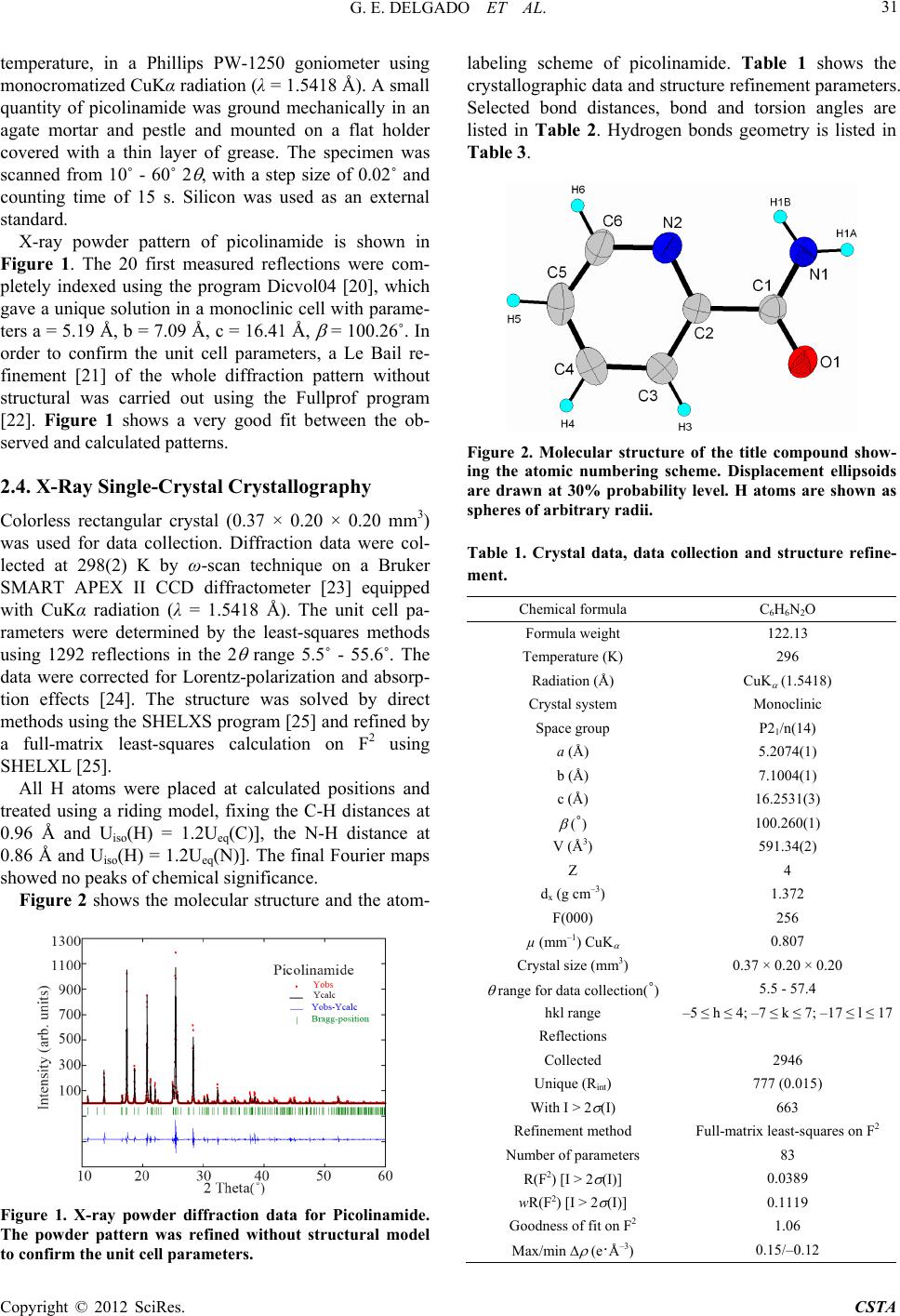 G. E. DELGADO ET AL. 31 temperature, in a Phillips PW-1250 goniometer using monocromatized CuKα radiation (λ = 1.5418 Å). A small quantity of picolinamide was ground mechanically in an agate mortar and pestle and mounted on a flat holder covered with a thin layer of grease. The specimen was scanned from 10˚ - 60˚ 2 , with a step size of 0.02˚ and counting time of 15 s. Silicon was used as an external standard. X-ray powder pattern of picolinamide is shown in Figure 1. The 20 first measured reflections were com- pletely indexed using the program Dicvol04 [20], which gave a unique solution in a monoclinic cell with parame- ters a = 5.19 Å, b = 7.09 Å, c = 16.41 Å, = 100.26˚. In order to confirm the unit cell parameters, a Le Bail re- finement [21] of the whole diffraction pattern without structural was carried out using the Fullprof program [22]. Figure 1 shows a very good fit between the ob- served and calculated patterns. 2.4. X-Ray Single-Crystal Crystallography Colorless rectangular crystal (0.37 × 0.20 × 0.20 mm3) was used for data collection. Diffraction data were col- lected at 298(2) K by ω-scan technique on a Bruker SMART APEX II CCD diffractometer [23] equipped with CuKα radiation (λ = 1.5418 Å). The unit cell pa- rameters were determined by the least-squares methods using 1292 reflections in the 2 range 5.5˚ - 55.6˚. The data were corrected for Lorentz-polarization and absorp- tion effects [24]. The structure was solved by direct methods using the SHELXS program [25] and refined by a full-matrix least-squares calculation on F2 using SHELXL [25]. All H atoms were placed at calculated positions and treated using a riding model, fixing the C-H distances at 0.96 Å and Uiso(H) = 1.2Ueq(C)], the N-H distance at 0.86 Å and Uiso(H) = 1.2Ueq(N)]. The final Fourier maps showed no peaks of chemical significance. Figure 2 shows the molecular structure and the atom- Figure 1. X-ray powder diffraction data for Picolinamide. The powder pattern was refined without structural model to confirm the unit cell parameters. labeling scheme of picolinamide. Table 1 shows the crystallographic data and structure refinement parameters. Selected bond distances, bond and torsion angles are listed in Table 2. Hydrogen bonds geometry is listed in Table 3. Figure 2. Molecular structure of the title compound show- ing the atomic numbering scheme. Displacement ellipsoids are drawn at 30% probability level. H atoms are shown as spheres of arbitrary radii. Table 1. Crystal data, data collection and structure refine- ment. Chemical formula C6H6N2O Formula weight 122.13 Temperature (K) 296 Radiation (Å) CuK (1.5418) Crystal system Monoclinic Space group P21/n(14) a (Å) 5.2074(1) b (Å) 7.1004(1) c (Å) 16.2531(3) (˚) 100.260(1) V (Å3) 591.34(2) Z 4 dx (g cm–3) 1.372 F(000) 256 µ (mm–1) CuK 0.807 Crystal size (mm3) 0.37 × 0.20 × 0.20 range for data collection(˚) 5.5 - 57.4 hkl range –5 ≤ h ≤ 4; –7 ≤ k ≤ 7; –17 ≤ l ≤ 17 Reflections Collected 2946 Unique (Rint) 777 (0.015) With I > 2 (I) 663 Refinement method Full-matrix least-squares on F2 Number of parameters 83 R(F2) [I > 2 (I)] 0.0389 wR(F2) [I > 2 (I)] 0.1119 Goodness of fit on F2 1.06 Max/min (e·Å–3) 0.15/–0.12 Copyright © 2012 SciRes. CSTA 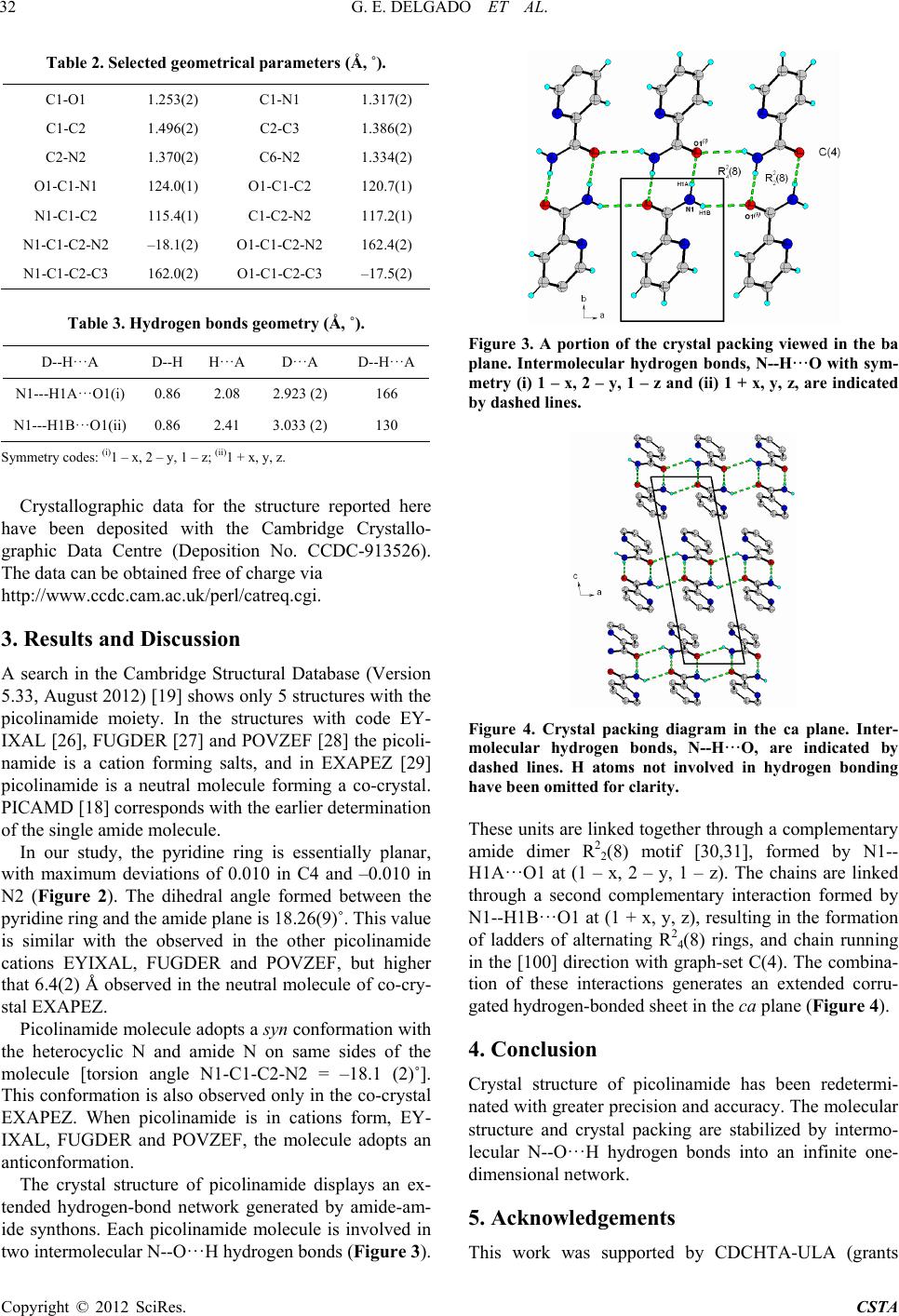 G. E. DELGADO ET AL. 32 Table 2. Selected geometrical parameters (Å, ˚). C1-O1 1.253(2) C1-N1 1.317(2) C1-C2 1.496(2) C2-C3 1.386(2) C2-N2 1.370(2) C6-N2 1.334(2) O1-C1-N1 124.0(1) O1-C1-C2 120.7(1) N1-C1-C2 115.4(1) C1-C2-N2 117.2(1) N1-C1-C2-N2 –18.1(2) O1-C1-C2-N2 162.4(2) N1-C1-C2-C3 162.0(2) O1-C1-C2-C3 –17.5(2) Table 3. Hydrogen bonds ge ome t ry (Å, ˚). D--H···A D--H H···A D···A D--H···A N1---H1A···O1(i) 0.86 2.08 2.923 (2) 166 N1---H1B···O1(ii) 0.86 2.41 3.033 (2) 130 Symmetry codes: (i)1 – x, 2 – y, 1 – z; (ii)1 + x, y, z. Crystallographic data for the structure reported here have been deposited with the Cambridge Crystallo- graphic Data Centre (Deposition No. CCDC-913526). The data can be obtained free of charge via http://www.ccdc.cam.ac.uk/perl/catreq.cgi. 3. Results and Discussion A search in the Cambridge Structural Database (Version 5.33, August 2012) [19] shows only 5 structures with the picolinamide moiety. In the structures with code EY- IXAL [26], FUGDER [27] and POVZEF [28] the picoli- namide is a cation forming salts, and in EXAPEZ [29] picolinamide is a neutral molecule forming a co-crystal. PICAMD [18] corresponds with the earlier determination of the single amide molecule. In our study, the pyridine ring is essentially planar, with maximum deviations of 0.010 in C4 and –0.010 in N2 (Figure 2). The dihedral angle formed between the pyridine ring and the amide plane is 18.26(9)˚. This value is similar with the observed in the other picolinamide cations EYIXAL, FUGDER and POVZEF, but higher that 6.4(2) Å observed in the neutral molecule of co-cry- stal EXAPEZ. Picolinamide molecule adopts a syn conformation with the heterocyclic N and amide N on same sides of the molecule [torsion angle N1-C1-C2-N2 = –18.1 (2)˚]. This conformation is also observed only in the co-crystal EXAPEZ. When picolinamide is in cations form, EY- IXAL, FUGDER and POVZEF, the molecule adopts an anticonformation. The crystal structure of picolinamide displays an ex- tended hydrogen-bond network generated by amide-am- ide synthons. Each picolinamide molecule is involved in two intermolecular N--O···H hydrogen bonds (Figure 3). Figure 3. A portion of the crystal packing viewed in the ba plane. Intermolecular hydrogen bonds, N--H···O with sym- metry (i) 1 – x, 2 – y, 1 – z and (ii) 1 + x, y, z, are indicated by dashed lines. Figure 4. Crystal packing diagram in the ca plane. Inter- molecular hydrogen bonds, N--H···O, are indicated by dashed lines. H atoms not involved in hydrogen bonding have been omitted for clarity. These units are linked together through a complementary amide dimer R22(8) motif [30,31], formed by N1-- H1A···O1 at (1 – x, 2 – y, 1 – z). The chains are linked through a second complementary interaction formed by N1--H1B···O1 at (1 + x, y, z), resulting in the formation of ladders of alternating R24(8) rings, and chain running in the [100] direction with graph-set C(4). The combina- tion of these interactions generates an extended corru- gated hydrogen-bonded sheet in the ca plane (Figure 4). 4. Conclusion Crystal structure of picolinamide has been redetermi- nated with greater precision and accuracy. The molecular structure and crystal packing are stabilized by intermo- lecular N--O···H hydrogen bonds into an infinite one- dimensional network. 5. Acknowledgements This work was supported by CDCHTA-ULA (grants Copyright © 2012 SciRes. CSTA 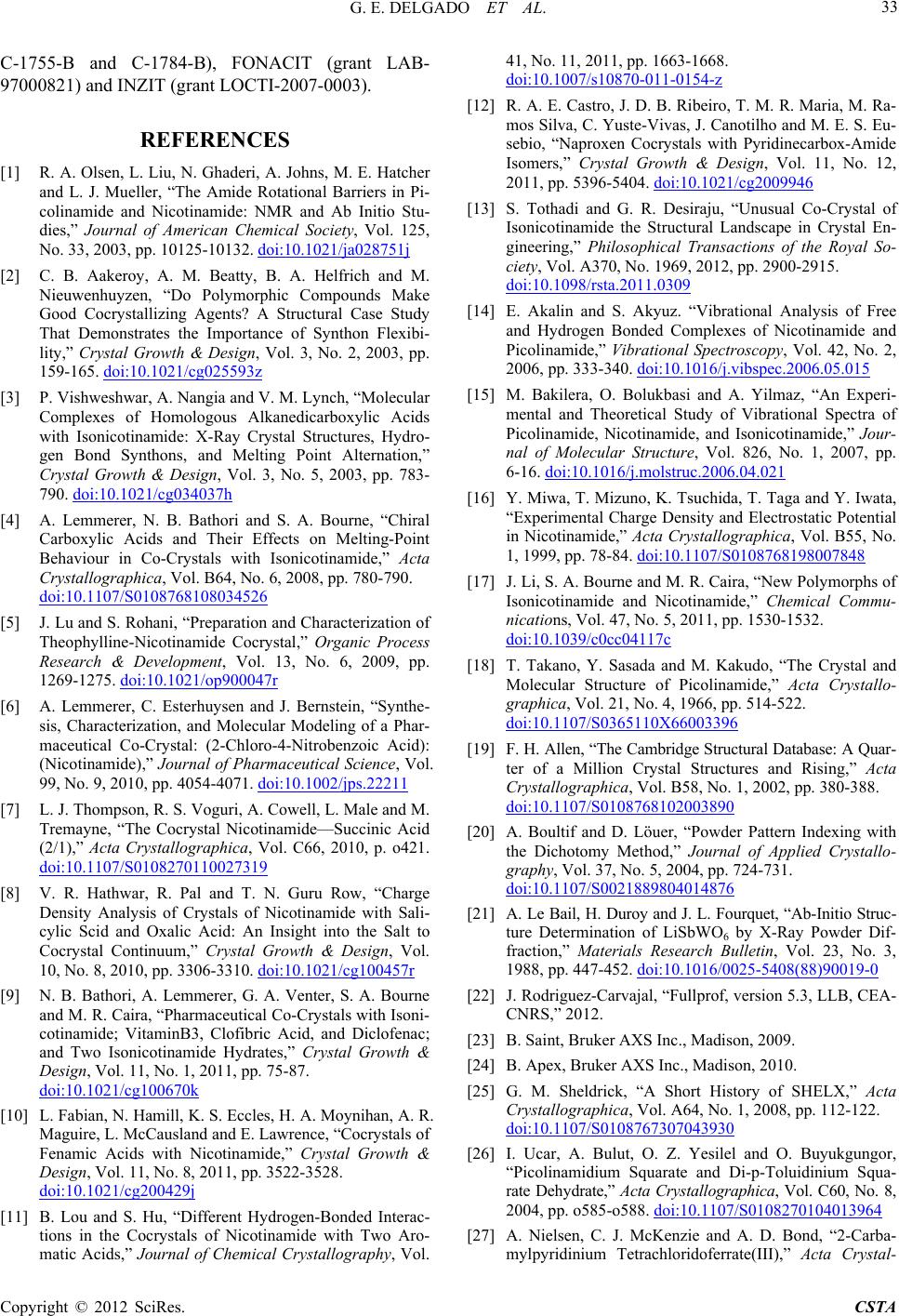 G. E. DELGADO ET AL. 33 C-1755-B and C-1784-B), FONACIT (grant LAB- 97000821) and INZIT (grant LOCTI-2007-0003). REFERENCES [1] R. A. Olsen, L. Liu, N. Ghaderi, A. Johns, M. E. Hatcher and L. J. Mueller, “The Amide Rotational Barriers in Pi- colinamide and Nicotinamide: NMR and Ab Initio Stu- dies,” Journal of American Chemical Society, Vol. 125, No. 33, 2003, pp. 10125-10132. doi:10.1021/ja028751j [2] C. B. Aakeroy, A. M. Beatty, B. A. Helfrich and M. Nieuwenhuyzen, “Do Polymorphic Compounds Make Good Cocrystallizing Agents? A Structural Case Study That Demonstrates the Importance of Synthon Flexibi- lity,” Crystal Growth & Design, Vol. 3, No. 2, 2003, pp. 159-165. doi:10.1021/cg025593z [3] P. Vishweshwar, A. Nangia and V. M. Lynch, “Molecular Complexes of Homologous Alkanedicarboxylic Acids with Isonicotinamide: X-Ray Crystal Structures, Hydro- gen Bond Synthons, and Melting Point Alternation,” Crystal Growth & Design, Vol. 3, No. 5, 2003, pp. 783- 790. doi:10.1021/cg034037h [4] A. Lemmerer, N. B. Bathori and S. A. Bourne, “Chiral Carboxylic Acids and Their Effects on Melting-Point Behaviour in Co-Crystals with Isonicotinamide,” Acta Crystallographica, Vol. B64, No. 6, 2008, pp. 780-790. doi:10.1107/S0108768108034526 [5] J. Lu and S. Rohani, “Preparation and Characterization of Theophylline-Nicotinamide Cocrystal,” Organic Process Research & Development, Vol. 13, No. 6, 2009, pp. 1269-1275. doi:10.1021/op900047r [6] A. Lemmerer, C. Esterhuysen and J. Bernstein, “Synthe- sis, Characterization, and Molecular Modeling of a Phar- maceutical Co-Crystal: (2-Chloro-4-Nitrobenzoic Acid): (Nicotinamide),” Journal of Pharmaceutical Science, Vol. 99, No. 9, 2010, pp. 4054-4071. doi:10.1002/jps.22211 [7] L. J. Thompson, R. S. Voguri, A. Cowell, L. Male and M. Tremayne, “The Cocrystal Nicotinamide—Succinic Acid (2/1),” Acta Crystallographica, Vol. C66, 2010, p. o421. doi:10.1107/S0108270110027319 [8] V. R. Hathwar, R. Pal and T. N. Guru Row, “Charge Density Analysis of Crystals of Nicotinamide with Sali- cylic Scid and Oxalic Acid: An Insight into the Salt to Cocrystal Continuum,” Crystal Growth & Design, Vol. 10, No. 8, 2010, pp. 3306-3310. doi:10.1021/cg100457r [9] N. B. Bathori, A. Lemmerer, G. A. Venter, S. A. Bourne and M. R. Caira, “Pharmaceutical Co-Crystals with Isoni- cotinamide; VitaminB3, Clofibric Acid, and Diclofenac; and Two Isonicotinamide Hydrates,” Crystal Growth & Design, Vol. 11, No. 1, 2011, pp. 75-87. doi:10.1021/cg100670k [10] L. Fabian, N. Hamill, K. S. Eccles, H. A. Moynihan, A. R. Maguire, L. McCausland and E. Lawrence, “Cocrystals of Fenamic Acids with Nicotinamide,” Crystal Growth & Design, Vol. 11, No. 8, 2011, pp. 3522-3528. doi:10.1021/cg200429j [11] B. Lou and S. Hu, “Different Hydrogen-Bonded Interac- tions in the Cocrystals of Nicotinamide with Two Aro- matic Acids,” Journal of Chemical Crystallography, Vol. 41, No. 11, 2011, pp. 1663-1668. doi:10.1007/s10870-011-0154-z [12] R. A. E. Castro, J. D. B. Ribeiro, T. M. R. Maria, M. Ra- mos Silva, C. Yuste-Vivas, J. Canotilho and M. E. S. Eu- sebio, “Naproxen Cocrystals with Pyridinecarbox-Amide Isomers,” Crystal Growth & Design, Vol. 11, No. 12, 2011, pp. 5396-5404. doi:10.1021/cg2009946 [13] S. Tothadi and G. R. Desiraju, “Unusual Co-Crystal of Isonicotinamide the Structural Landscape in Crystal En- gineering,” Philosophical Transactions of the Royal So- ciety, Vol. A370, No. 1969, 2012, pp. 2900-2915. doi:10.1098/rsta.2011.0309 [14] E. Akalin and S. Akyuz. “Vibrational Analysis of Free and Hydrogen Bonded Complexes of Nicotinamide and Picolinamide,” Vibrational Spectroscopy, Vol. 42, No. 2, 2006, pp. 333-340. doi:10.1016/j.vibspec.2006.05.015 [15] M. Bakilera, O. Bolukbasi and A. Yilmaz, “An Experi- mental and Theoretical Study of Vibrational Spectra of Picolinamide, Nicotinamide, and Isonicotinamide,” Jour- nal of Molecular Structure, Vol. 826, No. 1, 2007, pp. 6-16. doi:10.1016/j.molstruc.2006.04.021 [16] Y. Miwa, T. Mizuno, K. Tsuchida, T. Taga and Y. Iwata, “Experimental Charge Density and Electrostatic Potential in Nicotinamide,” Acta Crystallographica, Vol. B55, No. 1, 1999, pp. 78-84. doi:10.1107/S0108768198007848 [17] J. Li, S. A. Bourne and M. R. Caira, “New Polymorphs of Isonicotinamide and Nicotinamide,” Chemical Commu- nications, Vol. 47, No. 5, 2011, pp. 1530-1532. doi:10.1039/c0cc04117c [18] T. Takano, Y. Sasada and M. Kakudo, “The Crystal and Molecular Structure of Picolinamide,” Acta Crystallo- graphica, Vol. 21, No. 4, 1966, pp. 514-522. doi:10.1107/S0365110X66003396 [19] F. H. Allen, “The Cambridge Structural Database: A Quar- ter of a Million Crystal Structures and Rising,” Acta Crystallographica, Vol. B58, No. 1, 2002, pp. 380-388. doi:10.1107/S0108768102003890 [20] A. Boultif and D. Löuer, “Powder Pattern Indexing with the Dichotomy Method,” Journal of Applied Crystallo- graphy, Vol. 37, No. 5, 2004, pp. 724-731. doi:10.1107/S0021889804014876 [21] A. Le Bail, H. Duroy and J. L. Fourquet, “Ab-Initio Struc- ture Determination of LiSbWO6 by X-Ray Powder Dif- fraction,” Materials Research Bulletin, Vol. 23, No. 3, 1988, pp. 447-452. doi:10.1016/0025-5408(88)90019-0 [22] J. Rodriguez-Carvajal, “Fullprof, version 5.3, LLB, CEA- CNRS,” 2012. [23] B. Saint, Bruker AXS Inc., Madison, 2009. [24] B. Apex, Bruker AXS Inc., Madison, 2010. [25] G. M. Sheldrick, “A Short History of SHELX,” Acta Crystallographica, Vol. A64, No. 1, 2008, pp. 112-122. doi:10.1107/S0108767307043930 [26] I. Ucar, A. Bulut, O. Z. Yesilel and O. Buyukgungor, “Picolinamidium Squarate and Di-p-Toluidinium Squa- rate Dehydrate,” Acta Crystallographica, Vol. C60, No. 8, 2004, pp. o585-o588. doi:10.1107/S0108270104013964 [27] A. Nielsen, C. J. McKenzie and A. D. Bond, “2-Carba- mylpyridinium Tetrachloridoferrate(III),” Acta Crystal- Copyright © 2012 SciRes. CSTA  G. E. DELGADO ET AL. Copyright © 2012 SciRes. CSTA 34 lographica, Vol. E65, No. 11, 2009, p. m1359. doi:10.1107/S1600536809040148 [28] K. Gotoh, H. Nagoshi and H. Ishida, “Hydrogen-Bonded Structures of the Isomeric 2-, 3- and 4-Carbamoylpyrid- inium Hydrogen Chloranilates,” Acta Crystallographica, Vol. C65, No. 6, 2009, pp. o273-o277. doi:10.1107/S010827010901525X [29] S. Ghosh, P. P. Bag and C. M. Reddy, “Co-Crystals of Sulfamethazine with Some Carboxylic Acids and Amides: Co-Former Assisted Tautomerism in an Active Pharma- ceutical Ingredient and Hydrogen Bond Competition Study,” Crystal Growth & Design, Vol. 11, No. 8, 2011, pp. 3489-3503. doi:10.1021/cg200334m [30] M. C. Etter, “Encoding and Decoding Hydrogen-Bond Patterns of Organic Compounds,” Account of Chemical Research, Vol. 23, No. 4, 1990, pp. 120-126. doi:10.1021/ar00172a005 [31] M. C. Etter, J. C. MacDonald and J. Bernstein, “Graph- Set Analysis of Hydrogen-Bond Patterns in Organic Crys- tals,” Acta crystallographica, Vol. B46, No. 2, 1990, pp. 256-262. doi:10.1107/S0108768189012929 |