

Paper Menu >>
Journal Menu >>
 American Journal of Plant Sciences, 2012, 3, 1562-1567 http://dx.doi.org/10.4236/ajps.2012.311188 Published Online November 2012 (http://www.SciRP.org/journal/ajps) Season, Environment Stress and Refrigerated Storage Affect Genomic DNA Isolation of Tung Tree Lingling Zhang1,2,3, Yue Pan1,2,3, Jinm in Fu1,2, Junhua Peng1,2* 1Wuhan Botanical Garden, Chinese Academy of Sciences, Wuhan, China; 2Key Lab of Plant Germplasm Enhancement and Specialty Agriculture, Chinese Academy of Sciences, Wuhan, China; 3Graduate University of Chinese Academy of Sciences, Beijing, China. Email: *[email protected], *[email protected] Received August 11th, 2012; revised September 19th, 2012; accepted October 6th, 2012 ABSTRACT Many metabolites in leaf tissue disturbed plant genomic DNA isolation and they always varied when leaf was harvested from different environments. Objective of this study was to investigate whether season, environment stress and refriger- ated storage affect genomic DNA isolation of tung tree leaves. Five types of young leaves and two DNA isolation pro- tocols, the recycling CTAB protocol I and II, were adopted to carry out the experiment. Our results showed that both leaf type and protocol affected DNA isolation of tung tree. Using the recycling CTAB protocol II, though little DNA were obtained from three types of young leaves, though the other two have satisfying results. Whereas the recycling CTAB protocol I could produce high yield genomic DNA from all the five types of young leaves. All the detectable DNA samples in agarose gel electrophoresis were good templates for PCR reaction. Season, environment stress and refrigerated storage had a big effect on genomic DNA isolation of tung tree. The recycling CTAB protocol I was proved to be an effective and universal protocol for DNA isolation of tung tree. Five types of young leaves could all act as the tissue for isolation of genomic DNA, but the summer healthy young leaves without long-time refrigerated storage are the best. The optimal leaf tissue will benefit DNA isolation of plant species. Keywords: Tung Tree; Vernicia fordii; DNA Isolation; Season; Environment Stress; Storage 1. Introduction Plant often produces many metabolites, such as polysac- charides, polyphenols, protein and tannin. These com- pounds always interfere with DNA isolation and down- stream DNA-based experiments [1-6]. To date, lots of DNA isolation protocols have been published and aimed at eliminating the negative effects of biochemical com- ponents on DNA isolation [7-17]. However, DNA isola- tion is still a challenge for many plant species. Chemotypic heterogeneity among different species did not allowed the direct application of an extraction proto- col for a species to other species [17-19]. Metabolites in plant leaves were not stable, changing with many factors, such as leaf age [19-21], season [22-24], light intensity [25] and environmental stress [26,27]. Leaf age was re- ported to affect the properties of extracted DNA, which was inferred to be related with the accumulation of de- fense compounds during the leaf development [19]. En- vironmental factors such as light intensity, temperature and seasonal variance were reported to affect the produc- tion of polysaccharide [25], phenolic compounds [28,29] and other metabolites in leaves. However, up to now, little reports were about the effect of theses environ- mental factors on DNA isolation of plant species. Tung tree (Vernicia fordii), belonging to Euphor- biaceae, is an oil-producing plant with multiple uses es- pecially its potential in biodiesel production [30-32]. DNA extraction is essential for molecular genetic analy- ses and marker-assisted improvement of this oil-pro- ducing species. In present study, we found that season, environment stress and refrigerated storage all had big effect on genomic DNA isolation of tung tree. Besides, the recycling CTAB protocol I was proved to be a uni- versal method for DNA isolation of tung tree. 2. Materials and Protocols 2.1. Materials Young leaves were harvested from healthy tung tree at different seasons, in the autumn (early November in 2009), spring (early April in 2010) and summer (July in 2010), respectively (hereafter called autumn healthy leaves, spring healthy leaves and summer healthy leaves for short). Besides, leaves were also harvested from the tung tree that suffered from both transplant and water- drown stress in the summer of 2010 (hereafter called *Corresponding author. Copyright © 2012 SciRes. AJPS  Season, Environment Stress and Refrigerated Storage Affect Genomic DNA Isolation of Tung Tree 1563 summer stressed leaves for short). These four types of leaf tissues were ground into fine powder in liquid nitro- gen, and then 0.3 g of powders for each leaf tissue type were immediately used to isolate genomic DNA. Besides, some other summer healthy leaf tissues were stored in freezer for three months and then was used to isolate genomic DNA. Tissue powder was a mixture of several genotypes. Unless state, the leaf referred to the young leaf without the refrigerated storage in the text below. 2.2. Protocols Two protocols (Lingling Zhang et al., unpublished) were adopted to extract DNA from the five types of tung tree young leaves. The main procedures of recycling CTAB protocol I was followed. 1) Add 1.5 mL of wash buffer to 0.3 g of ground leaf sample. Mix well and place at the low temperature with occasional swirling for at least 15 min. Then centrifuge at 12,000 rpm for 15 min. Discard the supernatant. The tissue pretreatment was carried out for three times; 2) Add 0.5 mL of CTAB extraction buffer and re-suspend tissue pellet. Incubate at 65˚C for 40 min. Centrifuge at 12,000 rpm for 15 min. Transfer the super- natant to a new tube; 3) Add 200 μL Chloroform-isoa- mylalcohol (24:1). Mix well and let stand for several minutes. Centrifuge at 12,000 rpm for 15 min and trans- fer the supernatant to a new 1.5 mL tube carefully; 4) Precipitate the DNA by adding two volumes of cold ab- solute ethanol and incubate for 30 min at −20˚C. Transfer the DNA precipitation with a tip and wash it with 70% ethanol for 30 min, better for overnight. Then pour off the ethanol; 5) Dry the DNA and dissolve it in 100 μL T0.1E. Add 1 µL of 10 μg/mL ribonuclease. Incubate the DNA solution at 37˚C for 30 min, and then incubate at 65˚C for 15 min. This DNA sample was termed as the first DNA (1st DNA); 6) Similarly, treat the residual pel- lets in the above step 2) for other three cycles of the step 2) to the step 5). Correspondingly, the resulted DNA samples were termed as the second DNA (2nd DNA), third DNA (3rd DNA) and fourth DNA (4th DNA), re- spectively. The recycling CTAB protocol II shared all the procedure of the recycling CTAB protocol I except that the tissue pretreatment was only carry out for one time. 2.3. DNA Isolation and Assessment The quality and yield of extracted DNA were determined by agarose gel electrophoresis. Concentration of DNA samples was assessed as the following. 2 μL of DNA solution for each sample was added into 48 μL of steril- ized water (mix well). Then 5 μL of the diluted DNA solution plus 5 μL of 2.5X loading buffer was loaded into 0.8% agrose gel and run in 0.5X TBE buffer (pH 8.0) at room temperature. The gel was visualized with ethidum bormide staining. DNA quantity for each lane/sample was estimated by comparing band intensity with the concentration-known λ DNA standards (Promega, Madi- son, WI, USA). DNA concentration was calculated using formula, concentration = [quantity (ng) of 5 μL diluted DNA/5 μL × dilution factor (50/2 = 25)]. 2.4. Polymerase Chain Reaction Amplification Coding region cloning of fad2 gene was performed using PCR approach. On the basis of GenBank sequence AF525534, sequences of primers for PCR amplification are 5’-GATGGGTGCTGGTGGCAGAATGTCA-3’ and 5’-CCAGAACTTCCAAGCCCTTCACTTTTG -3’ [33], which amplify an approximately 1.2 kb fragment encom- passing the entire coding region of fad2 gene. The PCR were performed in a 25 μL reaction volume (60 ng tem- plate DNA, 0.4 μM specific primers, 1 U Taq DNA po- lymerase, 0.2 mM dNTP mixture, 1 × PCR buffer, 0.15 mM Mg2+) using BIORAD-My Cycle Thermocyclers (Bio-Rad Laboratories, Inc., California, USA) with the following program: pre-denaturation at 94˚C for 4 min; 30 cycles of denaturation at 94˚C for 45 s, annealing at 65˚C for 45 s, extension at 72˚C for 60 s; and a final ex- tension step at 72˚C for 10 min, then hold on for 15 min at 4˚C. Then PCR products were separated in 1% agarose gels and the gel was visualized with ethidium bromide staining. 3. Results 3.1. Comparisons of DNA Samples Isolated From Five Types of Leaves Using the Protocol I Using agarose gel electrophoresis and λDNA of known concentration as standards, concentration of DNA sam- ples produced by the protocol I were determined. As shown in table 1, yield of the 1st DNA varied for the five types of young leaves, about 200 ng/μL both for spring healthy leaf and summer healthy leaf, 100 ng/μL for summer stressed leaf, but little DNA for summer healthy leaf with three-month refrigerated storage and autumn healthy leaf (Figure 1). As shown in Figure 1, the 2nd, 3rd and 4th DNA for each type of leaf were all obviously detectable in agrose gel electrophoresis, the concentra- tion of these DNA samples ranging from 300 ng/μL to 500 ng/μL (Table 1). These data indicated that the pro- tocol I could be successfully applied to different types of young leaves without significant differences. 3.2. Comparisons of DNA Samples Isolated from Five Types of Leaves Using the Protocol II However using the recycling CTAB method II, DNA samples extracted from the fve types of leaves greatly i Copyright © 2012 SciRes. AJPS 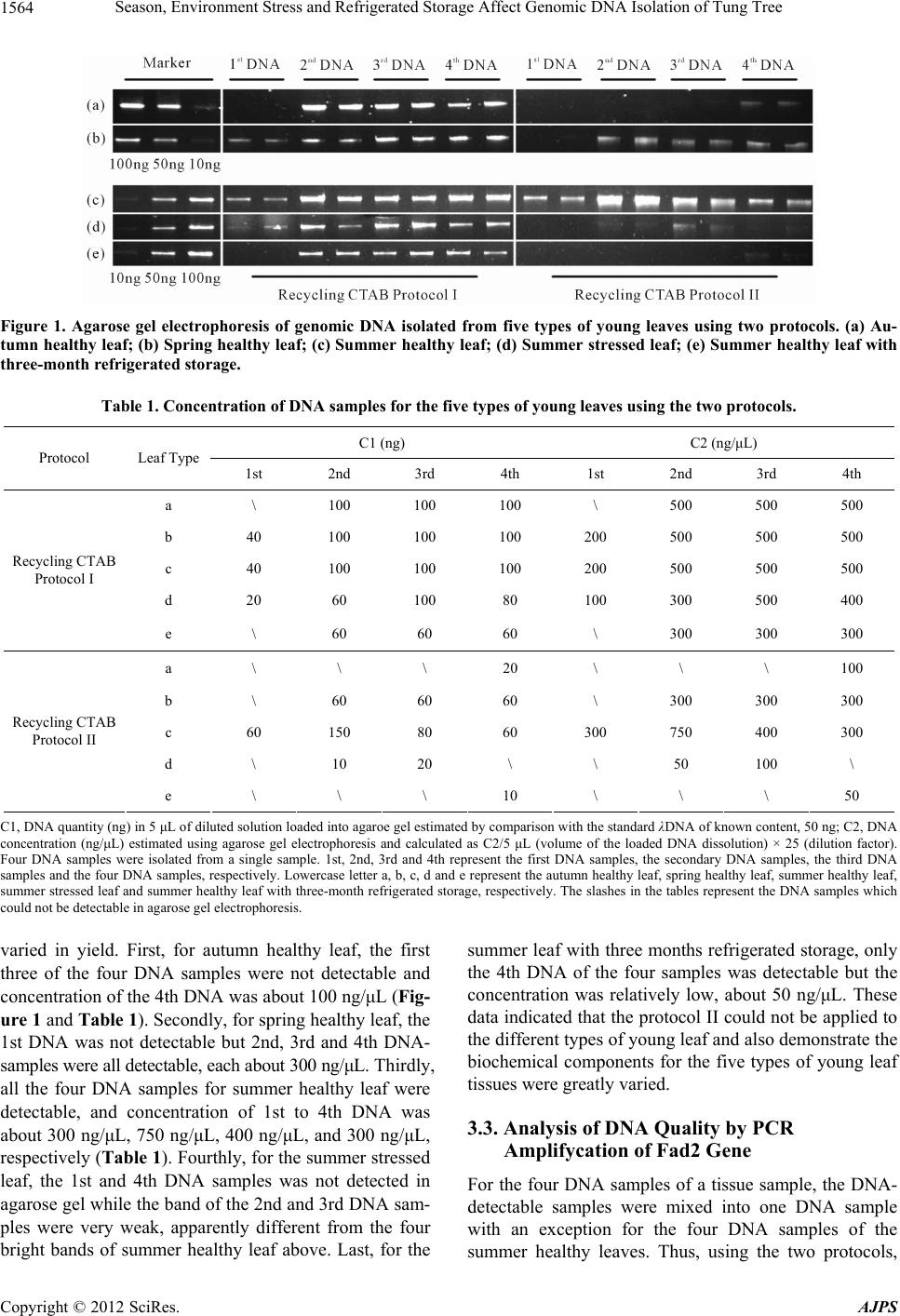 Season, Environment Stress and Refrigerated Storage Affect Genomic DNA Isolation of Tung Tree Copyright © 2012 SciRes. AJPS 1564 Figure 1. Agarose gel electrophoresis of genomic DNA isolated from five types of young leaves using two protocols. (a) Au- tumn healthy leaf; (b) Spring healthy leaf; (c) Summer healthy leaf; (d) Summer stressed leaf; (e) Summer healthy leaf with three-month refrigerated storage. Table 1. Concentration of DNA samples for the five types of young leaves using the two protocols. C1 (ng) C2 (ng/μL) Protocol Leaf Type 1st 2nd 3rd 4th 1st 2nd 3rd 4th a \ 100 100 100 \ 500 500 500 b 40 100 100 100 200 500 500 500 c 40 100 100 100 200 500 500 500 d 20 60 100 80 100 300 500 400 Recycling CTAB Protocol I e \ 60 60 60 \ 300 300 300 a \ \ \ 20 \ \ \ 100 b \ 60 60 60 \ 300 300 300 c 60 150 80 60 300 750 400 300 d \ 10 20 \ \ 50 100 \ Recycling CTAB Protocol II e \ \ \ 10 \ \ \ 50 C1, DNA quantity (ng) in 5 μL of diluted solution loaded into agaroe gel estimated by comparison with the standard λDNA of known content, 50 ng; C2, DNA concentration (ng/μL) estimated using agarose gel electrophoresis and calculated as C2/5 μL (volume of the loaded DNA dissolution) × 25 (dilution factor). Four DNA samples were isolated from a single sample. 1st, 2nd, 3rd and 4th represent the first DNA samples, the secondary DNA samples, the third DNA samples and the four DNA samples, respectively. Lowercase letter a, b, c, d and e represent the autumn healthy leaf, spring healthy leaf, summer healthy leaf, summer stressed leaf and summer healthy leaf with three-month refrigerated storage, respectively. The slashes in the tables represent the DNA samples which could not be detectable in agarose gel electrophoresis. summer leaf with three months refrigerated storage, only the 4th DNA of the four samples was detectable but the concentration was relatively low, about 50 ng/μL. These data indicated that the protocol II could not be applied to the different types of young leaf and also demonstrate the biochemical components for the five types of young leaf tissues were greatly varied. varied in yield. First, for autumn healthy leaf, the first three of the four DNA samples were not detectable and concentration of the 4th DNA was about 100 ng/μL (Fig- ure 1 and Table 1). Secondly, for spring healthy leaf, the 1st DNA was not detectable but 2nd, 3rd and 4th DNA- samples were all detectable, each about 300 ng/μL. Thirdly, all the four DNA samples for summer healthy leaf were detectable, and concentration of 1st to 4th DNA was about 300 ng/μL, 750 ng/μL, 400 ng/μL, and 300 ng/μL, respectively (Tabl e 1). Fourthly, for the summer stressed leaf, the 1st and 4th DNA samples was not detected in agarose gel while the band of the 2nd and 3rd DNA sam- ples were very weak, apparently different from the four bright bands of summer healthy leaf above. Last, for the 3.3. Analysis of DNA Quality by PCR Amplifycation of Fad2 Gene For the four DNA samples of a tissue sample, the DNA- detectable samples were mixed into one DNA sample with an exception for the four DNA samples of the summer healthy leaves. Thus, using the two protocols, 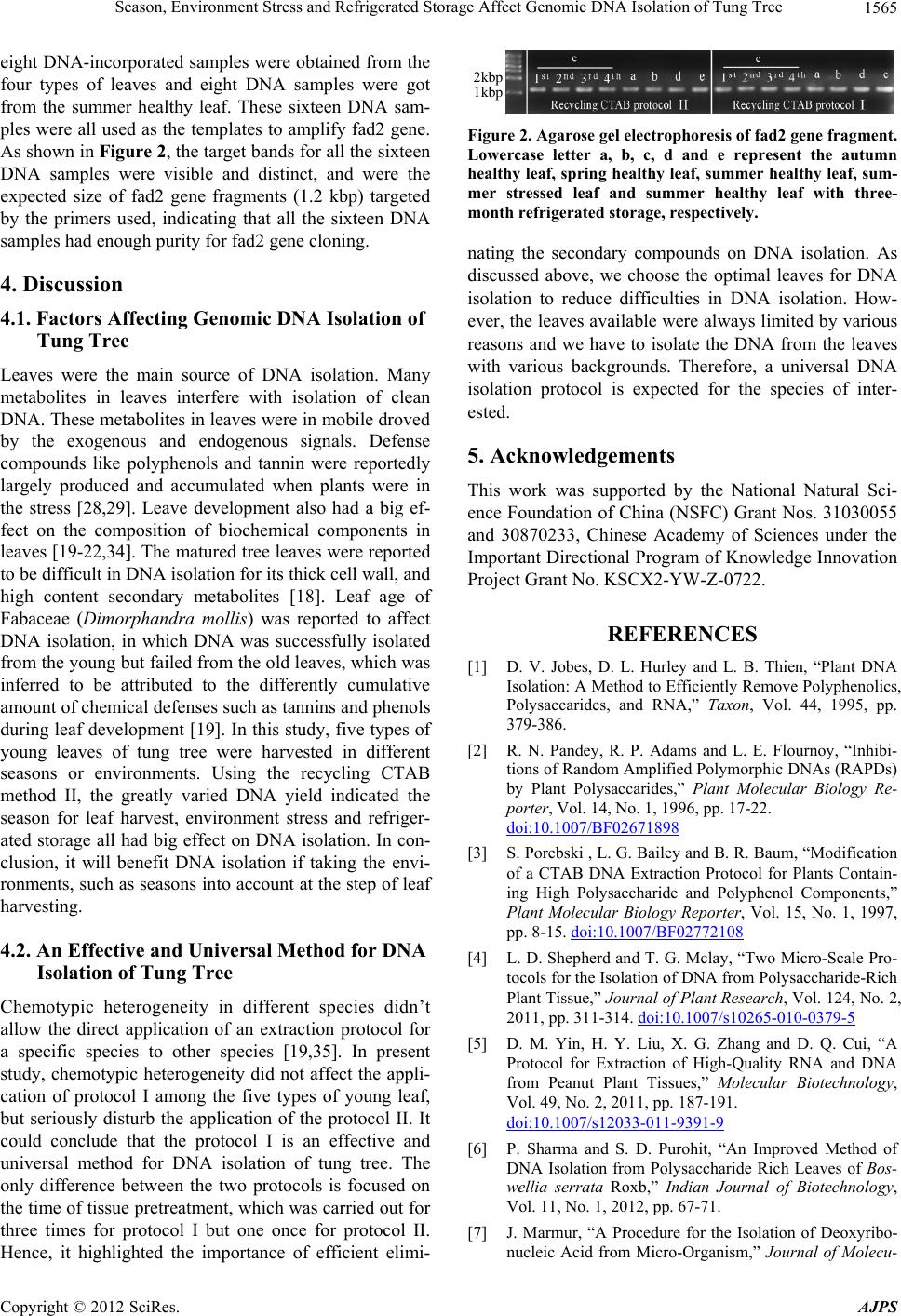 Season, Environment Stress and Refrigerated Storage Affect Genomic DNA Isolation of Tung Tree 1565 eight DNA-incorporated samples were obtained from the four types of leaves and eight DNA samples were got from the summer healthy leaf. These sixteen DNA sam- ples were all used as the templates to amplify fad2 gene. As shown in Figure 2, the target bands for all the sixteen DNA samples were visible and distinct, and were the expected size of fad2 gene fragments (1.2 kbp) targeted by the primers used, indicating that all the sixteen DNA samples had enough purity for fad2 gene cloning. 4. Discussion 4.1. Factors Affecting Genomic DNA Isolation of Tung Tree Leaves were the main source of DNA isolation. Many metabolites in leaves interfere with isolation of clean DNA. These metabolites in leaves were in mobile droved by the exogenous and endogenous signals. Defense compounds like polyphenols and tannin were reportedly largely produced and accumulated when plants were in the stress [28,29]. Leave development also had a big ef- fect on the composition of biochemical components in leaves [19-22,34]. The matured tree leaves were reported to be difficult in DNA isolation for its thick cell wall, and high content secondary metabolites [18]. Leaf age of Fabaceae (Dimorphandra mollis) was reported to affect DNA isolation, in which DNA was successfully isolated from the young but failed from the old leaves, which was inferred to be attributed to the differently cumulative amount of chemical defenses such as tannins and phenols during leaf development [19]. In this study, five types of young leaves of tung tree were harvested in different seasons or environments. Using the recycling CTAB method II, the greatly varied DNA yield indicated the season for leaf harvest, environment stress and refriger- ated storage all had big effect on DNA isolation. In con- clusion, it will benefit DNA isolation if taking the envi- ronments, such as seasons into account at the step of leaf harvesting. 4.2. An Effective and Universal Method for DNA Isolation of Tung Tree Chemotypic heterogeneity in different species didn’t allow the direct application of an extraction protocol for a specific species to other species [19,35]. In present study, chemotypic heterogeneity did not affect the appli- cation of protocol I among the five types of young leaf, but seriously disturb the application of the protocol II. It could conclude that the protocol I is an effective and universal method for DNA isolation of tung tree. The only difference between the two protocols is focused on the time of tissue pretreatment, which was carried out for three times for protocol I but one once for protocol II. Hence, it highlighted the importance of efficient elimi- 2kbp 1kbp Figure 2. Agarose gel electrophoresis of fad2 gene fragment. Lowercase letter a, b, c, d and e represent the autumn healthy leaf, spring healthy leaf, summer healthy leaf, sum- mer stressed leaf and summer healthy leaf with three- month refrigerated storage, respectively. nating the secondary compounds on DNA isolation. As discussed above, we choose the optimal leaves for DNA isolation to reduce difficulties in DNA isolation. How- ever, the leaves available were always limited by various reasons and we have to isolate the DNA from the leaves with various backgrounds. Therefore, a universal DNA isolation protocol is expected for the species of inter- ested. 5. Acknowledgements This work was supported by the National Natural Sci- ence Foundation of China (NSFC) Grant Nos. 31030055 and 30870233, Chinese Academy of Sciences under the Important Directional Program of Knowledge Innovation Project Grant No. KSCX2-YW-Z-0722. REFERENCES [1] D. V. Jobes, D. L. Hurley and L. B. Thien, “Plant DNA Isolation: A Method to Efficiently Remove Polyphenolics, Polysaccarides, and RNA,” Taxon, Vol. 44, 1995, pp. 379-386. [2] R. N. Pandey, R. P. Adams and L. E. Flournoy, “Inhibi- tions of Random Amplified Polymorphic DNAs (RAPDs) by Plant Polysaccarides,” Plant Molecular Biology Re- porter, Vol. 14, No. 1, 1996, pp. 17-22. doi:10.1007/BF02671898 [3] S. Porebski , L. G. Bailey and B. R. Baum, “Modification of a CTAB DNA Extraction Protocol for Plants Contain- ing High Polysaccharide and Polyphenol Components,” Plant Molecular Biology Reporter, Vol. 15, No. 1, 1997, pp. 8-15. doi:10.1007/BF02772108 [4] L. D. Shepherd and T. G. Mclay, “Two Micro-Scale Pro- tocols for the Isolation of DNA from Polysaccharide-Rich Plant Tissue,” Journal of Plant Research, Vol. 124, No. 2, 2011, pp. 311-314. doi:10.1007/s10265-010-0379-5 [5] D. M. Yin, H. Y. Liu, X. G. Zhang and D. Q. Cui, “A Protocol for Extraction of High-Quality RNA and DNA from Peanut Plant Tissues,” Molecular Biotechnology, Vol. 49, No. 2, 2011, pp. 187-191. doi:10.1007/s12033-011-9391-9 [6] P. Sharma and S. D. Purohit, “An Improved Method of DNA Isolation from Polysaccharide Rich Leaves of Bos- wellia serrata Roxb,” Indian Journal of Biotechnology, Vol. 11, No. 1, 2012, pp. 67-71. [7] J. Marmur, “A Procedure for the Isolation of Deoxyribo- nucleic Acid from Micro-Organism,” Journal of Molecu- Copyright © 2012 SciRes. AJPS  Season, Environment Stress and Refrigerated Storage Affect Genomic DNA Isolation of Tung Tree 1566 lar Biology, Vol. 3, No, 2, 1961, pp. 208-218. [8] M. G. Murray and W. F. Thompson, “Rapid Isolation of High Molecular Weight Plant DNA,” Nucleic Acids Re- search, Vol. 8, No. 19, 1980, pp. 4321-4325. doi:10.1093/nar/8.19.4321 [9] J. J. Doyle and J. L. Doyle, “Isolation of Plant DNA from Fresh Tissue,” Focus, Vol. 12, No. 1, 1990, pp. 13-15. [10] D. Goldenberger, I. Perschil, M. Ritzler and M. Altwegg, “A Simple Universal DNA Extraction Procedure Using SDS and Proteinase K Is Compatible with Directly PCR Amplification,” Genome Research, Vol. 4, 1995, pp. 368- 370. [11] S. M. Aljanabi and I. Martinez, “Universal and Rapid Salt-Extraction of High Quality Genomic DNA for PCR- Based Techniques,” Nucleic Acids Research, Vol. 25, No. 22, 1997, pp. 4692-4693. doi:10.1093/nar/25.22.4692 [12] N. Stein, G. Herren and B. Keller, “A New DNA Extrac- tion Method for High-Throughput Marker Analysis in a Large-Gemone such as Triticum aestivum,” Plant Breed- ing, Vol. 120, No. 4, 2001, pp. 354-356. doi:10.1046/j.1439-0523.2001.00615.x [13] K. L. Hill-Ambroz, G. L. Brown-Guedira and J. P. Fellers, “Modified Rapid DNA Extraction Protocol for High throught Microsatellite Analysis in Wheat,” Crop Science, Vol. 42, No. 6, 2002, pp. 2088-2091. doi:10. 2135/cropsci2002.2088 [14] R. J. Mogg and J. M. Bond, “A Cheap, Reliable and Rapid Method of Extracting High-Quality DNA from Plants,” Molecular Ecology Notes, Vol. 3, No. 4, 2003, pp. 666-668. doi:10.1046/j.1471-8286.2003.00548.x [15] G. C. Allen, M. A. Flores-Vergara, S. Krasynanski, S. Kumar and W. F. Thompson, “A Modified Protocol for Rapid DNA Isolation from Plant Tissues Using Cetyl- trimethylammonium Bromide,” Nature Protocol, Vol. 1, No. 5, 2006, pp. 2320-2325. [16] J. Amani, R. Kazemi, A. R. Abbasi and A. H. Salmanian, “A Simple and Rapid Leaf Genomic DNA Extraction Method for Polymerase Chain Reaction Analysis,” Ira- nian Journal of Biotechnology, Vol. 9, No. 1, 2011, pp. 69-71. [17] H. A. Souza, L. A. Muller, R. L. Brandao and M. B. Lovato, “Isolation of High Quality and Polysaccharide- Free DNA from Leaves of Dimorphandra mollis (Legu- minosae), a Tree from the Brazilian Cerrado,” Genetic Molecular Research, Vol. 11, No. 1, 2012, pp. 756-764. [18] F. Lefort and G. C. Douglas, “An Efficient Micro-Method of DNA Isolation from Mature Leaves of Four Hardwood Tree Species Acer, Fraxinus, Prunus and Quercu,” An- nual Forest Science, Vol. 56, No. 3, 1999, pp. 259-263. doi:10.1051/forest: 19990308 [19] P. A. Moreira and D. A. Oliveira, “Leaf Age Affects the Quality of DNA Extracted from Dimorphandra mollis (Fabaceae), a Tropical Tree Species from the Cerrado Region of Brazil,” Genetic Molecular Research, Vol. 10, No. 1, 2011, pp. 353-358. doi:10.4238/vol10-1gmr1030 [20] N. M. Van Dam, R. Verpoorte and E. Van der Meljden, “Extreme Difference in Pyrrolizidine Alkaloid Levels between Leaves of cynoglossum officinale,” Phytochem- istry, Vol. 37, No. 4, 1994, pp. 1013-1016. doi:10.1016/S0031-9422(00)89519-9 [21] M. A. Azmat, I. A. Khan and H. M. N. Cheema, “Extrac- tion of DNA Suitable for PCR Applications from Mature Leaves of Mangifera indica L.,” Journal of Zhejiang Uni- versity Science B., Vol. 13, No. 4, 2012, pp. 239-243. doi:10.1631/jzus.B1199194 [22] Z. Liu, S. B. Carpenter, W. J. Bourgeois, Y. Yu, R. J. Constantin, M. J. Falcon and J. C. Adams, “Variations in the Secondary Metabolite Camptothecin in Relation to Tissue Age and Season in Camptotheca acuminate,” Tree Physiology, Vol. 18, No. 4, 1998, pp. 265-270. [23] F. Vallejo, F. A. Tomas-Barberan and C. Garcia-Viguera, “Effect of Climatic and Sulphur Fertilization Conditions, on Phenolic Compounds and Vitamin C, in the Inflores- cences of Eight Broccoli Cultivars,” European Food Re- search and Technology, Vol. 216, No. 5, 2003, pp. 395- 401. doi:10.1007/s00217-003-0664-9 [24] M. C. Shih, C. M Chang, S. M. Kang and M. L. Tsai, “Effect of Different Parts (Leaf, Stem and Stalk) and Season (Summer and Winter) on the Chemical Composi- tions and Antioxidant Activity of Moringa oleifera,” In- ternational Journal of Molecular Sciences, Vol. 12, No. 9, 2011, pp. 6077-6088. doi:10.3390/ijms12096077 [25] Z. Yang, L. L. Geng, W. Wei and J. Zhang, “Combined Effects of Temperature, Light Intensity, and Nitrogen Concentration on the Growth and Polysaccharide Content of Microcystis aeruginosa in Batch Culture,” Biochemical Systematics and Ecology, Vol. 41, 2012, pp. 130-135. doi:10.1016/j.bse.2011.12.015 [26] N. Benhamou, “Elicitor-Induced Plant Defence Path- ways,” Trends Plant Science, Vol. 1, No. 7, 1996, pp. 233-240. doi:10.1016/1360-1385(96)86901-9 [27] F. Bourgaud, A. Gravot, S. Milesi and E. Gontier, “Pro- duction of Plant Secondary Metabolites: A Historial Per- spective,” Plant Science, Vol. 161, No. 5, 2001, pp. 839- 851. doi:10.1016/S0168-9452(01)00490-3 [28] T. Siatka and M. Kasparova, “Seasonal Variation in Total Phenolic and Flavonoid Content and DPPH Scavenging Activity of Bellis perennis L. Flowers,” Molecular, Vol. 15, No. 12, 2010, pp. 9450-9461. [29] A. S. Rodrigues, M. R. Perez-Gregorio, M. S. Garcia- Falcon, J. Simal-Gandara and D. P. F. Almeida, “Effect of Meteorological Conditions on Antioxidant Flavonoids in Portuguese Cultivars of White and Red Onions,” Food Chemistry, Vol. 124, No. 1, 2011, pp. 303-308. doi:10.1016/j.foodchem.2010.06.037 [30] J. Y. Park, D. K. Kim, Z. M. Wang, P. Lu, S. C. Park and J. S. Lee, “Production and Characterization of Biodiesel from Tung Oil,” Applied Biochemistry and Biotechnology, Vol. 148, No. 6, 2008, pp. 109-117. doi:10.1007/s12010-007-8082-2 [31] Q. Shang, W. Jiang, H. F. Lu and B. Liang, “Properties of Tung Oil Biodiesel and Its Blends with 0# Diesel,” Bio- resource Technology, Vol. 101, No. 2, 2010, pp. 826-828. doi:10.1016/j.biortech.2009.08.047 [32] Y. H. Chen, J. H. Chen and Y. M. Luo, “Complementary Biodiesel Combination from Tung and Medium-Chain Copyright © 2012 SciRes. AJPS  Season, Environment Stress and Refrigerated Storage Affect Genomic DNA Isolation of Tung Tree Copyright © 2012 SciRes. AJPS 1567 Fatty Acid Oils,” Renewable Energy, Vol. 44, 2012, pp. 305-310. doi:10.1016/j.renene.2012.01.098 [33] J. M. Dyer, D. C. Chapital, J. C. Kuan, R. T. Mullen, C. Turner, T. A. McKeon and A.B. Pepperman, “Molecular Analysis of a Bifunctional Fatty Acid Conjugase/De- saturase from Tung: Implications for the Evolution of Plant Fatty Acid Diversity,” Plant Physiology, Vol. 130, No. 4, 2002, pp. 2027-2038. doi:10.1104/pp.102.010835 [34] Y. Chen and T. M. Poland, “Interactive Influence of Leaf Age, Light Intensity, and Girdling on Green Ash Foliar Chemistry and Emerald Ash Border Development,” Jour- nal of Chemical Ecology, Vol. 35, 2009, pp. 806-815. [35] L. Drabkova, J. Kirschner and C.Vlcek, “Comparison of Seven DNA Extraction and Amplification Protocols in Historical Herbarium Specimens of Juncaceae,” Plant Molecular Reporter, Vol. 20, No. 2, 2002, pp. 161-175. doi:10.1007/BF02799431. |