

Paper Menu >>
Journal Menu >>
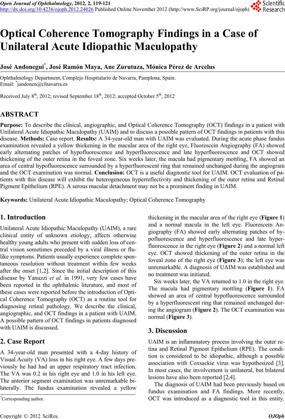 Open Journal of Ophthalmology, 2012, 2, 119-121 http://dx.doi.org/10.4236/ojoph.2012.24026 Published Online November 2012 (http://www.SciRP.org/journal/ojoph) 119 Optical Coherence Tomography Findings in a Case of Unilateral Acute Idiopathic Maculopathy José Andonegui*, José Ramón Maya, Ane Zurutuza, Mónica Pérez de Arcelus Ophthalmology Department, Complejo Hospitalario de Navarra, Pamplona, Spain. Email: *[email protected] Received July 8th, 2012; revised September 18th, 2012; accepted October 5th, 2012 ABSTRACT Purpose: To describe the clinical, angiographic, and Optical Coherence Tomography (OCT) findings in a patient with Unilateral Acute Idiopathic Maculopathy (UAIM) and to discuss a possible pattern of OCT findings in patients with this disease. Methods: Case r eport. Results: A 34-year-old man with UAIM was evaluated. During the acute phase fundus examination revealed a yellow thickening in the macular area of the right eye, Fluorescein Angiography (FA) showed early alternating patches of hypofluorescence and hyperfluorescence and late hyperfluorescence and OCT showed thickening of the outer retina in the foveal zone. Six weeks later, the macula had pigmentary mottling, FA showed an area of central hypofluorescence surrounded by a hyperfluorescent ring that remained unchanged during the angiogram and the OCT examination was normal. Conclusion: OCT is a useful diagnostic tool for UAIM. OCT evaluation of pa- tients with this disease will exhibit the heterogeneous hyperreflectivity and thickening of the outer retina and Retinal Pigment Epithelium (RPE). A serous macular detachment may not be a prominent finding in UAIM. Keywords: Unilateral Acute Idiopathic Maculopathy; Optical Coherence Tomography 1. Introduction Unilateral Acute Idiopathic Maculopathy (UAIM), a rare clinical entity of unknown etiology, affects otherwise healthy young adults who present with sudden loss of cen- tral vision sometimes preceded by a viral illness or flu- like symptom s. Pat ient s usual ly experience complete spon- taneous resolution without treatment within few weeks after the onset [1,2]. Since the initial description of this disease by Yanuzzi et al. in 1991, very few cases have been reported in the ophthalmic literature, and most of these cases were reported befo r e the introductio n of Opti- cal Coherence Tomography (OCT) as a routine tool for diagnosing retinal pathology. We describe the clinical, angiographic, and OCT findings in a patient with UAIM. A possible pattern of OCT findings in patients diagnosed with UAIM is discussed. 2. Case Report A 34-year-old man presented with a 4-day history of Visual Acuity (VA) loss in his right eye. A few days pre- viously he had had an upper respiratory tract infection. The VA was 0.2 in his right eye and 1.0 in his left eye. The anterior segment examination was unremarkable bi- laterally. The fundus examination revealed a yellow thickening in the macular area of the right eye (Figure 1) and a normal macula in the left eye. Fluorescein An- giography (FA) showed early alternating patches of hy- pofluorescence and hyperfluorescence and late hyper- fluorescence in the right eye (Figure 2) and a normal left eye. OCT showed thickening of the outer retina in the foveal zone of the right eye (Figure 3); the left eye was unremarkable. A diagnosis of UAIM was established and no treatment was initiated. Six weeks later, the VA returned to 1.0 in th e right eye. The macula had pigmentary mottling (Figure 1). FA showed an area of central hypofluorescence surrounded by a hyperfluorescent ring that remained unchanged dur- ing the angiogram (Figure 2). The OCT examination was normal (Figure 3). 3. Discussion UAIM is an inflammatory process involving the outer re- tina and Retinal Pigment Epithelium (RPE). The condi- tion is considered to be idiopathic, although a possible association with Coxsackie virus was hypothesized [3]. In most cases, the involvement is unilateral, but bilateral lesions have also been report ed [2,4] . The diagnosis of UAIM had been previously based on fundus examination and FA findings. More recently, OCT was introduced as a diagnostic tool in this entity. *Corresponding a uthor. Copyright © 2012 SciRes. OJOph 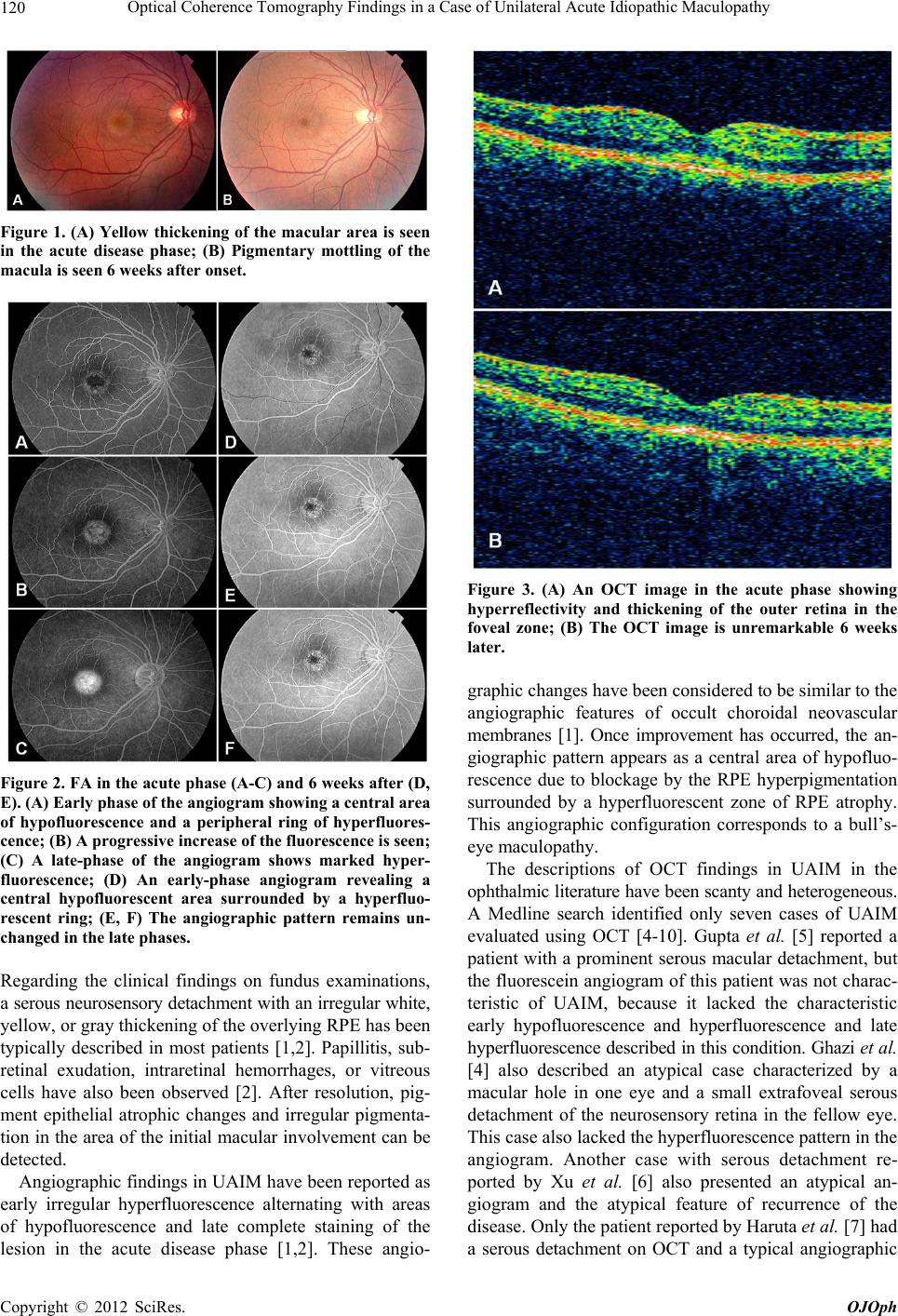 Optical Coherence Tomograph y Findings in a Cas e of Unilateral Acute Idiopathic Maculopathy 120 Figure 1. (A) Yellow thickening of the macular area is seen in the acute disease phase; (B) Pigmentary mottling of the macula is seen 6 weeks after onset. Figure 2. FA in the acute phase (A-C) and 6 weeks after (D, E). (A) Early phase of the angiogram showing a central area of hypofluorescence and a peripheral ring of hyperfluores- cence; (B) A progressive increase of the fluorescence is seen; (C) A late-phase of the angiogram shows marked hyper- fluorescence; (D) An early-phase angiogram revealing a central hypofluorescent area surrounded by a hyperfluo- rescent ring; (E, F) The angiographic pattern remains un- changed in the late phases. Regarding the clinical findings on fundus examinations, a serous neurosensory detachment with an irregular white, yellow, or gray thickening of the overlying RPE has been typically described in most patients [1,2]. Papillitis, sub- retinal exudation, intraretinal hemorrhages, or vitreous cells have also been observed [2]. After resolution, pig- ment epithelial atrophic changes and irregular pigmenta- tion in the area of the in itial macular involvement can be detected. Angiographic findings in UAIM have been reported as early irregular hyperfluorescence alternating with areas of hypofluorescence and late complete staining of the lesion in the acute disease phase [1,2]. These angio- Figure 3. (A) An OCT image in the acute phase showing hyperreflectivity and thickening of the outer retina in the foveal zone; (B) The OCT image is unremarkable 6 weeks later. graphic changes have been considered to be similar to the angiographic features of occult choroidal neovascular membranes [1]. Once improvement has occurred, the an- giographic pattern appears as a central area of hypofluo- rescence due to blockage by the RPE hyperpigmentation surrounded by a hyperfluorescent zone of RPE atrophy. This angiographic configuration corresponds to a bull’s- eye maculopathy. The descriptions of OCT findings in UAIM in the ophthalmic literature have been scanty and heterogeneous. A Medline search identified only seven cases of UAIM evaluated using OCT [4-10]. Gupta et al. [5] reported a patient with a prominent serous macular detachment, but the fluorescein angiogram of this patient was not charac- teristic of UAIM, because it lacked the characteristic early hypofluorescence and hyperfluorescence and late hyperfluorescen ce described in this con dition. Ghazi et al. [4] also described an atypical case characterized by a macular hole in one eye and a small extrafoveal serous detachment of the neurosensory retina in the fellow eye. This case also lacked the hyperfluorescence pattern in the angiogram. Another case with serous detachment re- ported by Xu et al. [6] also presented an atypical an- giogram and the atypical feature of recurrence of the disease. Only the patient reported by Haruta et al. [7] had a serous detachment on OCT and a typical angiographic Copyright © 2012 SciRes. OJOph 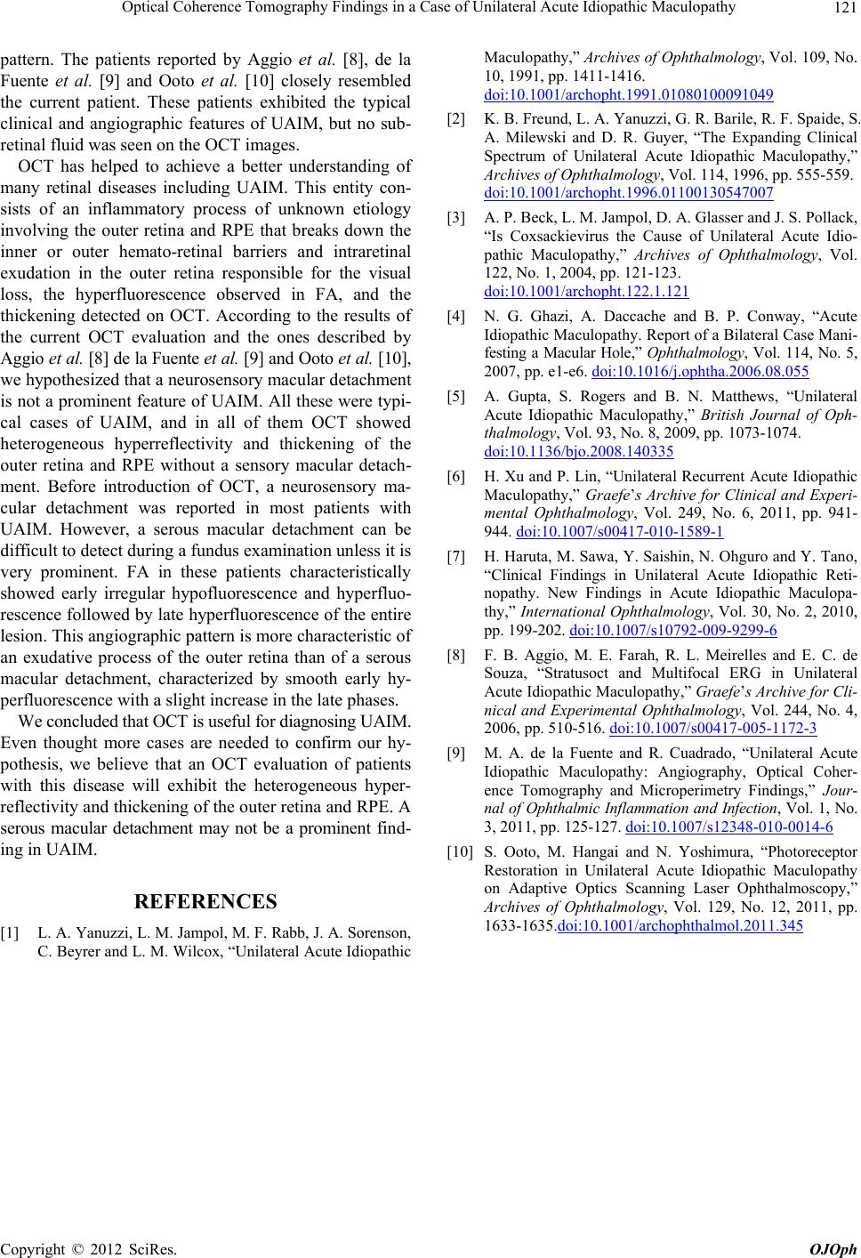 Optical Coherence Tomograph y Findings in a Cas e of Unilateral Acute Idiopathic Maculopathy Copyright © 2012 SciRes. OJOph 121 pattern. The patients reported by Aggio et al. [8], de la Fuente et al. [9] and Ooto et al. [10] closely resembled the current patient. These patients exhibited the typical clinical and angiographic features of UAIM, but no sub- retinal fluid was seen on the OCT images. OCT has helped to achieve a better understanding of many retinal diseases including UAIM. This entity con- sists of an inflammatory process of unknown etiology involving the outer retina and RPE that breaks down the inner or outer hemato-retinal barriers and intraretinal exudation in the outer retina responsible for the visual loss, the hyperfluorescence observed in FA, and the thickening detected on OCT. According to the results of the current OCT evaluation and the ones described by Aggio et al. [8] de la Fuente et al. [9] and Ooto et al. [10], we hypothesized that a neurosensory macular detachment is not a prominent feature of UAIM. All these were typi- cal cases of UAIM, and in all of them OCT showed heterogeneous hyperreflectivity and thickening of the outer retina and RPE without a sensory macular detach- ment. Before introduction of OCT, a neurosensory ma- cular detachment was reported in most patients with UAIM. However, a serous macular detachment can be difficult to detect during a fundus examination unless it is very prominent. FA in these patients characteristically showed early irregular hypofluorescence and hyperfluo- rescence followed by late hyperfluorescence of the entire lesion. This angiographic pa ttern is more characteristic of an exudative process of the outer retina than of a serous macular detachment, characterized by smooth early hy- perfluorescence with a slight increase in the late phases. We concluded that OCT is useful for di agnosi ng UAIM . Even thought more cases are needed to confirm our hy- pothesis, we believe that an OCT evaluation of patients with this disease will exhibit the heterogeneous hyper- reflectivity and thickening of the outer retin a and RPE. A serous macular detachment may not be a prominent find- ing in UAIM. REFERENCES [1] L. A. Yanuzzi, L. M. Jampol, M. F. Rabb, J. A. Sorenson, C. Beyrer and L. M. Wilcox, “Unilateral Acute Idiopathic Maculopathy,” Archives of Ophthalmology, Vol. 109, No. 10, 1991, pp. 1411-1416. doi:10.1001/archopht.1991.01080100091049 [2] K. B. Freund, L. A. Yanuzzi, G. R. Barile, R. F. Spaide, S. A. Milewski and D. R. Guyer, “The Expanding Clinical Spectrum of Unilateral Acute Idiopathic Maculopathy,” Archives of Ophthalmology, Vol. 114, 1996, pp. 555-559. doi:10.1001/archopht.1996.01100130547007 [3] A. P. Beck, L. M. Jampol, D. A. Glasser and J. S. Pollack, “Is Coxsackievirus the Cause of Unilateral Acute Idio- pathic Maculopathy,” Archives of Ophthalmology, Vol. 122, No. 1, 2004, pp. 121-123. doi:10.1001/archopht.122.1.121 [4] N. G. Ghazi, A. Daccache and B. P. Conway, “Acute Idiopathic Maculopathy. Report of a Bilateral Case Mani- festing a Macular Hole,” Ophthalmology, Vol. 114, No. 5, 2007, pp. e1-e6. doi:10.1016/j.ophtha.2006.08.055 [5] A. Gupta, S. Rogers and B. N. Matthews, “Unilateral Acute Idiopathic Maculopathy,” British Journal of Oph- thalmology, Vol. 93, No. 8, 2009, pp. 1073-1074. doi:10.1136/bjo.2008.140335 [6] H. Xu and P. Lin, “Unilateral Recurre nt Acute Idiopat hic Maculopathy,” Graefe’s Archive for Clinical and Experi- mental Ophthalmology, Vol. 249, No. 6, 2011, pp. 941- 944. doi:10.1007/s00417-010-1589-1 [7] H. Haruta, M. Sawa, Y. Saishin, N. Ohguro and Y. Tano, “Clinical Findings in Unilateral Acute Idiopathic Reti- nopathy. New Findings in Acute Idiopathic Maculopa- thy,” International Ophthalmology, Vol. 30, No. 2, 2010, pp. 199-202. doi:10.1007/s10792-009-9299-6 [8] F. B. Aggio, M. E. Farah, R. L. Meirelles and E. C. de Souza, “Stratusoct and Multifocal ERG in Unilateral Acute Idiopathic Maculopathy,” Graefe’s Archive for Cli- nical and Experimental Ophthalmology, Vol. 244, No. 4, 2006, pp. 510-516. doi:10.1007/s00417-005-1172-3 [9] M. A. de la Fuente and R. Cuadrado, “Unilateral Acute Idiopathic Maculopathy: Angiography, Optical Coher- ence Tomography and Microperimetry Findings,” Jour- nal of Ophthalmic Inflammatio n and Infection, Vol. 1, No. 3, 2011, pp. 125-127. doi:10.1007/s12348-010-0014-6 [10] S. Ooto, M. Hangai and N. Yoshimura, “Photoreceptor Restoration in Unilateral Acute Idiopathic Maculopathy on Adaptive Optics Scanning Laser Ophthalmoscopy,” Archives of Ophthalmology, Vol. 129, No. 12, 2011, pp. 1633-1635.doi:10.1001/archophthalmol.2011.345 |