

Paper Menu >>
Journal Menu >>
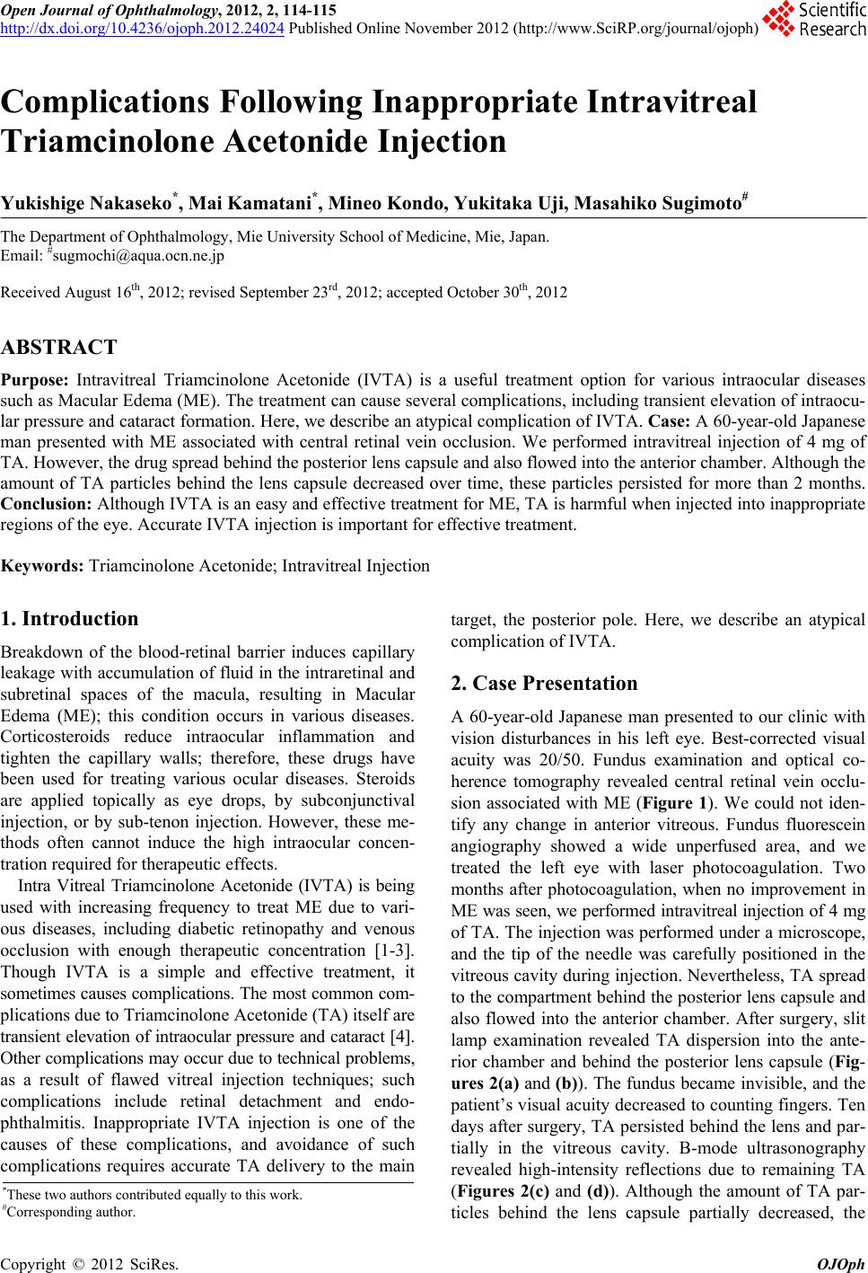 Open Journal of Ophthalmology, 2012, 2, 114-115 http://dx.doi.org/10.4236/ojoph.2012.24024 Published Online November 2012 (http://www.SciRP.org/journal/ojoph) Complications Following Inappropriate Intravitreal Triamcinolone Acetonide Injection Yukishige Nakaseko*, Mai Kamatani*, Mineo Kondo, Yukitaka Uji, Masahiko Sugimoto# The Department of Ophthalmology, Mie University School of Medicine, Mie, Japan. Email: #[email protected] Received August 16th, 2012; revised September 23rd, 2012; accepted October 30th, 2012 ABSTRACT Purpose: Intravitreal Triamcinolone Acetonide (IVTA) is a useful treatment option for various intraocular diseases such as Macular Edema (ME). The treatment can cause several complications, including tran sient elevation of intraocu- lar pressure and cataract formation. Here, we describe an atypical complication of IVTA. Case: A 60-year-old Japanese man presented with ME associated with central retinal vein occlusion. We performed intravitreal injection of 4 mg of TA. However, the drug spread behind the posterior lens capsule and also flowed into the anterior chamber. Althou gh the amount of TA particles behind the lens capsule decreased over time, these particles persisted for more than 2 months. Conclusion: Although IVTA is an easy and effective treatment for ME, TA is harmful when injected into inappropr iate regions of the eye. Accurate IVTA injection is important for effective treatment. Keywords: Triamci nol on e Ac et oni de; I nt ra vi treal Inject io n 1. Introduction Breakdown of the blood-retinal barrier induces capillary leakage with accumulation of fluid in the intraretinal and subretinal spaces of the macula, resulting in Macular Edema (ME); this condition occurs in various diseases. Corticosteroids reduce intraocular inflammation and tighten the capillary walls; therefore, these drugs have been used for treating various ocular diseases. Steroids are applied topically as eye drops, by subconjunctival injection, or by sub-tenon injection. However, these me- thods often cannot induce the high intraocular concen- tration required for thera peutic effects. Intra Vitreal Triamcinolone Acetonide (IVTA) is being used with increasing frequency to treat ME due to vari- ous diseases, including diabetic retinopathy and venous occlusion with enough therapeutic concentration [1-3]. Though IVTA is a simple and effective treatment, it sometimes causes co mplications. The most c ommon com- plications due to Triamcinolone Acetonide (TA) itself are transient elevation of intraocular pressure and cataract [4]. Other complications may occur due to technical problems, as a result of flawed vitreal injection techniques; such complications include retinal detachment and endo- phthalmitis. Inappropriate IVTA injection is one of the causes of these complications, and avoidance of such complications requires accurate TA delivery to the main target, the posterior pole. Here, we describe an atypical complication of IVTA. 2. Case Presentation A 60-year-old Japanese man presented to our clinic with vision disturbances in his left eye. Best-corrected visual acuity was 20/50. Fundus examination and optical co- herence tomography revealed central retinal vein occlu- sion associated with ME (Figure 1). We could not iden- tify any change in anterior vitreous. Fundus fluorescein angiography showed a wide unperfused area, and we treated the left eye with laser photocoagulation. Two months after photocoagulation, when no improvement in ME was seen, we performed intravitreal injection of 4 mg of TA. The injection was performed under a microscope, and the tip of the needle was carefully positioned in the vitreous cavity during injection. Nevertheless, TA spread to the compartment behind the po sterior lens capsule and also flowed into the anterior chamber. After surgery, slit lamp examination revealed TA dispersion into the ante- rior chamber and behind the posterior lens capsule (Fig- ures 2(a) and (b)). The fundus became invisible, and the patient’s visual acuity decreased to counting fingers. Ten days after surgery, TA persisted behind the lens and par- tially in the vitreous cavity. B-mode ultrasonography revealed high-intensity reflections due to remaining TA (Figures 2(c) and (d)). Although the amount of TA par- ticles behind the lens capsule partially decreased, the *These two authors contributed equally to this work. #Corresponding author. Copyright © 2012 SciRes. OJOph 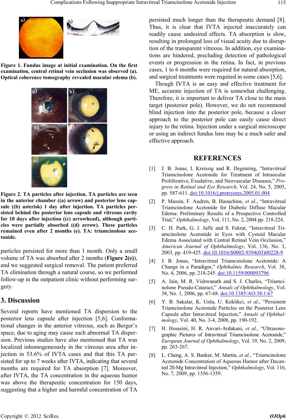 Complications Following Inappropriate Intravitreal Triamcinolone Acetonide Injection 115 Figure 1. Fundus image at initial examination. On the first examination, central retinal vein occlusion was observed (a). Optical coherence tomography revealed macular edema (b). Figure 2. TA particles after injection. TA particles are seen in the anterior chamber ((a) arrow) and posterior lens cap- sule ((b) asterisk) 1 day after injection. TA particles per- sisted behind the posterior lens capsule and vitreous cavity for 10 days after injection ((c) arrowhead), although parti- cles were partially absorbed ((d) arrow). These particles remained even after 2 months (e). TA: triamcinolone ace- tonide. particles persisted for more than 1 month. Only a small volume of TA was absorbed after 2 months (Figure 2(e)), and we suggested surgical removal. The patient preferred TA elimination through a natural course, so we performed follow-up in the outpatient clinic without performing su r- gery. 3. Discussion Several reports have mentioned TA dispersion to the posterior lens capsule after injection [5,6]. Conforma- tional changes in the anterior vitreous, such as Berger’s space, due to aging may cause such abnormal TA disper- sion. Previous studies have also mentioned that TA was localized inhomogeneously in the vitreous area after in- jection in 53.6% of IVTA cases and that this TA per- sisted for up to 7 weeks after IVTA, indicating that several months are required for TA absorption [7]. Moreover, after IVTA, the TA concentration in the aqueous humor was above the therapeutic concentration for 150 days, suggesting that a higher and harmful concentration of TA persisted much longer than the therapeutic demand [8]. Thus, it is clear that IVTA injected inaccurately can readily cause undesired effects. TA absorption is slow, resulting in prolong ed loss of visual acuity due to disrup- tion of the transparent vitreous. In addition , eye examina- tions are hindered, precluding detection of pathological events or progression in the retina. In fact, in previous cases, 1 to 6 months were required for natural absorption, and surgical treatments were required in some cases [5,6]. Though IVTA is an easy and effective treatment for ME, accurate injection of TA is somewhat challenging. Therefore, it is important to deliv er TA close to the main target (posterior pole). However, we do not recommend blind injection into the posterior pole, because a closer approach to the posterior pole can easily cause direct injury to the retina. Injection under a surg ical microscope or using an indirect fundus lens may be a much safer and effective approach. REFERENCES [1] J. B. Jonas, I. Kreissig and R. Degenring, “Intravitreal Triamcinolone Acetonide for Treatment of Intraocular Proliferative, Exudative, and Neovascular Diseases,” Pro- gress in Retinal and Eye Research, Vol. 24, No. 5, 2005, pp. 587-611. doi:10.1016/j.preteyeres.2005.01.004 [2] P. Massin, F. Audren, B. Haouchine, et al., “Intravitreal Triamcinolone Acetonide for Diabetic Diffuse Macular Edema: Preliminary Results of a Prospective Controlled Trial,” Ophthalmology, Vol. 111, No. 2, 2004 pp. 218-224. [3] C. H. Park, G. J. Jaffe and S. Fekrat, “Intravitreal Tri- amcinolone Acetonide in Eyes with Cystoid Macular Edema Associated with Central Retinal Vein Occlusion,” American Journal of Ophthalmology, Vol. 136, No. 3, 2003, pp. 419-425. doi:10.1016/S0002-9394(03)00228-9 [4] J. B. Jonas, “Intravitreal Triamcinolone Acetonide: A Change in a Paradigm,” Ophthalmic Research, Vol. 38, No. 4, 2006, pp. 218-245. doi:10.1159/000093796 [5] A. Jain, M. R. Vishwanath and S. J. Charles, “Triamci- nolone Pseudo-Cataract,” Annals of Ophthalmology, Vol. 38, No. 1, 2006, pp. 67-68. doi:10.1385/AO:38:1:67 [6] Y. B. Sakalar, K. Unlu, U. Keklikci, et al., “Persistent Triamcinolone Acetonide Particles on the Posterior Lens Capsule after Intravitreal Injection,” Annals of Ophthal- mology, Vol. 40, No. 3-4, 2008, pp. 190-192. [7] H. Hosseini, H. R. Anvari-Ardakani, et al., “Ultrasono- graphic Pictures of Intravitreal Triamcinolone Acetonide,” European Journal of Ophthalmology, Vol. 19, No. 2, 2009, pp. 263-267. [8] L. Cheng, A. S. Banker, M. Martin, et al., “Triamcinolone Acetonide Concentration of Aqueous Humor after Decan- ted 20-Mg Intravitreal Injection,” Ophthalmology, Vol. 116, No. 7, 2009, pp. 1356-1359. Copyright © 2012 SciRes. OJOph |