

Paper Menu >>
Journal Menu >>
 Advances in Anthropology 2012. Vol.2, No.4, 214-220 Published Online November 2012 in SciRes (http://www.SciRP.org/journal/aa) http://dx.doi.org/10.4236/aa.2012.24023 Copyright © 2012 SciRes. 214 A New Biomarker for Hepatocellular Damage: Plasma Cell-Free DNA* Zhi Yan1, Yingli He1, Qian Li1, Meiling Cui1, Ke Wang1, Tianyan Chen2, Hongli Liu2, Ying-Ren Zhao2# 1Department of Infectious Diseases, The First Affiliated Hospital of Medical College, Xi’an Jiaotong University, Xi’an, China 2Hepatology Institution, The First Affiliated Hospital of Medical College, Xi’an Jiaotong University, Xi’an, China Email: #[email protected] Received August 16th, 2012; revised September 20th, 2012; accepted October 6th, 2012 Background: Accumulating evidence has suggested that cell-free DNA (cf-DNA) enters the circulation following cell apoptosis or necrosis. An increased level of cf-DNA fragments has been found in the blood of mice with drug-induced liver damage. We sought to determine the role of cf-DNA in hepatocellular damage. Methods: Plasma samples were collected from 204 patients with hepatitis. The patients were di- vided into three groups according to liver pathologic characteristics: with chronic hepatitis (CH) and compensated liver cirrhosis (LC) (the group 1); with decompensated liver cirrhosis (DLC) (the group 2); with liver failure (LF), acute hepatitis (AH) and hepatocellular carcinoma (HCC) (the group 3). The cf-DNA was extracted with the phenol/chloroform/isoamyl alcohol (PCI) method and the plasma cf-DNA was quantified using real-time polymerase chain reaction (rt-PCR) for β-globin. The cf-DNA copies were converted to log2 values for comparison. Results: Cf-DNA was detected in all the 3 groups. The group 3 had a significantly higher cf-DNA level than the other two groups (17.70 ± 1.79, P = 0.002). The level of plasma cf-DNA was correlated with the baseline aniline transaminase (ALT) and aspertate transaminase (AST) activities (P < 0.005). The cf-DNA concentration in patients with cirrhosis was correlated with the model of end-stage liver disease-Na (MELD-Na) score and the ALT and AST activities. Correlation of the cf-DNA level with laboratory parameters, such as bilirubin and international normalized ratio (INR), were found in patients with high cf-DNA levels (cf-DNA > 19.5), or with severe hepatocellular damage (ALT > 500 U/L). Conclusion: Plasma cell-free DNA may be a new promising, independent, non-inva- sive biomarker for hepatocellular damage. Keywords: Hepatocellular Damage; Cell-Free DNA Introduction The World Health Organization reports that cirrhosis and primary liver cancer caused 783,000 and 619,000 death, respec- tively, in 2002 (WHO, 2003). Most of these deaths in both de- veloping and developed countries are attributed to hepadnavirus infection. Hepadnavirus infection may cause acute hepatitis, chronic hepatitis, cirrhosis, and hepatocellular carcinoma. Liver biopsy has long been used as the gold standard for the clinical evaluation of chronic hepatitis. However, invasion and sam- pling error make biopsy a flawed benchmark. Recently noninvasive marker tests such as liver function tests and coagulation tests have been widely adopted to assess the severity of acute and chronic liver injury. The model for end- stage liver disease (MELD) score is used to rank patients awaiting liver transplants based on 3 laboratory variations in- cluding international normalized ratio (INR), serum creatine and total bilirubin (Kamath, 2007). The MELD-Na score, cre- ated as an accurate predictor of survival in patients with ad- vanced liver diseases, provides better prognostic accuracy than the MELD score (Hsu, 2010). However, all the above tests cannot provide specific or direct measurements of the liver function. For example, elevated serum levels of the baseline aniline transaminase (ALT) and aspertate transaminase (AST) are nonspecific indicators of hepatocellular damage; hyper- bilirubinemia may not be detected in the cases of moderate to severe hepatocellular damage; albumin has a long half-life time and cannot immediately reflect changes in hepatic synthesis; the prolonged prothrombin time is not a distinctive feature of liver diseases (Pratt & Schiff, 2007; Dufour, 2000) . Therefore, one easy, direct, specific marker is needed to assist physicians in the diagnosis and treatment of patients with hepatitis. An elevated level of circulating cell-free DNA (cf-DNA) has been detected in patients under pathologic conditions such as cancer, trauma, stroke, pregnancy, autoimmune disorders, and solid organ transplant (Tokuhisa, 2007; Kamat, 2010; Lam, 2003; Lau, 2002; Holdenrieder, 2006; Lui, 2002). Roth et al. have also found that the cf-DNA level is elevated in the serum of liver failure patients (Roth, 2009). There are two origins of the circulating cf-DNA in patients under pathologic conditions: the DNA fragments enter the blood following cell death; they are actively released by living cells (Van der Vaart, 2007). However, the origin and mechanism of the increased cf-DNA are still uncertain. Liver cell death can be attributed to apop- tosis or necrosis or a combination of the two. Apoptosis is a prominent feature of acute and chronic hepatocellular damage. *Objectives: None of the authors has any potential financial conflict of in- terest related to this manuscript. #Corresponding author.  Z. YAN ET AL. Moreover, the cf-DNA clearance mechanism is poorly under- stood. Emlen et al. have produced evidence suggesting that the liver may be the major organ for the removal of cf-DNA (Em- len, 1978). Another research has reported that circulating DNA has a short half-life time of 16.3 min in plasma (Lo, 1999). We hypothesized that cf-DNA could be another non-invasive marker of hepatocellular damage. The aim of our research was to evaluate the role of cf-DNA as a biomarker of hepatocellular damage and to investigate the effect of the impaired liver and renal function on the cf-DNA level in plasma. Materials and Methods Plasma Samples 204 archived plasma samples were obtained from 204 pa- tients on the first day when they visited the First Affiliated Hospital of Medical School of Xi’an Jiaotong University (China). All the patients were diagnosed as having viral, drug- induced or unexplained hepatitis without immunologic diseases. According to their pathological characteristics, patients were divided into 3 groups: the group 1 (60) including patients with chronic hepatitis (CH) and compensated liver cirrhosis (LC), the group 2 (66) including patients with decompensated liver cirrhosis (DLC) and the group 3 (78) including patients with liver failure (LF), acute hepatitis (AH), and hepatocellular car- cinoma (HCC). Written informed consent was obtained directly from each patient before peripheral blood was collected. Blood samples (3 ml) were drawn into tubes containing ethylenedia- mine tetra-acetic acid (EDTA). Plasma fractions were separated within 2 hours according to a two-step centrifugation procedure: at 1600 × g for 10 min and at 16,000 × g for 10 min. The plasma fractions were stored at −80˚C until further process- ing. DNA Extraction from Plasma Samples Cell-free DNA was extracted with the golden phenol/chloro- form/isoamyl alcohol (PCI) method. 300 μl plasma was mixed with a lysis solution and proteinase K. The mixture was incu- bated at 56˚C for 2 h and then heat denatured at 95˚C for 10 min. 1200 μl of phenol: chloroform: isoamyl alcohol (25:24:1) was added to the mixture. The top aqueous layer, after cen- trifugation at 12,000 rpm for 15 min, was mixed with 100% ethanol and precipitated at −20˚C overnight. The precipitates were washed with 70% ethanol and dissolved with distilled water. All the cell-free DNA samples were stored at −20˚C until quantification. Quantification of Cell-Free DNA Quantification of cell-free DNA (β-globin) was performed using real-time polymerase chain reaction (rt-PCR) with SYBR Green I (Applied Biosystem 7500). The forward primer was ACACAACTGTGTTCACTAGC and the reverse primer was CAACTTCATCCACGTTCACC. The thermal cycling protocol was as follows: 95˚C for 1 min, followed by 40 cycles at 95˚C for 30 s, at 57˚C for 20 s and at 72˚C for 32 s. A standard curve was created and the DNA concentration, expressed as genome equivalents per milliliter (GE/ml), was calculated using the following equation: DNA PCREXT CQ VV1V where C is the target concentration in plasma (GE/ml), Q is the target quantity (copies), VDNA is the total volume of DNA extraction (60 µl), VPCR is the volume of DNA used per PCR reaction (9.5 µl), and Vext is the volume of plasma used to ex- tract DNA (300 µl). All samples were run in triplicate. Statistical Analysis All statistical procedures were performed using SPSS16.0 statistical software. The plasma cf-DNA concentrations of the three hepatitis groups were compared with the nonparametric Mann-Whitney U test and the LSD (least significant differ- ence)-t test. The Kruskal-Wallis H test was used to compare the laboratory parameters among the three groups, and the non- parametric Spearman test was applied to determine the bivariate correlation. In all tests, P < 0.05 was considered statistically significant. Results Our cohort included 146 patients with HBV infection, 33 with HCV infection and 25 with unexplained, drug-induced, hepatitis A virus (HAV) or hepatitis E virus (HEV) infection. Table 1 shows the information of the patients, their cf-DNA concentrations and the results of all the laboratory tests, in- cluding liver function tests, renal function tests, sodium test, blood routine tests and coagulation tests. Cell-free DNA was detected in all patients. Cf-DNA concen- tration mainly ranges from 15 to 20 (Figure 1(a)). Patients with cf-DNA < 15 were mainly from the group 2 while patients with cf-DNA > 20 were mainly from the groups 2 and 3. As shown in Figure 1(b), the highest mean concentration of cf-DNA was observed in the group 3 (17.70 ± 1.79), followed by the group 1 (17.13 ± 1.12) and group 2 (16.69 ± 1.93) (P = 0.002) with statistic significance. The cf-DNA concentration was found to have no correlation with age, sex, etiology and diagnosis of hepatitis. The cf-DNA concentration was positively correlated with ALT activity, AST activity and white blood cell (WBC) count (r = 0.183, 0.267 and 0.156, respectively; P = 0.009, 0.000 and 0.028, respectively). Correlation of cf-DNA with Laboratory Parameters in Cirrhosis Patients The MELD score and MELD-Na score of patients with compensated and decompensated cirrhosis, excluding patients with HCC and liver failure, were calculated. The circulating cf-DNA concentration was negatively correlated with the MELD-Na score (r = −0.256, P = 0.036), but showed no corre- lation with the MELD score (P > 0.05). The cf-DNA concen- tration was also found to be correlated with ALT activity, AST activity and INR (r = 0.353, 0.336 and −0.282, respectively and P = 0.001, 0.002 and 0.010, respectively) (Figure 2). Correlations of cf-DNA with Laboratory Parameters at cf-DNA > 19.5 We further included the patients with a high cf-DNA con- centration (cf-DNA > 19.5) into the Spearman-test, using 19.5 as the baseline concentration. It was found that the high cf-DNA concentration was significantly correlated with clinical parameters indicating hepatocellular damage and liver function, Copyright © 2012 SciRes. 215 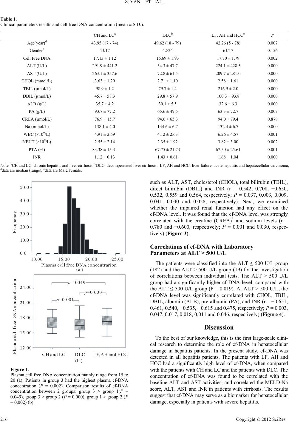 Z. YAN ET AL. Copyright © 2012 SciRes. 216 Table 1. Clinical parameters results and cell free DNA concentration (mean ± S.D.). CH and LCa DLCb LF, AH and HCCc P Age(year)d 43.95 (17 - 74) 49.62 (18 - 79) 42.26 (5 - 78) 0.007 Gendere 43/17 42/24 61/17 0.156 Cell Free DNA 17.13 ± 1.12 16.69 ± 1.93 17.70 ± 1.79 0.002 ALT (U/L) 291.9 ± 441.2 54.3 ± 47.7 224.1 ± 428.5 0.000 AST (U/L) 263.1 ± 357.6 72.8 ± 61.5 209.7 ± 281.0 0.000 CHOL (mmol/L) 3.63 ± 1.29 2.71 ± 1.10 2.58 ± 1.61 0.000 TBIL (µmol/L) 98.9 ± 1.2 79.7 ± 1.4 216.9 ± 2.0 0.000 DBIL (µmol/L) 45.7 ± 58.3 29.8 ± 57.9 100.3 ± 93.8 0.000 ALB (g/L) 35.7 ± 4.2 30.1 ± 5.5 32.6 ± 6.3 0.000 PA (g/L) 93.7 ± 77.2 65.6 ± 49.5 63.3 ± 72.7 0.007 CREA (µmol/L) 76.9 ± 15.7 94.6 ± 65.3 94.0 ± 79.4 0.878 Na (mmol/L) 138.1 ± 4.0 134.6 ± 6.7 132.4 ± 6.7 0.000 WBC (×109/L) 4.91 ± 2.69 4.12 ± 2.63 6.26 ± 4.57 0.001 NEUT (×109/L) 2.55 ± 2.14 2.35 ± 1.92 3.82 ± 3.00 0.002 PTA (%) 83.38 ± 15.31 67.75 ± 21.73 67.50 ± 25.61 0.001 INR 1.12 ± 0.13 1.43 ± 0.61 1.68 ± 1.04 0.000 Note: aCH and LC: chronic hepatitis and liver cirrhosis; bDLC: decompensated liver cirrhosis; cLF, AH and HCC: liver failure, acute hepatitis and hepatocellular carcinoma; ddata are median (range); edata are Male/Female. such as ALT, AST, cholesterol (CHOL), total bilirubin (TBIL), direct bilirubin (DBIL) and INR (r = 0.542, 0.708, −0.650, 0.532, 0.559 and 0.564, respectively; P = 0.037, 0.003, 0.009, 0.041, 0.030 and 0.028, respectively). Next, we examined whether the impaired renal function had any effect on the cf-DNA level. It was found that the cf-DNA level was strongly correlated with the creatine (CREA)3 and sodium levels (r = 0.780 and −0.600, respectively; P = 0.001 and 0.030, respec- tively) (Figure 3). Correlations of cf-DNA with Laboratory Parameters at ALT > 500 U/L The patients were classified into the ALT ≤ 500 U/L group (182) and the ALT > 500 U/L group (19) for the investigation of correlations between individual tests. The ALT > 500 U/L group had a significantly higher cf-DNA level, compared with the ALT ≤ 500 U/L group (P = 0.019). At ALT > 500 U/L, the cf-DNA level was significantly correlated with CHOL, TBIL, DBIL, albumin (ALB), pre-albumin (PA), and INR (r = −0.651, 0.461, 0.540, −0.535, −0.615 and 0.475, respectively; P = 0.003, 0.047, 0.017, 0.018, 0.011 and 0.046, respectively) (Figure 4). Discussion To the best of our knowledge, this is the first large-scale clini- cal research to determine the role of cf-DNA in hepatocellular damage in hepatitis patients. In the present study, cf-DNA was detected in all hepatitis patients. The patients with LF, AH and HCC had a significantly high level of cf-DNA, when compared with the patients with CH and LC and the patients with DLC. The concentration of cf-DNA was found to be correlated with the baseline ALT and AST activities, and correlated the MELD-Na score, ALT, AST and INR in patients with cirrhosis. The results suggest that cf-DNA may serve as a biomarker for hepatocellular damage, especially in patients with severe hepatitis. Figure 1. Plasma cell free DNA concentration mainly range from 15 to 20 (a); Patients in group 3 had the highest plasma cf-DNA concentration (P = 0.002). Comparison results of cf-DNA concentration between 2 groups: group 3 > group 1(P = 0.049), group 3 > group 2 (P = 0.000), group 1 > group 2 (P = 0.002) (b). 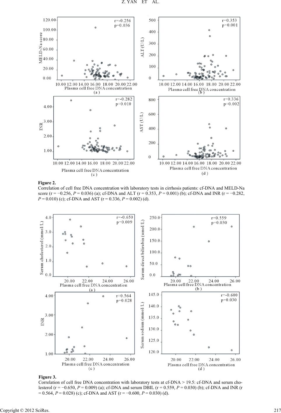 Z. YAN ET AL. Figure 2. Correlation of cell free DNA concentration with laboratory tests in cirrhosis patients: cf-DNA and MELD-Na score (r = −0.256, P = 0.036) (a); cf-DNA and ALT (r = 0.353, P = 0.001) (b); cf-DNA and INR (r = −0.282, P = 0.010) (c); cf-DNA and AST (r = 0.336, P = 0.002) (d). Figure 3. Correlation of cell free DNA concentration with laboratory tests at cf-DNA > 19.5: cf-DNA and serum cho- lesterol (r = −0.650, P = 0.009) (a); cf-DNA and serum DBIL (r = 0.559, P = 0.030) (b); cf-DNA and INR (r = 0.564, P = 0.028) (c); cf-DNA and AST (r = −0.600, P = 0.030) (d). Copyright © 2012 SciRes. 217  Z. YAN ET AL. Figure 4. Correlation of cell free DNA concentration with laboratory tests at ALT > 500 U/L: cf-DNA and serum cholesterol (r = −0.651, P = 0.003) (a); cf-DNA and serum DBIL (r = 0.540, P = 0.017) (b); cf-DNA and pre-albumin (r = −0.615, P = 0.011) (c); cf-DNA and albumin (r = −0.535, P = 0.018) (d). The elevated cf-DNA level may reflect the disturbance of equilibrium between the release and clearance of circulating cf-DNA. The origin of cf-DNA has been studied for approxi- mately 30 years; however, the underlying mechanism is still unclear. Sabine Jahr et al. have detected circulating cf-DNA in the plasma of cancer patients and identified it as a hallmark of necrosis and apoptosis (Jahr, 2001). Philippe Anker et al. have corroborated the spontaneous release of DNA by lymphocytes in vitro (Anker, 1975). Our results revealed a significant eleva- tion of the cf-DNA concentration in patients with severe heap- tocellular damage and an association between cf-DNA and WBC. Probably, cell death and inflammation are both the ori- gins of cf-DNA in patients with hepatitis. Emlen et al. have found that the liver is the major organ for removal of circulat- ing ssDNA (Emlen, 1978). Botezatu et al. have detected male- specific DNA sequences in the urine of females who had been transfused with male blood (Botezatu, 2000). Maybe impaired liver or kidney function is another explanation for the elevation of the cf-DNA concentration. The key finding of this study is that although not all indi- viduals showed an elevated cf-DNA concentration, the mean cf-DNA concentration was significantly elevated in the group 3 patients with severe hepatocellular damage and poor hepatic function, when compared with those in the other two groups. This study possessed potential confounding factors that cannot be ruled out. For example, some of the patients were compli- cated with upper gastrointestinal tract bleeding, or ascites. We could not match all the confounding factors in our study groups. To date, ALT activity, AST activity and bilirubin have been generally used by clinicians to assess the severity of liver injury. Aminotransferases and bilirubin are sensitive indicators of hepatocellular damage. The highest elevations of the ami- notransferases level occur in disorders associated with exten- sive hepatocellular damage. In our study, circulating cf-DNA concentrations were found to be correlated with the baseline ALT and AST activities. The liver is the major site of synthesis of cholesterol, protein and blood coagulation factors. Our re- sults show that cf-DNA increased with the severity of the im- paired liver function. Interestingly, cf-DNA was found to be correlated with aminotransferases when patients with cirrhosis were considered as a whole, but to show no correlation with aminotransferases in patients with chronic hepatitis. These data support the hypothesis that the mechanism underlying the ele- vation of the cf-DNA level in the plasma is not only associated with hepatocellular damage but also with impaired liver func- tion. Hepatorenal syndrome (HRS) is a fatal complication of de- compensated cirrhosis. HRS may be manifested as impaired renal function, ascites and edema. Sodium retention plays a fundamental role in the formation of ascites and edema and is the first manifestation of renal impairment in patients with cir- rhosis. Creatinine is a marker for decreased renal perfusion. Based on the relations between cf-DNA and serum sodium and CREA at cf-DNA > 19.5, we predicted that kidney might play a role in the elevation of the circulating cf-DNA. We also estab- lished the relationship between cf-DNA and WBC, which is in agreement with the finding of a previous study [6]. Cf-DNA originates from cell death; however, little hepatic cells were reserved in patients with cirrhosis. Based on this Copyright © 2012 SciRes. 218  Z. YAN ET AL. knowledge, we found a moderately negative correlation be- tween cf-DNA and MELD-Na score. MELD-Na score has been introduced as a predictor of mortality and can provide better prognostic accuracy than MELD-score (Kamath, 2007). There- fore, Cf-DNA can be selected as non-invasive assessment marker for survival and prognosis of patients with hepatic dis- eases. Lee et al. found that most cf-DNA in the serum samples was generated during the process of clotting in the original collec- tion tubes (Lee, 2001). Fong L. et al. performed a comparative study of 7 cf-DNA isolation methods, and their results showed that PCI was a highly efficient method for cf-DNA isolation, compared with QIAamp DNA blood kit (Fong, 2009). Jung et al. described that plasma cf-DNA did not change after blood samples were stored at room temperature for 8 h or at 4˚C for 24 h before being processed (Jung, 2003). Based on the above findings, in this study, blood samples were collected into EDTA-tubes (Lam, 2004) to exclude contaminants from lyses cells during clotting and residual cells were removed from plasma within 2 hrs after blood collection using a two-step centrifugation method. In conclusion, cf-DNA can immediately provide easy and direct measurement of hepatocellular damage. The plasma cell- free DNA concentration may be a new promising non-invasive independent biomarker for hepatocellular damage. Impaired liver and kidney may play a role in the elevation of the plasma cf-DNA level. These findings may provide valuable informa- tion for further studies on the mechanism, origin and kinetics of the circulating cf-DNA associated with hepatocellular damage. Acknowledgements Grant support was provided by the Major National Science and Technology Projects for Infectious Diseases (11th Five Year, China) (Project Code 2008ZX10002-007). REFERENCES Anker, P., Stroun, M., & Maurice, P. A. (1975). Spontaneous release of DNA by human blood lymphocytes as shown in an in vitro system. Cancer Research, 35, 2375-2382. Botezatu, I., Serdyuk, O., Potapova, G., Shelepov, V., Alechina, R., Molyaka, Y. et al. (2000). Genetic analysis of DNA excreted in urine: A new approach for detecting specific genomic DNA sequences from cells dying in an organism. Clinical Chemistry, 46, 1078-1084. Dufour, D. R., Lott, J. A., Nolte, F. S., Gretch, D. R., Koff, R. S., & Seeff, L. B. (2000). Diagnosis and monitoring of hepatic injury. II. Recommendations for use of laboratory tests in screening, diagnosis, and monitoring. Clinical Chemistry, 46, 2050-2068. Emlen, W., & Mannik, M. (1978). Kinetics and mechanisms for re- moval of circulating single-stranded DNA in mice. Journal of Ex- perimental Medicine, 147, 684-699. doi:10.1084/jem.147.3.684 Fong, S. L., Zhang, J. T., Lim, C. K., Eu, K. W., & Liu, Y. (2009). Comparison of 7 methods for extracting cell-free DNA from serum samples of colorectal cancer patients. Clinical Chemistry, 55, 587- 589. doi:10.1373/clinchem.2008.110122 Holdenrieder, S., Eichhorn, P., Beuers, U., Samtleben, W., Schoener- marck, U., Zachoval, R. et al. (2006). Nucleosomal DNA fragments in autoimmune diseases. Annals of the New York Academy of Sci- ences, 1075, 318-327. doi:10.1196/annals.1368.043 Hsu, C. Y., Lin, H. C., Huang, Y. H., Su, C. W., Lee, F. Y., Huo, T. I. et al. (2010). Comparison of the model for end-stage liver disease (MELD), MELD-Na and MELDNa for outcome prediction in pa- tients with acute decompensated hepatitis. Digestive and Liver Dis- ease, 42, 137-142. doi:10.1016/j.dld.2009.06.004 Jahr, S., Hentze, H., Englisch, S., Hardt, D., Fackelmayer, F. O., Hesch, R. D., et al. (2001). DNA fragments in the blood plasma of cancer patients: Quantitations and evidence for their origin from apoptotic and necrotic cells. Cancer Research, 61, 1659-1665. Jung, M., Klotzek, S., Lewandowski, M., Fleischhacker, M., & Jung K. (2003). Changes in concentration of DNA in serum and plasma dur- ing storage of blood samples. Clinical Chemistry, 49 , 1028-1029. doi:10.1373/49.6.1028 Kamat, A. A., Baldwin, M., Urbauer, D., Dang, D., Han, L. Y., Godwin, A. et al. (2010). Plasma cell-free DNA in ovarian cancer: An inde- pendent prognostic biomarker. Cancer, 116, 1918-1925. doi:10.1002/cncr.24997 Kamath, P. S., & Kim, W. R. (2007). The model for end-stage liver disease (MELD). Hepatology, 45, 797-805. doi:10.1002/hep.21563 Lam, N. Y., Rainer, T. H., Chan, L. Y., Joynt, G. M., & Lo, Y. M. (2003). Time course of early and late changes in plasma DNA in trauma patients. Clinical Chemistry, 49 , 1286-1291. doi:10.1373/49.8.1286 Lam, N. Y., Rainer, T. H., Chiu, R. W., & Lo, Y. M. (2004). EDTA is a better anticoagulant than heparin or citrate for delayed blood proc- essing for plasma DNA analysis. Clinical Chemistry, 50, 256-257. doi:10.1373/clinchem.2003.026013 Lau, T. W., Leung, T. N., Chan, L. Y., Lau, T. K., Chan, K. C., Tam, W. H. et al. (2002). Fetal DNA clearance from maternal plasma is im- paired in preeclampsia. Clinical C hemistry, 48, 2141-2146. Lee, T. H., Montalvo, L., Chrebtow, V., & Busch, M. P. (2001). Quan- titation of genomic DNA in plasma and serum samples: Higher con- centrations of genomic DNA found in serum than in plasma. Trans- fusion, 41, 276-282. doi:10.1046/j.1537-2995.2001.41020276.x Lo, Y. M., Zhang, J., Leung, T. N., Lau, T. K., Chang, A. M., & Hjelm, N. M. (1999). Rapid clearance of fetal DNA from maternal plasma. The American journal of Human Genetics, 64, 218-224. doi:10.1086/302205 Lui, Y. Y., Chik, K. W., Chiu, R.W., Ho, C. Y., Lam, C. W., & Lo, Y. M. (2002). Predominant hematopoietic origin of cell-free DNA in plasma and serum after sex-mismatched bone marrow transplantation. Clinical Chemistry, 48, 421-427. Pratt, D. S., &Kaplan, M. M. (2007). Laboratory tests. In E. R. Schiff, (Ed.), Schiff’s diseases of the liver (10th ed., pp. 19-54). Hoboken, NJ: Wiley-Blackwell. Roth, G. A., Lubsczyk, B. A., Pilz, J., Faybik, P., Hetz, H., & Krenn, C. G. (2009). Nucleosome serum levels in acute hepatic failure and MARS treatment. Transplant Proceedings, 41, 4207-4210. doi:10.1016/j.transproceed.2009.08.073 Tokuhisa, Y., Iizuka, N., Sakaida, I., Moribe, T., Fujita, N., Miura, T. et al. (2007). Circulating cell-free DNA as a predictive marker for dis- tant metastasis of hepatitis C virus-related hepatocellular carcinoma. British Journal of Cancer, 97, 1399-1403. doi:10.1038/sj.bjc.6604034 van der Vaart, M., & Pretorius, P. J. (2007). The origin of circulating free DNA. Clinical Chemistry, 53, 2215. doi:10.1373/clinchem.2007.092734 World Health Organization (2003). The world health report 2003— Shaping the future. URL (last checked 16 August 2012). http://www.who.int/whr/2003/en/ Copyright © 2012 SciRes. 219  Z. YAN ET AL. Abbreviations cf-DNA: cell-free DNA CH: chronic hepatitis LC: compensated liver cirrhosis DLC: decompensated liver cirrhosis LF: liver failure AH: acute hepatitis HCC: hepatocellular carcinoma CREA: creatine Copyright © 2012 SciRes. 220 |