

Paper Menu >>
Journal Menu >>
 Advances in Physical Education 2012. Vol.2, No.4, 139-143 Published Online November 2012 in SciRes (http://www.SciRP.org/journal/ape) http://dx.doi.org/10.4236/ape.2012.24024 Copyright © 2012 SciRes . 139 The Effects of Knee Joint Pain and Disorders on Knee Extension Strength and Walking Ability in the Female Elderly Hiroki Sugiu ra, Shinichi Dem ura Graduate School of Natural Science & Technology, Kanazawa Unive rs i ty, Kanazawa, Japan Email: [email protected], [email protected] Received August 4th, 2012; revised September 8th, 20 12 ; a cce pte d S ep tember 20th, 2012 This study examines the differences in leg strength and walking ability among groups with different knee problems. The participants were 328 elderly females (60 - 94 years old; mean age 76.1 years; SD = 6.2) who were classified into three groups: those without knee pain or a knee disorder, those with knee pain, and those with a knee disorder. The subjects took knee extension strength and 12 meter maximum effort walk tests. Knee extension strength was significantly lower in the groups with knee pain and a knee dis- order than in the group without pain or a knee disorder. Walking speed was significantly slower in the group with a knee disorder than in the other two groups. In conclusion, the female elderly with knee pain or a knee disorder are inferior in knee extension strength and walking ability. In addition, the elderly with a knee disorder are inferior in walking ability to the elderly with knee pain. Keywords: Knee Joint Pain; Knee Joint Disorder; Knee Extension Strength; Gait; Female Elderly Introduction Walking is the most basic movement in daily life (Pratt, 1994). Leg strength, balance and leg joint functions are impor- tant factors related to walking. These physical functions de- crease with age, and thus walking ability also decreases with age. As a result, the frequency of daily life activities is limited markedly, and there is also a decrease in the ability to perform activities of daily life (ADL) (Jochanan et al., 2009; Hurley et al., 1998). In addition, a decrease in walking ability also in- creases fall occurrences (Mary & Tinetti, 2003), which there- fore greatly reduces quality of life (Sato et al., 2007; Suzuki et al., 2002). Hence, it is very important to prevent a decrease in walking functions in order to maintain independent daily life in the elderly. Knee joints have the maximum load capacity of all the leg joints, and they play a very important role in walking (Kuro- kawa et al., 2001). Recently, more knee disorders have been found in the elderly (Peat et al., 2001). Tennant et al. (1995) reported that 8% of the elderly have them. Leg strength de- creases with age in the elderly years (Frontera et al., 1991; Murrary et al., 1985), and the elderly with leg joint disorders are greatly limited in walking, due to both the knee disorder and a decrease in leg strength (Zeni & Higginson, 2009; Zoltan et al., 2006). Hence, the active mass of the elderly with knee dis- orders decreases markedly, subsequently causing a rapid de- crease in leg strength (Kirsten, 2009). Kirsten (2009) reported that subjects with a knee disorder were inferior in walking speed, stance stability and accelerating force. Berman et al. (1987) and Andriacchi et al. (1982) re- ported that the elderly requiring knee arthroplasty are inferior in walking speed, stance time, step length and cadence, as com- pared to the general elderly. On the other hand, there are many older people with subjec- tive knee pain, even though they do not have a serious knee disorder (Zoltan et al., 2006; Al-Zaharni & Bakheit, 2002; Peat et al., 2001). They can be regarded as an auxiliary group to people with a knee disorder. Alindon et al. (1992) and Urwin et al. (1998) reported that about 20% of the elderly had knee pain. Sugiura & Demura (2012) reported that the group with knee pain was inferior in knee extension strength to the group with- out knee pain, and the group with pain in both knees was infe- rior in stride length and step length to the group without knee pain. From the above, it is considered that the elderly with a knee disorder or knee pain are inferior in leg strength and walking ability to those without a knee disorder or pain. On the other hand, it is assumed that knee disorder participants with a spe- cific knee disorder are inferior in leg strength and/or walking ability to the people with knee pain, who are the reserve group of subjects with a knee disorder. The prevalence of knee pain and knee disorders is high in the female elderly (Oida & Nakamura, 2008; Peat et al., 2001). This study examines the differences in knee extension strength and walking ability among female elderly, who are divided into three groups: those without knee pain or a knee disorder, those with knee pain, and those with knee disorders. Methods Subjects Subjects consisted of 328 female elderly people (60 - 94 years old; mean age 76.1 years; SD = 6.2), who which classi- fied into the following three groups: 168 persons without knee pain or disorder (knee non-pain and disorder group), 116 per- sons with subjective knee pain (one knee pain group, n = 75; both knees in pain group, n = 41), and 44 persons with a knee disorder (one knee disorder group, n = 21; both knees disorder group, n = 23). Forty persons in the knee pain group and 11 persons in the knee disorder group had right knee pain. The knee pain and the knee disorder were grouped by Japanese 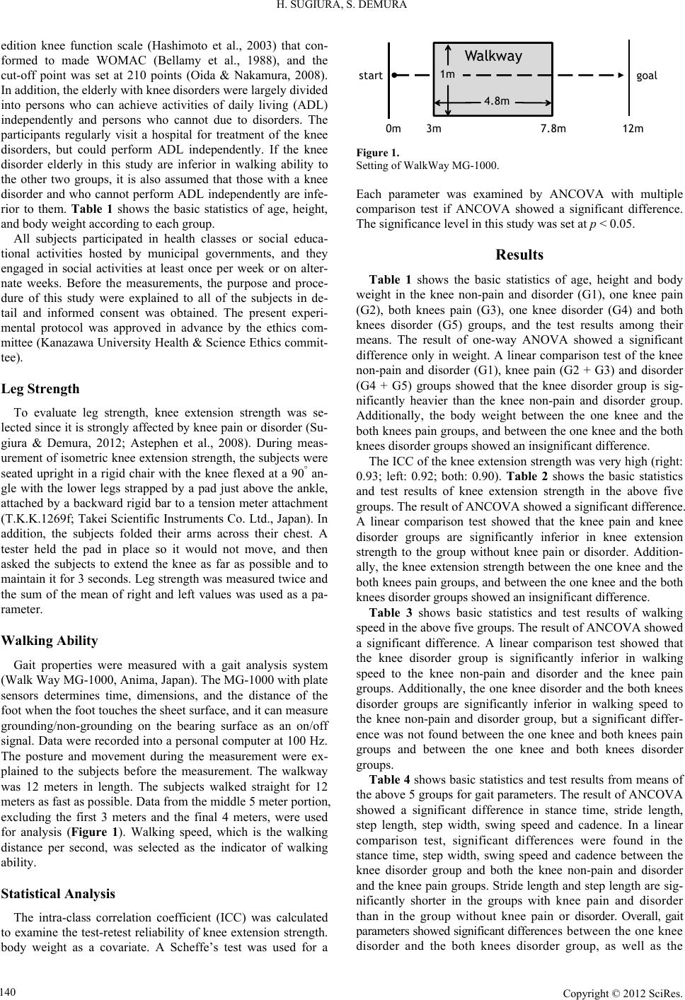 H. SUGIURA, S. DEMURA edition knee function scale (Hashimoto et al., 2003) that con- formed to made WOMAC (Bellamy et al., 1988), and the cut-off point was set at 210 points (Oida & Nakamura, 2008). In addition, the elderly with knee disorders were largely divided into persons who can achieve activities of daily living (ADL) independently and persons who cannot due to disorders. The participants regularly visit a hospital for treatment of the knee disorders, but could perform ADL independently. If the knee disorder elderly in this study are inferior in walking ability to the other two groups, it is also assumed that those with a knee disorder and who cannot perform ADL independently are infe- rior to them. Table 1 shows the basic statistics of age, height, and body weight according to each group. All subjects participated in health classes or social educa- tional activities hosted by municipal governments, and they engaged in social activities at least once per week or on alter- nate weeks. Before the measurements, the purpose and proce- dure of this study were explained to all of the subjects in de- tail and informed consent was obtained. The present experi- mental protocol was approved in advance by the ethics com- mittee (Kanazawa University Health & Science Ethics commit- tee). Leg Strength To evaluate leg strength, knee extension strength was se- lected since it is strongly affected by knee pain or disorder (Su- giura & Demura, 2012; Astephen et al., 2008). During meas- urement of isometric knee extension strength, the subjects were seated upright in a rigid chair with the knee flexed at a 90° an- gle with the lower legs strapped by a pad just above the ankle, attached by a backward rigid bar to a tension meter attachment (T.K.K.1269f; Takei Scientific Instruments Co. Ltd., Japan). In addition, the subjects folded their arms across their chest. A tester held the pad in place so it would not move, and then asked the subjects to extend the knee as far as possible and to maintain it for 3 seconds. Leg strength was measured twice and the sum of the mean of right and left values was used as a pa- rameter. Walking Ability Gait properties were measured with a gait analysis system (Walk Way MG-1000, Anima, Japan). The MG-1000 with plate sensors determines time, dimensions, and the distance of the foot when the foot touches the sheet surface, and it can measure grounding/non-grounding on the bearing surface as an on/off signal. Data were recorded into a personal computer at 100 Hz. The posture and movement during the measurement were ex- plained to the subjects before the measurement. The walkway was 12 meters in length. The subjects walked straight for 12 meters as fast as possible. Data from the middle 5 meter portion, excluding the first 3 meters and the final 4 meters, were used for analysis (Figure 1). Walking speed, which is the walking distance per second, was selected as the indicator of walking ability. Statistical Analysis The intra-class correlation coefficient (ICC) was calculated to examine the test-retest reliability of knee extension strength. body weight as a covariate. A Scheffe’s test was used for a 0m 3m7.8m12m Walkway 4.8m goalstart 1m Figure 1. Setting of WalkWay MG-1000. Each parameter was examined by ANCOVA with multiple comparison test if ANCOVA showed a significant difference. The significance level in this study was set at p < 0.05. Results Table 1 shows the basic statistics of age, height and body weight in the knee non-pain and disorder (G1), one knee pain (G2), both knees pain (G3), one knee disorder (G4) and both knees disorder (G5) groups, and the test results among their means. The result of one-way ANOVA showed a significant difference only in weight. A linear comparison test of the knee non-pain and disorder (G1), knee pain (G2 + G3) and disorder (G4 + G5) groups showed that the knee disorder group is sig- nificantly heavier than the knee non-pain and disorder group. Additionally, the body weight between the one knee and the both knees pain groups, and between the one knee and the both knees disorder groups showed an insignificant difference. The ICC of the knee extension strength was very high (right: 0.93; left: 0.92; both: 0.90). Table 2 shows the basic statistics and test results of knee extension strength in the above five groups. The result of ANCOVA showed a significant difference. A linear comparison test showed that the knee pain and knee disorder groups are significantly inferior in knee extension strength to the group without knee pain or disorder. Addition- ally, the knee extension strength between the one knee and the both knees pain groups, and between the one knee and the both knees disorder groups showed an insignificant difference. Table 3 shows basic statistics and test results of walking speed in the above five groups. The result of ANCOVA showed a significant difference. A linear comparison test showed that the knee disorder group is significantly inferior in walking speed to the knee non-pain and disorder and the knee pain groups. Additionally, the one knee disorder and the both knees disorder groups are significantly inferior in walking speed to the knee non-pain and disorder group, but a significant differ- ence was not found between the one knee and both knees pain groups and between the one knee and both knees disorder groups. Table 4 shows basic statistics and test results from means of the above 5 groups for gait parameters. The result of ANCOVA showed a significant difference in stance time, stride length, step length, step width, swing speed and cadence. In a linear comparison test, significant differences were found in the stance time, step width, swing speed and cadence between the knee disorder group and both the knee non-pain and disorder and the knee pain groups. Stride length and step length are sig- nificantly shorter in the groups with knee pain and disorder than in the group without knee pain or disorder. Overall, gait parameters showed significant differences between the one knee disorder and the both knees disorder group, as well as the Copyright © 2012 SciRe s . 140 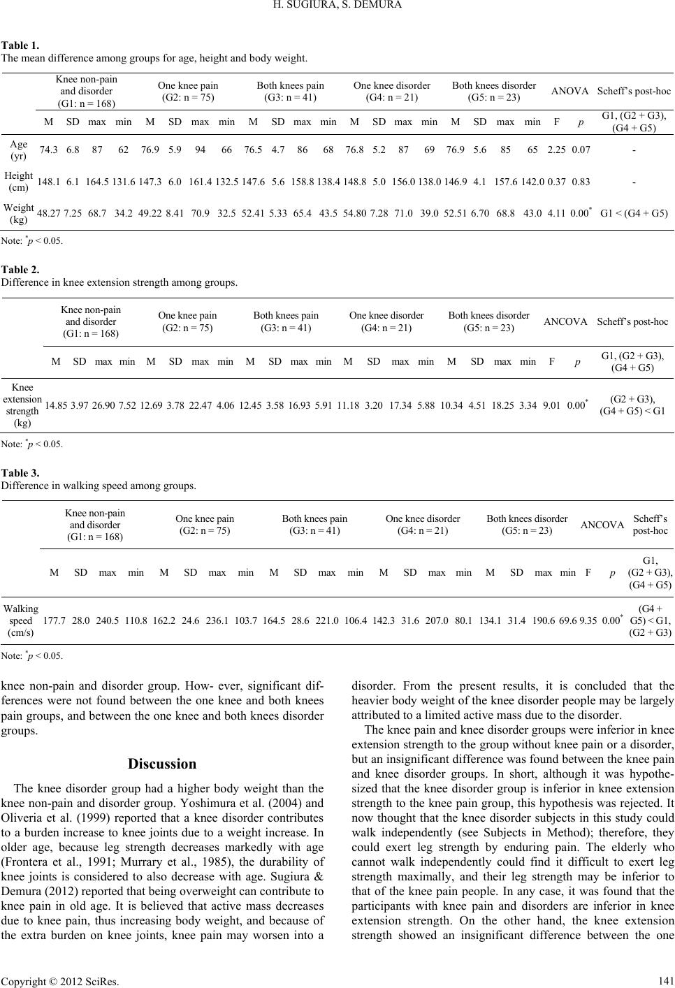 H. SUGIURA, S. DEMURA Copyright © 2012 SciRe s . 141 Table 1. The mean difference am ong groups for age, height and body weight. Knee non-pain and disorder (G1: n = 168) One knee pain (G2: n = 75) Both knees pain (G3: n = 41) One knee disorder (G4: n = 21) Both knees disorder (G5: n = 23) ANOVA Scheff’s post-hoc M SD max min M SD max min M SD max min MSD max min MSD max min F p G1, (G2 + G3), (G4 + G5) Age (yr) 74.3 6.8 87 62 76.9 5.9 94 66 76.5 4.7866876.85.2876976.95.685 65 2.25 0.07 - Height (cm) 148.1 6.1 164.5 131.6 147.3 6.0 161.4 132.5 147.6 5.6158.8138.4148.85.0156.0138.0146.94.1157.6 142.0 0.37 0.83 - Weight (kg) 48.27 7.25 68.7 34.2 49.22 8.41 70.9 32.5 52.41 5.3365.443.554.807.2871.039.052.516.7068.8 43.0 4.11 0.00* G1 < (G4 + G5) Note: *p < 0.05. Table 2. Difference in knee extension str ength among groups. Knee non-pain and disorder (G1: n = 168) One knee pain (G2: n = 75) Both knees pain (G3: n = 41) One knee disorder (G4: n = 21) Both knees disorder (G5: n = 23) ANCOVA Scheff’s post-hoc M SD max min M SD max min M SDmax min MSD max minMSD max min F p G1, (G2 + G3), (G4 + G5) Knee extension strength (kg) 14.85 3.97 26.90 7.52 12.69 3.78 22.47 4. 06 12. 45 3.5816.935.9111.183.2017.345.8810.344.5118.25 3.34 9.01 0.00* (G2 + G3), (G4 + G5) < G 1 Note: *p < 0.05. Table 3. Difference in walking speed among groups. Knee non-pain and disorder (G1: n = 168) One knee pain (G2: n = 75) Both knees pain (G3: n = 41) One knee disorder (G4: n = 21) Both knees disorder (G5: n = 23) ANCOVA Scheff’s post-hoc M SD max min MSD max min MSD max min MSD max min MSD max min F p G1, (G2 + G3), (G4 + G5) Walking speed (cm/s) 177. 7 28.0 240.5 110.8 162. 224.6 236.1 103.7 164. 528.6221. 0106.4142.331.6207.080.1134.131.4 190.6 69.6 9.35 0.00*(G4 + G5) < G1, (G2 + G3) Note: *p < 0.05. knee non-pain and disorder group. How- ever, significant dif- ferences were not found between the one knee and both knees pain groups, and between the one knee and both knees disorder groups. Discussion The knee disorder group had a higher body weight than the knee non-pain and disorder group. Yoshimura et al. (2004) and Oliveria et al. (1999) reported that a knee disorder contributes to a burden increase to knee joints due to a weight increase. In older age, because leg strength decreases markedly with age (Frontera et al., 1991; Murrary et al., 1985), the durability of knee joints is considered to also decrease with age. Sugiura & Demura (2012) reported that being overweight can contribute to knee pain in old age. It is believed that active mass decreases due to knee pain, thus increasing body weight, and because of the extra burden on knee joints, knee pain may worsen into a disorder. From the present results, it is concluded that the heavier body weight of the knee disorder people may be largely attributed to a limited active mass due to the disorder. The knee pain and knee disorder groups were inferior in knee extension strength to the group without knee pain or a disorder, but an insignificant difference was found between the knee pain and knee disorder groups. In short, although it was hypothe- sized that the knee disorder group is inferior in knee extension strength to the knee pain group, this hypothesis was rejected. It now thought that the knee disorder subjects in this study could walk independently (see Subjects in Method); therefore, they could exert leg strength by enduring pain. The elderly who cannot walk independently could find it difficult to exert leg strength maximally, and their leg strength may be inferior to that of the knee pain people. In any case, it was found that the participants with knee pain and disorders are inferior in knee extension strength. On the other hand, the knee extension strength showed an insignifican difference between the one t 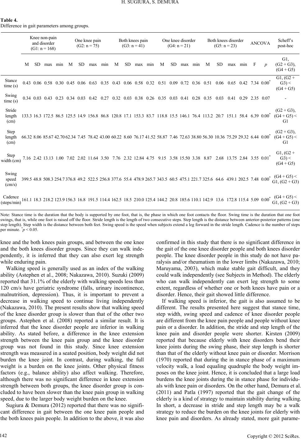 H. SUGIURA, S. DEMURA Table 4. Difference in gait parameters among groups. Knee non-pain and disorder (G1: n = 168) One knee pain (G2: n = 75) Both knees pain (G3: n = 41) O ne knee disorde r (G4: n = 21) Both knees disorder (G5: n = 23) ANCOVA Scheff’s post-hoc M SD max min M SD max min MSDmax min MSD max min MSD max min F p G1, (G2 + G3), (G4 + G5) Stance time (s) 0.43 0.06 0.58 0.30 0.450.06 0.63 0.35 0.430.060.580.320.510.090.720.360.510.060.65 0.42 7.34 0.00* G1, (G2 + G3) < (G4 + G5) Swing time (s) 0.34 0.03 0.43 0.23 0.340.03 0.42 0.27 0.320.030.380.260.350.030.410.280.350.030.41 0.29 2.35 0.07 - Stride length (cm) 133.3 16.3 172.5 86.5 125.514.9 156.8 86.8 120.817.1153.383.7118.815.5146.176.4113. 220.7151.1 58.4 6.39 0.00* (G2 + G3), (G4 + G5) < G1 Step length (cm) 66.32 8.06 85.67 42.70 62.347.45 78.42 43.00 60.228.6076.1741.5258.877.4672.6338.8056.3010.3675.29 29.32 6.44 0.00* (G2 + G3), (G4 + G5) < G1 Step width (cm) 7.16 2.42 13.13 1. 00 7.022.02 11.64 3.50 7.762.3212.844.759.153.5815. 503.388.872.6813.75 2.84 3.55 0.01* G1, (G2 + G3) < (G4 + G5) Swing speed (cm/s) 399.5 48.8 508.3 254.7 376. 849.2 522.5 256.8 377.655.4478.9265.7343.560.5475.1221.7325.664. 6439. 1 202.5 7.48 0.00* (G4 + G5) < G1, (G2 + G3) Cadence (steps/min) 161.1 18.3 218.2 123.9 156. 316.8 191.5 114.4 162.518.5210.0125.4144.220.8185.6110.1142.913. 6172. 8 115.4 5.09 0.00* (G4 + G 5) < G1, (G2 + G3) Note: Stance time is the duration that th e body is supported b y one foot, that is, the phase in which one foot contacts the flo or. Swing time is the duration th at one foot swings, that is , whi le one foot is rai sed off the floor. St rid e length is th e length of t wo cons ecut iv e steps . St ep l ength is the distance between anterior-posterior patterns (one step length ). St ep widt h is the dist ance between bot h feet . Swing speed is the speed when subjects extend a leg forward in the stride length. Cadence is the number of steps per minute . *p < 0.05. knee and the both knees pain groups, and between the one knee and the both knees disorder groups. Since they can walk inde- pendently, it is inferred that they can also exert leg strength while enduring pain. Walking speed is generally used as an index of the walking ability (Astephen et al., 2008; Nakazawa, 2010). Suzuki (2009) reported that 31.1% of the elderly with walking speeds less than 120 cm/s have geriatric syndrome (falls, urinary incontinence, malnutrition, depression). Thus, it is important to prevent a decrease in walking speed to continue living independently (Nakazawa, 2010). The present results show that walking speed of the knee disorder group is slower than that of the other two groups. Astephen et al. (2008) reported a similar result. It is inferred that the knee disorder people are inferior in walking ability. As stated before, a difference in the knee extension strength between the knee pain group and the knee disorder group was not found in this study. Since knee extension strength was measured in a seated position, body weight did not burden the knee joint. In contrast, during walking, the full weight is a burden on the knee joints. Other physical fitness factors (e.g., balance ability) also affect walking. Therefore, although there was no significant difference in knee extension strength between both groups, the knee disorder group is con- cluded to have been slower than the knee pain group in walking speed, due to the larger body weight burden on the knee. Sugiura & Demura (2012) reported that there was no signifi- cant difference in gait between the one knee pain people and the both knees pain people. In addition to the above, it was also confirmed in this study that there is no significant difference in the gait of the one knee disorder people and both knees disorder people. The knee disorder people in this study do not have pa- ralysis and/or rheumatism in the lower limbs (Nakazawa, 2010; Maruyama, 2003), which make stable gait difficult, and they could walk independently (see Subjects in Method). The elderly who can walk independently can exert leg strength to some extent, regardless of whether one or both knees have pain or a disorder. Hence, their gait showed little difference. If walking speed is inferior, the gait is also assumed to be different. The results presented here suggest that stance time, step width, swing speed and cadence of knee disorder people are different from the knee pain people and people without knee pain or a disorder. In addition, the stride and step length of the knee pain and disorder people were shorter. Kirsten (2009) reported that because elderly with knee disorders bend their knee joints during the swing phase, their step length is shorter than that of the elderly without knee pain or disorder. Morrison (1970) reported that during the in stance phase of a maximum velocity walk, a load equaling quadruple the body weight im- poses on the knee joint. Hence, it is concluded that a large load burdens the knee joints during the in stance phase for individu- als with knee pain or disorders. On the other hand, Demura et al. (2011) and Patla (1997) reported that the gait change of the elderly is a kind of strategy to maintain stability during walking. In short, a decrease in stride and step length may be a walk strategy to reduce the burden on the knee joints for elderly with knee pain and disorders. As already stated, more gait parame- Copyright © 2012 SciRe s . 142  H. SUGIURA, S. DEMURA ters (stance time, step width, swing speed and cadence) showed significant differences between the knee disorder group and the knee pain group. It is inferred that the knee disorder individuals establish a strategy to maintain walking stability, rather than trying to walk quickly. It is necessary to further examine ability to achieve activities of daily living other than walking in the female elderly with knee pain and disorder. Conclusion In conclusion, the female elderly with knee pain and/or dis- orders are inferior in knee extension strength and walk ability to the elderly without knee pain or disorder. In addition, the fe- male elderly with knee disorders are inferior in walking ability to the elderly with knee pain. REFERENCES Alindon, T. E., Snow, S., Cooper, C., & Dieppe, P. A. (1992). Patterns of osteoarthritis of the knee joint in the community: The importance of the patellofemoral joint. Annals of the Rheumatic Diseases, 51, 844-849. doi:10.1136/ard.51.7.844 Al-Zaharni, K. S., & Bakheit, A. M. (2002). A study of the gait charac- teristics of patients with chronic osteoarthritis of the knee. Disability and Rehabilitation , 24, 275-280. doi:10.1080/09638280110087098 Andriacchi, T., Galante, J., & Fermier, R. (1982). The influence of total knee replacement design on walking and stair-climbing. Journal of Bone and Joint Surgery -Mericana Volume, 64, 1328-1335. Astephen, J. L., Deluzio, K. J., Caldwell, G. E., & Dunbar, M. J. (2008). Biomechanical changes at the hip, knee, and ankle joints during gait are associated with knee osteoarthritis severity. Journal of Ortho- paedic Research, 26, 332-341. doi:10.1002/jor.20496 Bellamy, N., Buchanan, W. W., Goldsmith, C. H., Campbell, J., & Stitt, L. W. (1988). Validation study of WOMAC: A health status instru- ment for measuring clinically important patient relevant outcomes to antirheumatic drug therapy in patients with osteoarthritis of the hip or knee. Journal of Rheu ma t o lo g y , 15, 1833-1840. Berman, A. T., Zarro, V. J., Bosacco, S. J., & Israelite, C. (1987). Quantitative gait analysis after unilateral or bilateral knee replace- ment. Journal of Bone and Joint Surgery-Mericana Volume, 69, 1340-1345. Demura, S., Yamaji, S., & Sato, S. (2011). Gait and fall characteristics of the elderly. Journal of Joint Surgery, 30, 100-107. Frontera, W. R., Hughes, V. A., Lutz, K. J., & Evans, W. J. (1991). A cross-sectional study of muscle strength and mass in 45- to 78-year-old men and women. Journal of Applied Physiology, 71, 644-650. Hashimoto, H., Hanyu, T., Clement, S., & Elizabeth, L. (2003). The development of Japanese edition knee function scale—A comparison with WOMAC (Western Ontario and McMaster Universities) os- teoarthritis index. Journal of Orthopaedic Science, 77, 22-23. Hurley, M. V., Rees, J., & Newham, D. J. (1998). Quadriceps function, proprioceptive acuity and functional performance in healthy young, middle-aged and eld erly subjects. Age and Ageing, 27, 55-62. doi:10.1093/ageing/27.1.55 Jochanan, S., Robert, H. R., Aaron, C., Eliana, E. M., & Jeremy, M. J. (2009). Physical acti vity, function, and longevity among the very old. Archives of Internal Medicine, 169, 1476-1483. doi:10.1001/archinternmed.2009.248 Kirsten, G. N. (2009). Gait analysis by observation. Tokyo: Igaku- Shoin. Kurokawa, Y., Takahashi, M., & Turumi T. (2001). Physiotherapy of leg joint disorders. Tokyo: Mitsuwa Shoten. Maruyama, H. (2003). The gait. Tokyo: The Society of Physical The- rapy Science. Mary, E., & Tinetti, M. D. (2003). Preventing falls in elderly persons. New England Journal of Medci ne , 348, 42-49. doi:10.1056/NEJMcp020719 Morrison, J. B. (1970). The mechanics of the knee joint in relation to normal walking. Journal of Biomechan i c al Engineering, 3, 51-61. doi:10.1016/0021-9290(70)90050-3 Nakazawa, K. (2010). Neurorehabilitation of walking. Tokyo: Kyo- rin-Shoin. Oida, Y., & Nakamura, N. (2008). Exercise learning support manual for the knee joint ache prevention and reduction. Tokyo: Sunlife Plan. Oliveria, S. A., Fels o n , D. T., Ciril lo , P. A., Re ed , J. I. , & Walker, A. M. (1999). Body weight, body mass index, and incident symptomatic osteoarthritis of the hand, hip and knee. Epidemiology, 10, 161-166. doi:10.1097/00001648-199903000-00013 Patla, A. E. (1997). Understanding the roles of vision in the control of human locomotion. Gait Posture, 5, 54-69. doi:10.1016/S0966-6362(96)01109-5 Peat, G., McCarney, R., & Croft, P. (2001). Knee pain and osteoarthri- tis in older adults: A review of community burden and current use of primary health care. Annals of the Rheumatic Diseases, 60, 91-97. doi:10.1136/ard.60.2.91 Pratt, D. J. (1994). Some aspects of modern orthotics. Physiological Measurement, 15, 1-27. doi:10.1088/0967-3334/15/1/001 Sato, D., Kaneda, K., Wakabayashi, H., & Nomura, T. (2007). The water exercise improves health-related quality of life of frail elderly people at day service facility. Quality of Life Research, 16, 1577- 1585. doi:10.1007/s11136-007-9269-2 Sugiura, H., & Demura, S. (2012). Effect of subjective knee joint pain on knee extension strength, gait and ADL score in the female elderly. Gazzetta Medica Italiana, 171, 533-540. Suzuki, M., Ohyama, N., Yamada, K., & Kanamori, M. (2002) The relationship between fear of falling, activities of daily living and quality of life among elderly individuals. Nursing and Health Sci- ences, 4, 155-161. doi:10.1046/j.1442-2018.2002.00123.x Suzuki, T. (2009). The physical characteristic of the elderly. Health Care, 51, 148-153. Tennant, A., Fear, J., Pickering, A., Hillman, M., Cutts, A., & Cham- berlain, M. A. (1995). Prevalence of knee problems in the population aged 55 years and over: Identifying the need for arthroplasty. British Medical Journal, 310, 1291-1293. doi:10.1136/bmj.310.6990.1291 Urwin, M., Symmons, D., Allison, T., Brammah, T., Busby, H., & Roxby, M. (1998). Estimating the burden of musculoskeletal disor- ders in the community: The comparative prevalence of symptoms at different anatomical sites, and the relation to social deprivation. An- nals of Rheumatology, 5 7 , 649-655. doi:10.1136/ard.57.11.649 Yoshimura, N., Nishioka, S., Kinoshita, H., Hori, N., Nishioka, T., Ryujin, M., Mantani, Y., Miyake, M., David, C., & Cyrus, C. (2004). Risk factors for knee osteoarthritis in Japanese women: Heavy weight, previous joint injuries, and occupational activities. Journal of Rheumatology, 31, 157-162. Zeni Jr., J. A., & Higginson, J. S. (2009). Differences in gait parameters between healthy subjects and persons with moderate and severe knee osteoarthritis: A result of altered walking speed? Clinical Biome- chanics, 24, 372-3 78. doi:10.1016/j.clinbiomech.2009.02.001 Zoltan, B., Robert, P., Arpad, I., & Rita, M. K. (2006). The influence of walking speed on gait parameters in healthy people and in patients with osteoarthritis. Knee Surgery, Sports Traumatology, Arthroscopy, 14, 612-662. doi:10.1007/s00167-005-0005-6 Copyright © 2012 SciRe s . 143 |