

Paper Menu >>
Journal Menu >>
 Open Journal of Endocrine and Metabolic Diseases, 2012, 2, 58-62 http://dx.doi.org/10.4236/ojemd.2012.24009 Published Online November 2012 (http://www.SciRP.org/journal/ojemd) Prognostic Value of N-Terminal Pro-Brain Natriuretic Peptide in Acute Pulmonary Embolism Abdelhakem Selem*, Hanan Radwan, Abdelaziz M. Gomaa Cardiology Department, Ibn Sina Faculty of Medicine, Zagazig University, Zagazig, Egypt Email: *[email protected] Received August 3, 2012; revised September 5, 2012; accepted October 6, 2012 ABSTRACT Patients with pulmonary embolism (PE) have a high risk of death and it is important to recognize factors associated with high mortality. N-Terminal pro-Brain Natriuretic Peptide (NT-pro BNP) has recently emerged as a promising biomarker for risk assessment in acute pulmonary embolism (PE). The aim of this study is to detect the in hospital prognostic value of NT-pro BNP in patients with acute (PE). Methods: This study included 64 patients diagnosed as (PE) with the mean age of 59.1 ± 16.5 years, 40 patients of them (62.5%) were male. All patients were subjected to 12 leads ECG. X-ray chest, laboratory tests including D-Dimer, troponin I, NT-pro BNP, Doppler ultrasound for the ve- nous system of both lower limbs, Echocardiograhy and 64 multislices CT pulmonary angiography. Results: According to the admission level of NT-pro BNP our patients were divided into two groups: group I included 22 patients with nor- mal NT-pro BNP (less than 300 pg/ml), and group II included 42 patients with elevated NT-pro BNP (more than or equal 300 pg/ml). Patients in group II were found to have a significantly higher incidence of heart failure (28.6% vs 4.6%, p = 0.025), impaired kidney function (serum creatinine was 1.7 ± 0.6 vs 1.1 ± 0.2, p = 0.018), tachypnea (85.7% vs 54.5%, p = 0.006) and cardiogenic shock (26.2% vs 0%, p = 0.014) but a significantly lower incidence of chest pain (21.4% vs 45.5%, p = 0.04) and lower left ventricular ejection fraction (51.3% ± 16.9% vs 67.3% ± 12.8%, p = 0.043) compared to group I. There were a significantly higher treatment with thrombolytic therapy (35.7% vs 9.1%, p =0.021) and positive inotropics (35.71% vs 4.55%, p = 0.006) in group II compared to group I. Also group II had a higher need for mechanical ventilation (26.12% vs 4.55%, p = 0.04) and a longer in hospital stay (19.5 ± 10.3 vs 5.3 ± 4.5, p = 0.001) than group I. The in hospital mortality was significantly higher in group II compared to group I (19.05% vs 0.0%, p = 0.042). Conclusion: Elevated NT-pro BNP levels in patients with (PE) are associated with worse short term prognosis in terms of higher morbidity and mortality and it could be used as a valuable prognostic parameter and good indicator for the need of more aggressive therapy. Keywords: Pulmonary Embolism; N-Terminal Pro-Brain Natriuretic Peptide 1. Introduction Pulmonary embolism is a common and serious disease with an incidence rate in hospitalized patients above 5% [1]. It has a high morbidity and mortality both early and late, the overall mortality is 7% to 11% while late mor- tality is 15% at three months and reaches 50% in patients presenting with cardiogenic shock on admission [2]. Patients with hemodynamic instability at presentation have a high mortality rate [3]. Hemodynamically stable patients with RV dysfunction have high mortality. How- ever; those patients are more difficult to recognize [4]. PE is caused by either inherited or acquired risk fac- tors. Combination of thrombophilia and acquired risk factors often precipitate overt thrombosis. The two most common genetic causes of thrombophilia are factor V Leiden and prothrombin gene mutation while the most common acquired thrombophilia is anti-phospholipid syndrome. The common acquired causes of PE include: advanced age, personal or family history, recent surgery, trauma, or immobility, congestive heart failure, acute infection, pregnancy, oral contraceptive pills and COPD [5]. Dyspnea is the most common symptom of PE. Other symptoms include chest pain, cough, hemoptysis and syncope. Tachycardia is the most frequent sign of PE. Other signs include tachypnea, left parasternal upleft, tricuspid regurgitation murmer, accentuated pulmonary second sound, and evidence of DVT [5]. Right ventricular dysfunction from PE results from a combination of increased wall stress and cardiac ische- mia [6]. Myocradial wall stress is a potent stimulus for increased synthesis and secretion of BNP, which gives *Corresponding author. C opyright © 2012 SciRes. OJEMD 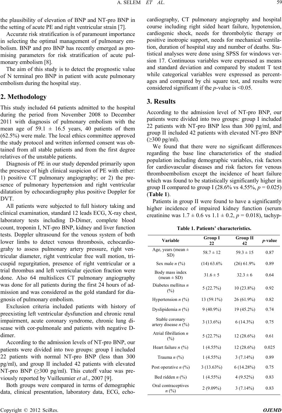 A. SELEM ET AL. 59 the plausibility of elevation of BNP and NT-pro BNP in the setting of acute PE and right ventricular strain [7]. Accurate risk stratification is of paramount importance in selecting the optimal management of pulmonary em- bolism. BNP and pro BNP has recently emerged as pro- mising parameters for risk stratification of acute pul- monary embolism [8]. The aim of this study is to detect the prognostic value of N terminal pro BNP in patient with acute pulmonary embolism during the hospital stay. 2. Methodology This study included 64 patients admitted to the hospital during the period from November 2008 to December 2011 with diagnosis of pulmonary embolism with the mean age of 59.1 ± 16.5 years, 40 patients of them (62.5%) were male. The local ethics committee approved the study protocol and written informed consent was ob- tained from all stable patients and from the first degree relatives of the unstable patients. Diagnosis of PE in our study depended primarily upon the presence of high clinical suspicion of PE with either: 1) positive CT pulmonary angiography; or 2) the pre- sence of pulmonary hypertension and right ventricular dilatation by echocardiography plus positive Doppler for DVT. All patients were subjected to full history taking and clinical examination, standard 12 leads ECG, X-ray chest, laboratory tests including D-Dimer, complete blood count, troponin I, NT-pro BNP, kidney and liver function tests. Doppler ultrasound for the venous system of both lower limbs to detect venous thrombosis, echocardio- grahy to assess pulmonary artery pressure, right ven- tricular diameter, right ventricular free wall motion, tri- cuspid regurgitation, presence of right ventricular or a trial thrombus and left ventricular ejection fraction were done. Also 64 multislices CT pulmonary angiography was done for all patients during the first 24 hours of ad- mission and was considered as the gold standard for dia- gnosis of pulmonary embolism. Exclusion criteria included patients with history of preexisting left ventricular dysfunction and chronic renal impairment, acute coronary syndrome, chronic lung di- sease with cor-pulmonale and patients with negative D- dimer. According to the admission levels of NT-pro BNP, our patients were divided into two groups: group I included 22 patients with normal NT-pro BNP (less than 300 pg/ml), and group II included 42 patients with elevated NT-pro BNP (≥300 pg/ml). This cutoff value was pre- viously reported by Vuilleumier et al., 2007 [9]. Both groups were compared in terms of demographic data, clinical presentation, laboratory data, ECG, echo- cardiography, CT pulmonary angiography and hospital course including right sided heart failure, hypotension, cardiogenic shock, needs for thrombolytic therapy or positive inotropic support, needs for mechanical ventila- tion, duration of hospital stay and number of deaths. Sta- tistical analyses were done using SPSS for windows ver- sion 17. Continuous variables were expressed as means and standard deviation and compared by student T test while categorical variables were expressed as percent- ages and compared by chi square test, and results were considered significant if the p-value is <0.05. 3. Results According to the admission level of NT-pro BNP, our patients were divided into two groups: group I included 22 patients with NT-pro BNP less than 300 pg/ml, and group II included 42 patients with elevated NT-pro BNP (≥300 pg/ml). We found that there were no significant differences regarding the base line characteristics of the studied population including demographic variables, risk factors for cardiovascular diseases and risk factors for venous thromboembolism except the incidence of heart failure which was found to be statistically significantly higher in group II compared to group I (28.6% vs 4.55%, p = 0.025) (Table 1). Patients in group II were found to have a significantly higher incidence of impaired kidney function (serum creatinine was 1.7 ± 0.6 vs 1.1 ± 0.2, p = 0.018), tachyp- Table 1. Patients’ characteristics. Variable Group I 22 Group II 42 p-value Age, years (mean ± SD) 58.7 ± 12 59.3 ± 15 0.87 Sex male n (%) (14) 63.6% (26) 61.9%0.89 Body mass index (mean ± SD) 31.6 ± 5 32.3 ± 6 0.64 Diabetes mellitus n (%) 5 (22.7%) 10 (23.8%)0.92 Hypertension n (%)13 (59.1%) 26 (61.9%)0.82 Dyslipidemia n (%)9 (40.9%) 19 (45.2%)0.74 Stable coronary artery disease n (%)3 (13.6%) 6 (14.3%) 0.75 Atrial fibrillation n (%) 5 (22.7%) 12 (28.6%)0.61 Heart failure n (%) 1 (4.55%) 12 (28.6%)0.025 Trauma n (%) 1 (4.55%) 3 (7.14%) 0.89 Post operative n (%)3 (13.63%) 6 (14.28%)0.75 Bed ridden n (%) 1 (4.55%) 4 (9.52%) 0.83 Oral contraceptives n (%) 2 (9.09%) 3 (7.14%) 0.83 Copyright © 2012 SciRes. OJEMD 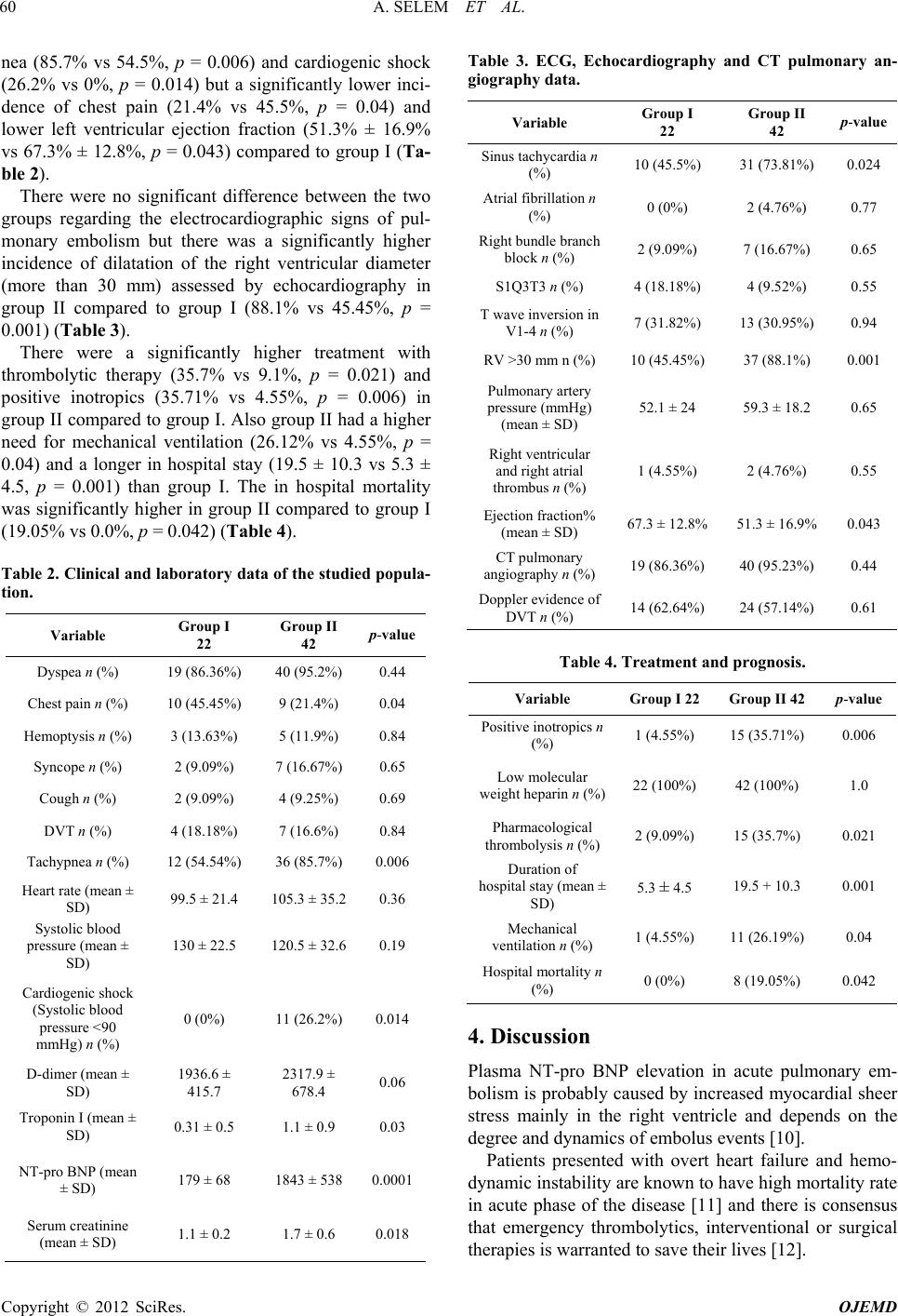 A. SELEM ET AL. 60 nea (85.7% vs 54.5%, p = 0.006) and cardiogenic shock (26.2% vs 0%, p = 0.014) but a significantly lower inci- dence of chest pain (21.4% vs 45.5%, p = 0.04) and lower left ventricular ejection fraction (51.3% ± 16.9% vs 67.3% ± 12.8%, p = 0.043) compared to group I (Ta- ble 2). There were no significant difference between the two groups regarding the electrocardiographic signs of pul- monary embolism but there was a significantly higher incidence of dilatation of the right ventricular diameter (more than 30 mm) assessed by echocardiography in group II compared to group I (88.1% vs 45.45%, p = 0.001) (Table 3). There were a significantly higher treatment with thrombolytic therapy (35.7% vs 9.1%, p = 0.021) and positive inotropics (35.71% vs 4.55%, p = 0.006) in group II compared to group I. Also group II had a higher need for mechanical ventilation (26.12% vs 4.55%, p = 0.04) and a longer in hospital stay (19.5 ± 10.3 vs 5.3 ± 4.5, p = 0.001) than group I. The in hospital mortality was significantly higher in group II compared to group I (19.05% vs 0.0%, p = 0.042) (Table 4). Table 2. Clinical and laboratory data of the studied pop ula- tion. Variable Group I 22 Group II 42 p-value Dyspea n (%) 19 (86.36%) 40 (95.2%) 0.44 Chest pain n (%) 10 (45.45%) 9 (21.4%) 0.04 Hemoptysis n (%) 3 (13.63%) 5 (11.9%) 0.84 Syncope n (%) 2 (9.09%) 7 (16.67%) 0.65 Cough n (%) 2 (9.09%) 4 (9.25%) 0.69 DVT n (%) 4 (18.18%) 7 (16.6%) 0.84 Tachypnea n (%) 12 (54.54%) 36 (85.7%) 0.006 Heart rate (mean ± SD) 99.5 ± 21.4 105.3 ± 35.2 0.36 Systolic blood pressure (mean ± SD) 130 ± 22.5 120.5 ± 32.6 0.19 Cardiogenic shock (Systolic blood pressure <90 mmHg) n (%) 0 (0%) 11 (26.2%) 0.014 D-dimer (mean ± SD) 1936.6 ± 415.7 2317.9 ± 678.4 0.06 Troponin I (mean ± SD) 0.31 ± 0.5 1.1 ± 0.9 0.03 NT-pro BNP (mean ± SD) 179 ± 68 1843 ± 538 0.0001 Serum creatinine (mean ± SD) 1.1 ± 0.2 1.7 ± 0.6 0.018 Table 3. ECG, Echocardiography and CT pulmonary an- giography data. Variable Group I 22 Group II 42 p-value Sinus tachycardia n (%) 10 (45.5%) 31 (73.81%) 0.024 Atrial fibrillation n (%) 0 (0%) 2 (4.76%) 0.77 Right bundle branch block n (%) 2 (9.09%) 7 (16.67%) 0.65 S1Q3T3 n (%) 4 (18.18%) 4 (9.52%) 0.55 T wave inversion in V1-4 n (%) 7 (31.82%) 13 (30.95%) 0.94 RV >30 mm n (%) 10 (45.45%) 37 (88.1%) 0.001 Pulmonary artery pressure (mmHg) (mean ± SD) 52.1 ± 24 59.3 ± 18.2 0.65 Right ventricular and right atrial thrombus n (%) 1 (4.55%) 2 (4.76%) 0.55 Ejection fraction% (mean ± SD) 67.3 ± 12.8% 51.3 ± 16.9%0.043 CT pulmonary angiography n (%) 19 (86.36%) 40 (95.23%) 0.44 Doppler evidence of DVT n (%) 14 (62.64%) 24 (57.14%) 0.61 Table 4. Treatment and prognosis. Variable Group I 22 Group II 42 p-value Positive inotropics n (%) 1 (4.55%) 15 (35.71%) 0.006 Low molecular weight heparin n (%)22 (100%) 42 (100%) 1.0 Pharmacological thrombolysis n (%) 2 (9.09%) 15 (35.7%) 0.021 Duration of hospital stay (mean ± SD) 5.3 ± 4.5 19.5 + 10.3 0.001 Mechanical ventilation n (%) 1 (4.55%) 11 (26.19%) 0.04 Hospital mortality n (%) 0 (0%) 8 (19.05%) 0.042 4. Discussion Plasma NT-pro BNP elevation in acute pulmonary em- bolism is probably caused by increased myocardial sheer stress mainly in the right ventricle and depends on the degree and dynamics of embolus events [10]. Patients presented with overt heart failure and hemo- dynamic instability are known to have high mortality rate in acute phase of the disease [11] and there is consensus that emergency thrombolytics, interventional or surgical therapies is warranted to save their lives [12]. Copyright © 2012 SciRes. OJEMD  A. SELEM ET AL. 61 We found that the most common clinical presentation in our study was dyspnea which was 86.4% in group I and 95% in group II, this was comparable to the result of Dores et al., 2011 who found that dyspnea was 83.3% in patients with less than median pro BNP and it was 87.1% among those with more than median pro BNP. The incidence of chest pain was significantly higher in group I compared to group II (45.5% vs 21.4%) while the incidence of tachypnea was higher in group II than group I (85.7% vs 54.5%) and this was comparable to the result obtained by Dorese et al., 2011 [13]. In our study we found elevated NT-pro BNP at ad- mission in patients with pulmonary embolism correlated significantly with worse in-hospital complications in- cluding the need for positive inotropics, the need for thrombolysis, the need for mechanical ventilation and duration of hospital stay as well as in-hospital mortality. The overall mortality rate in our patients was found to be 12.5% (0% within group I and 19.05% in group II, p-value 0.042) this result was consistent with keron [2] 2003 who reported that in-hospital mortality was 7% - 11%. Dores et al., 2011 [13] reported that the overall three months mortality was 15% while it was 50% for patients presented with cardiogenic shock. Recent meta analyses of 32 studies including 1172 pa- tients with pulmonary embolism demonstrated the abi- lity of NT-pro BNP to predict adverse effect and also concluded that in patient with higher NT-pro BNP, the concomitant elevation of Troponin I added a prognostic value [14]. N terminal pro BNP or troponin I combined with Echo- cardiography reliably identify patients with high risk of pulmonary embolism [8]. Agterof et al., 2010 [15] suggested that patients with PE who are hemodynamically stable and with low NT- pro BNP levels (less than 500 pg/ml) can be treated as outpatients, with no increase in complication or adverse events which if confirmed would bring considerable benefits in both clinical and economic terms. Cavallazi et al., 2008 [7] meta-analyzed 16 studies and concluded that BNP and NT-pro BNP are associated with right ventricular dysfunction in patients with acute PE and are significant predictor of all cause in hospital or short term mortality in these patients. If both troponin I and BNP levels were normal a low risk population free of adverse clinical outcome likely exists and right ventricular function in echocardiography will almost be normal and these patients may be suitable for short hospital stay [16] or even for outpatient ma- nagement [17]. In contrast patient with elevated cardiac markers may require immediate triage for ICU, urgent thrombolysis [18] and catheter embolectomy [19] or open surgical em- bolectomy [20]. Kostrubiec et al., 2007 [21] found that persistent ele- vation of NT-pro BNP 24 hours after diagnosis (reduc- tion of less than 50% from initial values) predicts higher mortality at thirty days. Serial measurements of NT-pro BNP may provide additional prognostic information com- pared to a single measurement at admission as well as being an indicator of therapeutic efficacy. Despite its proven relevance for prognostic assessment routine NT-pro BNP measurement is not universally ac- cepted strategy for patients with pulmonary embolism [13]. 5. Limitations of the Study 1) We studied a small number of patients because we are working in a low capacity general hospital and not a specified big cardiac center; 2) The cutoff value of NT pro BNP which we used in our analysis was relatively low, 300 pg/ml [9]. However, other authors used a higher different cutoff values of 500 pg/ml [10] 600 [21] and 1000 pg/ml [8]. 6. Conclusion Elevated NT-pro BNP levels in patients with pulmonary embolism are associated with worse short term prognosis in terms of higher morbidity and mortality and it could be used as a valuable prognostic parameter and good indicator for the need of more aggressive therapy. REFERENCES [1] J. A. Heit, L. J. Melton, C. M. Lohse, T. M. Petterson, M. D. Silverstein, D. N. Mohr and W. M. O’Fallon, “Inci- dence of Venous Thromboembolism in Hospitalized Pa- tients vs Community Residents,” Mayo Clinic Proceed- ings, 2001, pp. 1102-1110. doi:10.4065/76.11.1102 [2] C. Kearon, “Natural History of Venous Thromboembo- lism,” Circulation, Vol. 107, No. 23S1, 2003, pp. 22-30. doi:10.1161/01.CIR.0000078464.82671.78 [3] S. Z. Goldhaber, L. Visani and M. De Rosa, “Acute Pul- monary Embolism: Clinical Outcomes in the International Cooperative Pulmonary Embolism Registery,” Lancet, Vol. 353, No. 9162, 1999, pp. 1386-1389. doi:10.1016/S0140-6736(98)07534-5 [4] S. Konstantinides, “Pulmonary Embolism: Impact of Right Ventricular Dysfunction,” Current Opinion on Car- diology, Vol. 20, No. 6, 2005, pp. 496-501. doi:10.1097/01.hco.0000179818.65329.bb [5] S. Z. Glodhabber, “Pulmonary Embolism. Braunwald’s Heart Disease: A Textbook of Cardiovascular Medicine,” 9th Edition, Saunders, 2012. [6] S. Z. Goldhaber, “Pulmonary Embolism,” Lancet, Vol. 363, No. 9417, 2004, pp. 1295-1305. doi:10.1016/S0140-6736(04)16004-2 [7] R. Cavallazzi, A. Nair, T. Vasu and P. E. Marik, “Natriuretic Peptides in Acute Pulmonary Embolism: A Systematic Review Published Online,” Vol. 34, No. 12, 2008, pp. 2147-2156. Copyright © 2012 SciRes. OJEMD  A. SELEM ET AL. Copyright © 2012 SciRes. OJEMD 62 [8] L. Binder, B. Pieske, M. Olschewski, A. Geibel, B. Klos- termann, C. Reiner and S. Konstantinides, “N-Terminal Pro-Brain Natriuretic Peptide or Troponin Testing Fol- lowed by Echocardiography for Risk Stratification of Acute Pulmonary Embolism,” Circulation, Vol. 112, No. 11, 2005, pp. 1573-1579. doi:10.1161/CIRCULATIONAHA.105.552216 [9] N. Vuilleumier, M. Righini, A. Perrier, A. Rosset, N. Turck, J. C. Sanchez, H. Bounameaux, G. Le Gal, N. Mensi and D. Hochstrasser, “Correlation between Cardiac Biomarkers and Right Ventricular Enlargement on Chest CT in Non Massive Pulmonary Embolism,” Thombo Re- search, Vol. 121, No. 5, 2008, pp. 617-624. doi:10.1016/j.thromres.2007.07.003 [10] N. Kucher, G. Printzen and S. Z. Goldhaber, “Prognostic Role of Brain Natriuretic Peptide in Acute Pulmonary Embolism,” Circulation, Vol. 107, No. 20, 2003, pp. 2545-2547. doi:10.1161/01.CIR.0000074039.45523.BE [11] W. Kasper, S. Konstantinides, A. Geibel, M. Olschewski, F. Heinrich, K. D. Grosser, K. Rauber, S. Iversen, M. Redecker and J. Kienast, “Management Strategies and Determinants of Outcome in Acute Major Pulmonary Embolism: Results of a Multicenter Registry,” Journal of American College Cardiology, Vol. 30, No. 5, 1997, pp. 1165-1171. doi:10.1016/S0735-1097(97)00319-7 [12] “British Thoracic Society Guidelines for the Management of Suspected Acute Pulmonary Embolism,” Thorax, Vol. 58, No. 6, 2003, pp. 470-483. doi:10.1136/thorax.58.6.470 [13] H. Dores, C. Fonseca, S. Leal, I. Rosário, J. Abecasis, J. Monge, M. J. Correia, L. Bronze, A. Leitão, I. Arroja, A. Aleixo and A. Silva, “NT-pro BNP for Risk Stratification of Pulmonary Embolism,” Portuguese Journal of Cardi- ology, Vol. 30, No. 12, 2011, pp. 881-886. [14] J. C. Lega, Y. Lacasse, L. Lakhal and S. Provencher, “Na- triuretic Peptides and Troponins in Pulmonary Embolism: A Meta-Analysis,” Thorax, Vol. 64, No. 10, 2009, pp. 869-875. doi:10.1136/thx.2008.110965 [15] M. J. Agterof, R. E. Schutgens, R. J. Snijder, G. Epping, H. G. Peltenburg, E. F. Posthuma, J. A. Hardeman, R. van der Griend, T. Koster, M. H. Prins and D. H. Biesma, “Out of Hospital Treatment of Acute Pulmonary Embo- lism in Patients with a Low NT-pro BNP Level,” Journal of Thromb Haemost, Vol. 8, No. 6, 2010, pp. 1235-1241. doi:10.1111/j.1538-7836.2010.03831.x [16] Columbus Investigators, “Low-Molecular-Weight Hepa- rin in the Treatment of Patients with Venous Throm- boembolism,” New England Journal of Medicine, Vol. 337, No. 10, 1997, pp. 657-662. doi:10.1056/NEJM199709043371001 [17] J. H. Beer, M. Burger, S. Gretener, S. Bernard-Bagattini and H. Bounameaux, “Outpatient Treatment of Pulmo- nary Embolism Is Feasible and Safe in a Substantial Pro- portion of Patients,” Journal of Thromb Haemost, Vol. 1, No. 1, 2003, pp. 186-187. doi:10.1046/j.1538-7836.2003.00005.x [18] S. Konstantinides, A. Geibel, G. Heusel, F. Heinrich and W. Kasper, “Management Strategies and Prognosis of Pulmonary Embolism-3 Trial Investigators. Heparin Plus Alteplase Compared with Heparin Alone in Patients with Submassive Pulmonary Embolism,” New England Jour- nal of Medicine, Vol. 347, No. 15, 2002, pp. 1143-1150. doi:10.1056/NEJMoa021274 [19] T. Schmitz-Rode, U. Janssens, S. H. Duda, C. M. Erley and R. W. Günther, “Massive Pulmonary Embolism: Per- cutaneous Emergency Treatment by Pigtail Rotation Ca- theter,” Journal of American College Cardiology, Vol. 36, No. 2, 2000, pp. 375-380. doi:10.1016/S0735-1097(00)00734-8 [20] L. Aklog, C. S. Williams, J. G. Byrne and S. Z. Gold- haber, “Acute Pulmonary Embolectomy: A Contemporary Approach,” Circulati on, 2002, Vol. 105, No. 12, pp. 1416 - 1419. doi:10.1161/01.CIR.0000012526.21603.25 [21] M. Kostrubiec, P. Pruszczyk, A. Kaczynska and N. Ku- cher, “Persistent NT-pro BNP Elevation in Acute Pulmo- nary Embolism Predicts Early Death,” Clinica Chimica Acta, Vol. 382, No. 1-2, 2007, pp. 124-128. doi:10.1016/j.cca.2007.04.010 Abbreviations PE = pulmonary embolism; DVT = deep venous throm- bosis; RV = right ventricle; BNP = brain natriuretic pep- tide; NT-pro BNP = N terminal pro brain natriuretic pep- tide. |