

Paper Menu >>
Journal Menu >>
 Open Journal of Urology, 2012, 2, 219-222 http://dx.doi.org/10.4236/oju.2012.24039 Published Online November 2012 (http://www.SciRP.org/journal/oju) Misleading New Entity in Tumors of Kidney: A Case Report and Literature Review* El Fatemi Hinde1#, Hammas Nawal1, Hafid Imane1, Tazi Fadl2, Elfassi Jamal2, Amarti Afaf1 1Department of Pathology, Hassan II Teaching Hospital, Fez, Morocco 2Department of Urology, Hassan II Teaching Hospital, Fez, Morocco Email: #[email protected] Received July 16, 2012; revised August 24, 2012; accepted September 7, 2012 ABSTRACT Introduction: Mucinous tubular and spindle cell carcinomas (MTSCC’s) are recently described rare type of renal cell carcinoma (RCC). They may have morphological similarities to papillary RCC (papRCC) and in our case to carcinoma collecting ducts of Bellini. Case Report: We report a rare case of renal tumor, mucinous tubular and spindle cell carci- noma in a 56-year-old woman. The tumor, located in the left kidney, was well circumscribed. MTSCC’s are characte- rized by small, elongated tubules lined by cuboidal cells and/or cords of spindled cells separated by pale mucinous stroma. Discussion: MTSCC is a rare type of renal cell carcinoma. Pathologists should be aware of the histological spectrum of MTSCCs to ensure an accurate diagnosis. Careful attention to the presence of a spindle cell population may be helpful in the differential diagnosis in tumors with predominant compact tubular growth. Keywords: Renal Carcinoma; Mucinous Tubular and Spindle Cell Carcinoma; CD10; Immunohistochemistry 1. Introduction Mucinous tubular and spindle cell carcinoma (MTSCC) of the kidney is a novel neoplastic entity of renal cell carcinoma (RCC) that has only recently been recognized. It was first described in 1998 and specifically described by Parwani et al. [1] a series of MTSCCs as a separate entity. The recent World Health Organization classifica- tion of renal tumors officially included MTSCC as a separate category [2]. Because MTSCC has only recently been recognized, there are no more than 80 cases re- ported in the literature [1,3-5]. In the present report, we report a case of MTSCC that showed unusual morphol- ogic features of spindle cell predominance, psammoma- tous calcification, and depletion of extracellular mucin. 2. Case Report A 56-year-old woman had macroscopic hematuria for 1 month. She had no medical or family history of any ma- lignancy. All of his laboratory data were within the nor- mal ranges. Computed tomography revealed a huge renal mass in the left kidney (Figure 1). No metastatic lesions or lymph node enlargement were noted. The gross tumor had dimensions of 12.5 × 12.0 × 10 cm and was located in the inferior pole, confined to the renal parenchyma, and surrounded by a pseudocapsule. The tumor was well circumscribed and showed a variegated, grayish white cut surface with hemorrhagic and necrotic areas. Renal vein invasion and perinephric fat involvement were not identified. The nephrectomy specimen was extensively sampled. Sections were fixed in 10% neutral-buffered formalin and processed for light microscopy via conven- tional methods. Routine processing for histologic ex- amination included paraffin embedding, sectioning, and standard staining with hematoxylin and eosin safran. Morphologically, the tumor showed predominantly spin- dle cells arranged in fascicles composed of bland oval to spindle nuclei with fine chromatin and small nucleoli (Figure 2). There were tubular structures variably lined by small cuboidal cells with eosinophilic cytoplasm and focal psammoma bodies. Approximately 95% of the tu- mor was composed of spindle cell carcinoma cells that merged with areas of tubular structures. There was no significant atypia, increased mitotic activity, or vascular invasion identified. An interesting finding was focal psammomatous calcification. There was depletion of extracellular mucinous material throughout the sections (Figure 2). The scant mucinous material was not appre- ciated by hematoxylin and eosin stain but it required mucin stain to be recognized. Some foamy histiocytes and a inflammatory cells identified in the stroma with necrotic and hemorrahgic areas. Three diagnoses were suspected in these histological patterns: inflammatory pseudo-tumours, carcinoma of collecting ducts of bellini *The authors declare no conflict of interest. #Corresponding author. C opyright © 2012 SciRes. OJU 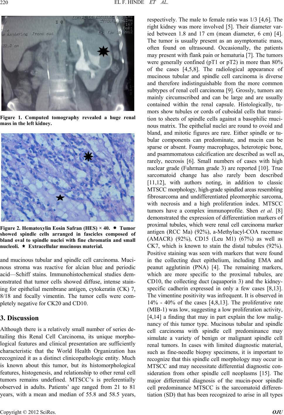 EL F. HINDE ET AL. 220 Figure 1. Computed tomography revealed a huge renal mass in the left kidney. Figure 2. Hematoxylin Eosin Safran (HES) × 40. Tumor showed spindle cells arranged in fascicles composed of bland oval to spindle nuclei with fine chromatin and small nucleoli. Extracellular mucinous material. and mucinous tubular and spindle cell carcinoma. Muci- nous stroma was reactive for alcian blue and periodic acid—Schiff stains. Immunohistochemical studies dem- onstrated that tumor cells showed diffuse, intense stain- ing for epithelial membrane antigen, cytokeratin (CK) 7, 8/18 and focally vimentin. The tumor cells were com- pletely negative for CK20 and CD10. 3. Discussion Although there is a relatively small number of series de- tailing this Renal Cell Carcinoma, its unique morpho- logical features and clinical presentation are sufficiently characteristic that the World Health Organization has recognized it as a distinct clinicopathologic entity. Much is known about this tumor, but its histomorphological features, histogenesis, and relationship to other renal cell tumors remains undefined. MTSCC’s is preferentially observed in adults. Patients’ age ranged from 21 to 81 years, with a mean and median of 55.8 and 58.5 years, respectively. The male to female ratio was 1/3 [4,6]. The right kidney was more involved [5]. Their diameter var- ied between 1.8 and 17 cm (mean diameter, 6 cm) [4]. The tumor is usually present as an asymptomatic mass, often found on ultrasound. Occasionally, the patients may present with flank pain or hematuria [7]. The tumors were generally confined (pT1 or pT2) in more than 80% of the cases [4,5,8]. The radiological appearance of mucinous tubular and spindle cell carcinoma is diverse and therefore indistinguishable from the more common subtypes of renal cell carcinoma [9]. Grossly, tumors are mainly circumscribed and can be large and are usually contained within the renal capsule. Histologically, tu- mors show tubules or cords of cuboidal cells that transi- tion to sheets of spindle cells against a basophilic muci- nous matrix. The epithelial nuclei are round to ovoid and bland, and mitotic figures are rare. Either spindle or tu- bular components can predominate, and mucin can be sparse or absent. Foamy macrophages, heterotopic bone, and psammomatous calcification are described as well as, rarely, necrosis [6]. Small numbers of cases with high nuclear grade (Fuhrman grade 3) are reported [10]. True sarcomatoid change has also rarely been described [11,12], with authors noting, in addition to classic MTSCC morphology, high-grade spindled areas resembling fibrosarcoma and undifferentiated pleomorphic sarcoma, with necrosis and a high proliferation index. MTSCC tumors have a complex immunoprofile. Shen et al. [8] demonstrated the expression of differentiation markers of proximal tubules, which were renal cell carcinoma marker antigen (RCC Ma) (92%), a-Methylacyl-COA racemase (AMACR) (92%), CD15 (Leu M1) (67%) as well as CK7, which is known to stain the distal tubules (92%). Positive staining was seen with markers that were found in the collecting duct epithelium, including EMA and peanut agglutinin (PNA) [4]. The remaining markers, which are more specific to the proximal tubules, are CD10, the collecting duct (aquaporin 3) and the kidney- specific cadherin expressed in only a few cases [8,13]. The vimentine positivity was infrequent. It is observed in 14% - 40% of the cases [4,8,13]. The proliferative rate (MIB-1) was low, suggesting a low proliferation activity, [4,14] a finding that may in part explain the low malig- nancy of this tumor type. Mucinous tubular and spindle cell carcinoma with spindle cell predominance may simulate a variety of benign or malignant spindle cell renal tumors. In cases with limited diagnostic material, such as fine-needle biopsy specimens, it is important to recognize that this spindle cell morphology may occur in MTSCC and may necessitate differential diagnostic con- sideration from other spindle cell neoplasms [15]. The major differential diagnosis of the mucin-poor spindle cell predominance MTSCC is the sarcomatoid differen- tiation (SD) that has been recognized to arise in all types Copyright © 2012 SciRes. OJU  EL F. HINDE ET AL. 221 of RCC since the Heidelberg classification of renal cell tumors was published in 1997 [16]. The presence of SD in RCC represents high-grade transformation that has a deleterious effect on prognosis. Tumors containing SD have a decreased 5-year survival rate from 79% to 22% in stage-matched patient cohorts; tumors containing more than 50% SD have an even worse prognosis [17]. Muci- nous tubular and spindle cell carcinoma lacks large, hy- perchromatic/pleomorphic nuclei or significant mitotic activity and had a much more uniform architectural pat- tern. Ki-67 can be helpful in demonstrating the low pro- liferation rate in MTSCC. The widening spectrum of MTSCC in recent years has led to the description of atypical, sarcomatoid and metastatic cases as well [10, 14]. It therefore seems more important to be aware of such morphological changes in MTSCC diagnosis and employ IHC analysis [18,19] However, there are also cases that are extremely similar to papRCC in the diffe- rential diagnosis [5,8]. Fluorescent in situ hybridization or comparative genomic hybridization studies may take the place of IHC investigation in the differential diagno- sis. These studies have reported multiple chromosomal losses including 1, 4, 6, 8, 11, 13, 14, 18, 22 instead of chromosomal additions [4,13,20,21]. The chromosome 3 losses that are typical for clear RCC have not been found. In summary, we report a case of mucin poor spindle cell predominance MTSCC with psammomatous calcifi- cation that expands the histologic spectrum encountered in these tumors that may overlap with other renal neo- plasms and can be a diagnostic dilemma. Awareness of variable morphologic variants of MTSCC is important to avoid a misdiagnosis. 4. Acknowledgements The authors thank Dr F. Mege-Lechevallier and Dr S. Collardeau Franchon for their help in the diagnosis. REFERENCES [1] A. V. Parwani, A. N. Husain, J. I. Epstein, J. B. Beckwith and P. Argani, “Lowgrade Myxoid Renal Epithelial Neo- plasms with Distal Nephron Differentiation,” Human Pa- thology, Vol. 32, No. 5, 2001, pp. 506-512. doi:10.1053/hupa.2001.24320 [2] J. N. Eble, G. Sauter, J. I. Epstein and I. A. Sesterhenn, “World Health Organization Classification of Tumors. Pathology and Genetics of Tumors of the Urinary System and Male Genital Organs,” International Agency for Re- search on Cancer (IARC), Lyon, 2004. [3] O. Hes, M. Hora, D. M. Perez-Montiel, S. Suster, R. Curik, L. Sokol, et al., “Spindle and Cuboidal Renal Cell Carcinoma, a Tumour Having Frequent Association with Nephrolithiasis: Report of 11 Cases including a Case with Hybrid Conventional Renal Cell Carcinoma/Spindle and Cuboidal Renal Cell Carcinoma Components,” Histopa- thology, Vol. 41, No. 6, 2002, pp. 549-555. doi:10.1046/j.1365-2559.2002.01515.x [4] C. Rakozy, G. E. Schmahl, S. Bogner and S. Storkel, “Low-Grade Tubularmucinous Renal Neoplasms: Mor- phologic, Immunohisto-Chemical, and Genetic Features,” Modern Pathology, Vol. 15, No. 11, 2002, pp. 1162-1171. doi:10.1097/01.MP.0000031709.40712.46 [5] G. P. Paner, J. R. Srigley, A. Radhakrishnan, et al., “Im- munohistochemical Analysis of Mucinous Tubular and Spindle Cell Carcinoma and Papillary Renal Cell Carci- noma of the Kidney: Significant Immunophenotypic Overlap Warrants Diagnostic Caution,” American Journal of Surgical Pathology, Vol. 30, No. 1, 2006, pp. 13-19. doi:10.1097/01.pas.0000180443.94645.50 [6] S. W. Fine, P. Argani, A. DeMarzo, et al., “Expanding the Histologic Spectrum of Mucinous Tubular and Spin- dle Cell Carcinoma of the Kidney,” American Journal of Surgical Pathology, Vol. 30, No. 12, 2006, pp. 1554-1560. doi:10.1097/01.pas.0000213271.15221.e3 [7] R. Srigley, “Mucinous Tubular and Spindle Cell Carci- noma,” In: J. N. Eblc, G. Sauter, J. I. Epstein and I. A. Sesterhenn, Eds., World Health Organization Classifica- tion of Tumours: Pathology and Genetics. Tumours of Urinary System and Male Genital Organs, Lyon, IARC Press, 2004, p. 40. [8] S. S. Shen, J. Y. Ro, P. Tamboli, et al., “Mucinous Tubu- lar and Spindle Cell Carcinoma of Kidney Is Probably a Variant of Papillary Renal Cell Carcinoma with Spindle Cell Features,” Annals of Diagn Pathology, Vol. 11, No. 1, 2007, pp. 13-21. doi:10.1016/j.anndiagpath.2006.09.005 [9] V. A. Sahni, M. S. Hirsch, C. A. Sadow and S. G. Si- lverman, “Mucinous Tubular and Spindle Cell Carcinoma of the Kidney: Imaging Features,” Cancer Imaging, Vol. 5, No. 12, 2012, pp. 66-71. doi:10.1102/1470-7330.2012.0008 [10] N. Kuroda, O. Hes, M. Michal, et al., “Mucinous Tubular and Spindle Cell Carcinoma with Fuhrman Nuclear Grade 3: A Histological, Immunohistochemical, Ultrastructural and FISH Study,” Histological Histopathology, Vol. 23, No. 12, 2008, pp. 1517-1523. [11] N. Pillay, P. K. Ramdial, K. Cooper, et al., “Mucinous Tubular and Spindle Cell Carcinoma with Aggressive Histomorphology—A Sarcomatoid Variant,” Human Pa- thology, Vol. 39, No. 6, 2008, pp. 966-969. doi:10.1016/j.humpath.2007.10.006 [12] S. Bulimbasic, D. Ljubanovic, R. Sima, et al., “Aggres- sive High-Grade Mucinous Tubular and Spindle Cell Carcinoma,” Human Pathology, 2009, Vol. 40, No. 6, pp. 906-907. doi:10.1016/j.humpath.2009.03.004 [13] S. Ferlicot, Y. Allory, E. Compérat, et al., “Mucinous Tubular and Spindle Cell Carcinoma: A Report of 15 Cases and Review of the Literature,” Virchows Archive, Vol. 477, No. 6, 2005, pp. 978-983. doi:10.1007/s00428-005-0036-x [14] P. Brandle, A. K. Lie, A. Bassarova, et al., “Genomic Aberrations in Mucinous Tubular and Spindle Cell Renal Cell Carcinomas,” Modern Pathology, Vol. 19, No. 2, 2006, pp. 186-194. doi:10.1038/modpathol.3800499 Copyright © 2012 SciRes. OJU  EL F. HINDE ET AL. Copyright © 2012 SciRes. OJU 222 [15] S. A. Gadre, S. K. Math, K. A. Elfeel and H. Farghaly, “Cytology of a Sarcomatoid Renal Cell Carcinoma with Unusual Coexpression of S-100 Protein: A Case Report, Review of the Literature and Cytologic-Histologic Corre- lation,” Diagn Cytopathology, Vol. 37, No. 3, 2009, pp. 195-198. doi:10.1002/dc.21003 [16] G. Kovacs, M. Akhtar, B. J. Beckwith, P. Bugert, C. S. Cooper, B. Delahunt, et al., “The Heidelberg Classifica- tion of Renal Cell Tumours,” Journal of Pathology, Vol. 183, No. 2, 1997, pp. 131-133. doi:10.1002/(SICI)1096-9896(199710)183:2<131::AID-P ATH931>3.0.CO;2-G [17] M. de Peralta-Venturina, H. Moch, M. Amin, P. Tamboli, S. Hailemariam, M. Mihatsch, et al., “Sarcomatoid Dif- ferentiation in Renal Cell Carcinoma: A Study of 101 Cases,” American Journal of Surgical Pathology, Vol. 25, No. 3, 2001, pp. 275-284. doi:10.1097/00000478-200103000-00001 [18] L. D. Truong and S. S. Shen, “Immunohistochemical Diagnosis of Renal Neoplasms,” Archive Pathological of Lab Medicine, Vol. 135, No. 12, 2011, pp. 92-109. [19] S. Sen, B. Sars, A. Simsir, E. Kmal and E. Gokmen, “Böbrekte Kitle Nedeniyle Yapılan Iğne Kor Biyopsileri ve Tanı Zorlukları,” Turk Patoloji Derg, Vol. 26, No. 2, 2010, pp. 5-18. [20] P. Cossu-Rocca, J. N. Eble, B. Delahunt, S. Zhang, G. Martignoni, M. Brunelli and L. Cheng, “Renal Mucinous Tubular and Spindle Carcinoma Lacks the Gains of Chro- mosomes 7 and 17 and Losses of Chromosome Y That Are Prevalent in Papillary Renal Cell Carcinoma,” Mod- ern Pathology, Vol. 19, No. 14, 2006, pp. 488-493. doi:10.1038/modpathol.3800565 [21] P. Argani, G. J. Netto and A. V. Parwani, “Papillary Re- nal Cell Carcinoma with Low-Grade Spindle Cell Foci: A Mimic of Mucinous Tubular and Spindle Cell Carci- noma,” American Journal of Surgical Pathology, Vol. 32, No. 9, 2008, pp. 1353-1359. doi:10.1097/PAS.0b013e31816a1c34 |