

Paper Menu >>
Journal Menu >>
 Journal of Biomaterials and Nanobiotechnology, 2012, 3, 528-532 http://dx.doi.org/10.4236/jbnb.2012.324054 Published Online October 2012 (http://www.SciRP.org/journal/jbnb) Antimicrobial Activity of Minocycline-Loaded Genipin-Crosslinked Nano-Fibrous Chitosan Mats for Guided Tissue Regeneration Peter A. Norowski1, Jegdish Babu2, Pradeep C. Adatrow3, Franklin Garcia-Godoy2, Warren O. Haggard1, Joel D. Bumgardner1 1Department of Biomedical Engineering, Herff College of Engineering, University of Memphis, Memphis, USA; 2Department of Bioscience Research, College of Dentistry, University of Tennessee Health Science Center, Memphis, USA; 3Department of Perio- dontology, College of Dentistry, University of Tennessee Health Science Center, Memphis, USA. Email: [email protected] Received August 8th, 2012; revised September 17th, 2012; accepted September 27th, 2012 ABSTRACT Antimicrobial delivery has been advocated for guided tissue regeneration (GTR) or guided bone regeneration (GBR) therapies involving patients with aggressive or unresolved periodontitis/peri-implantitis. Electrospun chitosan mem- branes demonstrate several advantages over traditional GTR barrier membranes because they stimulate healing, mimic the topology of the extracellular matrix, and allow for diffusion of nutrients and wastes into/out of the graft site, and were shown to stimulate bone formation in a rabbit calvarial critical-size defect model. Previously, we have shown im- provements in mechanical properties and degradation kinetics by crosslinking electrospun membranes with 5 mM or 10 mM genipin. We have also demonstrated the ability of elecrospun chitosan membranes to inhibit lippopolysaccharide (LPS)-induced monocyte activation. In this study, minocycline was incorporated into the chitosan membrane by passive absorption at 5 or 10 mg/mL. The minocycline-loaded membranes and control membranes (carrier only) were tested against Porphyromonas gingivalis (P. gingivalis) by repeated zone of inhibition (ZOI) measurements. Testing showed that uncrosslinked and genipin-crosslinked membranes have similar capacity to absorb aqueous solutions (swelling ratio 1.7 - 2.2). Minocycline loading resulted in bacterial inhibition for up to 8 days from crosslinked membranes (with 11 mm initial ZOI) whereas uncrosslinked membranes loaded with minocycline only inhibited bacteria for 4 days (with 8 mm initial ZOI). These in vitro results suggest that genipin-crosslinked electrospun chitosan membranes loaded with minocycline may be able to reduce early bacterial contamination of GTR graft sites. Keywords: Chitosan; Nanofiber; Genipin; Minocycline; Guided Tissue Regeneration 1. Introduction Bone graft materials are widely used to help repair bone defects in periodontitis. Guided bone regeneration (GBR) and guided tissue regeneration (GTR) barrier membranes are often used to help maintain space for newly forming bone and to prevent the infiltration of soft tissues into the bone graft space. However, a major problem with current GBR/GTR membranes is that they often become exposed and are susceptible to infection which can reduce amount of regenerated bone. Hence, there is a need for new method to prevent infection of GBR/GTR membranes and regenerating alveolar bone. Electrospun chitosan and other chitosan membrane materials have been advocated for GBR/GTR barrier membranes [1-3]. Chitosan is ad- vantageous because it has been reported to stimulate the healing of dental pulp wounds, [4] is osteogenic, and has been shown to inhibit lippopolysaccharide (LPS)-induced monocyte activation [5,6]. Electrospun material are ad- vantageous because they mimic the topology of the ex- tracellular matrix, [7] allow the diffusion of nutrients and waste, have a small enough pore size to limit cellular infiltration, and also have the capability to regenerate dense cortical bone in animal models [8]. We have developed electrospun chitosan membranes with genipin cross-linking (natural crosslinker) that have resulted in 12% reduction in mass loss over 16 weeks, and increased the tensile strength of the membranes three fold [9]. We have also demonstrated that the genipin crosslinked chitosan membranes were not cytotoxic to osteoblast or fibroblast cells, and did not cause monocyte activation. We have also shown the ability of genipin Copyright © 2012 SciRes. JBNB  Antimicrobial Activity of Minocycline-Loaded Genipin-Crosslinked Nano-Fibrous Chitosan Mats for Guided Tissue Regeneration 529 crosslinked electropun chitosan membranes to inhibit the LPS-induced release of nitric oxide (NO) from RAW 264.7 monocyte cells over a 3 day period [10]. NO ex- pression is elevated in the periodontal and gingival tis- sues of patients with periodontitis and its inhibition is a potential therapeutic target [11,12]. Because of the hydrophilic nature of chitosan and the high surface area of electrospun fibers, electrsopun chi- tosan membranes may also serve as an effective antibi- otic delivery vehicle. The ability of the membranes to prevent infection through the local delivery of antibiotics during bone healing of the site while also preventing soft tissue penetration may lead to a significant improvement in clinical bone healing in patients. Minocycline is com- monly used in periodontal therapy as an antimicrobial agent but it also has the ability to limit tissue destruction, by inhibition of tissue destroying enzymes such as colla- genase, MMP-2 and MMP-9 [13]. In this study, uncrosslinked and genipin crosslinked electrospun chitosan membranes were impregnated by immersion in 10 mg/mL minocycline or 5 mg/mL mino- cycline and tested against Porphyromonas gingivalis (P. gingivalis) by zone of inhibition. P. gingivalis is an im- portant and extensively studied periodontal pathogen involved in the pathogenesis of periodontitis. In addition, swelling was measured to assess the capacity of the elec- trospun membranes to absorb aqueous solutions. 2. Materials and Methods 2.1. Electrospinning Procedure Electrospun chitosan nanofibrous mats were fabricated as previously described (Norowski et al., in review). Briefly, a 5.50 wt% chitosan solution in 70(v/v)% trifluoroacetic acid and 30(v/v)% methylene chloride was mixed with genipin for 30 minutes prior to the start of electrospin- ning. The genipin concentrations investigated were 0, 5, or 10 mM. The solution was electrospun at 25 kV and the fibers were collected on a non-stick aluminum foil target (Reynolds wrap®) rotated at 8.4 RPM by an AC motor to ensure even and random distribution of fibers. After electrospinning, the nano-fibrous mat was put under vacuum overnight to remove residual solvent, removed from the foil, and then neutralized at room temperature in 5 M Na2CO3 (saturated solution) for 3 hours [14]. Mem- branes were sterilized by ethylene oxide gas. 2.2. Characterization of Nano-Fibrous Membrane 2.2.1. Swelling The swelling index of the nanofibrous membranes was determined by a swelling test. Swelling in phosphate buffered saline (PBS) was evaluated to estimate the amount of antibiotic solution that could be absorbed by the electrospun membranes. To determine the dry weight, membranes were maintained at 40˚C overnight in a dry- ing oven. After measuring the dry weight, membranes were submerged in PBS for 1 hr (±15 minutes) to ensure complete swelling (n = 4 - 5). Swelling index was calcu- lated by (WtWET - WtDRY)/WtDRY. 2.2.2. Minocy cli n e Loadi ng Minocycline was loaded into chitosan nano-fibrous mem- branes by passive absorption. Pre-cut, pre-sterilized cir- cular specimens (10 mm diameter), were submersed in minocycline solution (10 or 5 mg/mL in de-ionized water) for 15 minutes. Negative controls were submersed in de- ionized water only. Minocycline solutions were weighed before and after membrane swelling to determine the amount of antibiotic solution absorbed. 2.2.3. P. gingivalis Zone of Inhibition (ZOI) The model periodontal pathogen used in this study was Porphyromonas gingivalis (ATCC No. 33277) which was originally isolated from human gingival sulcus. Bac- teria were maintained as frozen stock cultures and grown anaerobically at 37˚C in trypticase soy broth (BD BBL, Franklin Lakes, NJ, USA) supplemented with 1 g of yeast extract per liter, 5 mg of hemin per liter, and 1 mg of menadione per liter. After 72 hours of growth, bacteria were collected and resuspended to contain 1 × 10⁷ cells/ml. A suspension (0.5 ml) of this stock suspension was spread on a blood agar plate (BD BBL, Franklin Lakes, NJ, USA) and the electrospun chitosan mats loaded with minocycline were placed onto the agar and incubated in an anaerobic jar with an anaerobic pack. Plates were checked development of a ZOI by sequentially placing membranes on freshly seeded bacterial lawns and re- cording ZOI at days 1, 4, 6, 8 and 11 (n = 5). Zones were measured in mm. For swelling and ZOI tests, statistical differences were detected by ANOVA with Tukey’s test used for post-hoc analysis (α = 0.05). 3. Results 3.1. Swelling Swelling experiments demonstrated that the electrospun membranes have a swelling ratio 2.24 ± 0.57, 1.71 ± 0.47, and 1.85 ± 0.41 for uncrosslinked, 5 mM crosslinked and 10 mM genipin-crosslinked membranes, respectively. The amount of swelling that occurred was not signifi- cantly affected by crosslinking (p = 0.29). Although swelling was allowed to occur for 1 hour to ensure com- plete swelling, the membranes appeared to be fully hy- drated within 5 minutes (data not shown). Copyright © 2012 SciRes. JBNB 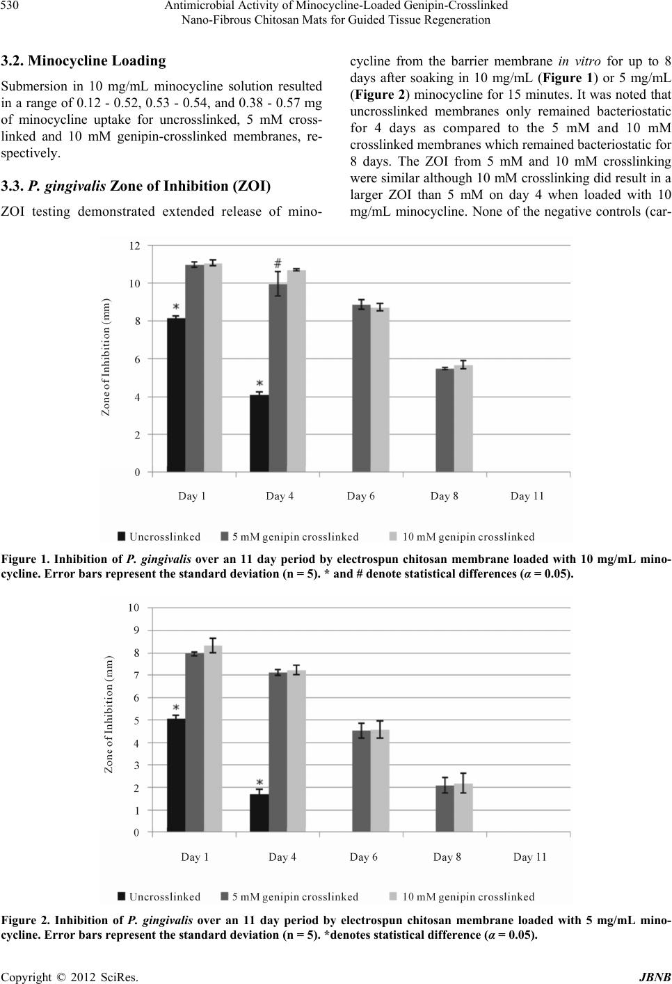 Antimicrobial Activity of Minocycline-Loaded Genipin-Crosslinked Nano-Fibrous Chitosan Mats for Guided Tissue Regeneration Copyright © 2012 SciRes. JBNB 530 3.2. Minocycline Loading Submersion in 10 mg/mL minocycline solution resulted in a range of 0.12 - 0.52, 0.53 - 0.54, and 0.38 - 0.57 mg of minocycline uptake for uncrosslinked, 5 mM cross- linked and 10 mM genipin-crosslinked membranes, re- spectively. 3.3. P. gingivalis Zone of Inhibition (ZOI) ZOI testing demonstrated extended release of mino- cycline from the barrier membrane in vitro for up to 8 days after soaking in 10 mg/mL (Figure 1) or 5 mg/mL (Figure 2) minocycline for 15 minutes. It was noted that uncrosslinked membranes only remained bacteriostatic for 4 days as compared to the 5 mM and 10 mM crosslinked membranes which remained bacteriostatic for 8 days. The ZOI from 5 mM and 10 mM crosslinking were similar although 10 mM crosslinking did result in a larger ZOI than 5 mM on day 4 when loaded with 10 mg/mL minocycline. None of the negative controls (car- Figure 1. Inhibition of P. gingivalis over an 11 day period by electrospun chitosan membrane loaded with 10 mg/mL mino- cycline. Error bars represent the standard deviation (n = 5). * and # denote statistical differences (α = 0.05). Figure 2. Inhibition of P. gingivalis over an 11 day period by electrospun chitosan membrane loaded with 5 mg/mL mino- ycline. Error bars represent the standard deviation (n = 5). *denotes statistical difference (α = 0.05). c  Antimicrobial Activity of Minocycline-Loaded Genipin-Crosslinked Nano-Fibrous Chitosan Mats for Guided Tissue Regeneration 531 rier only) produced zones of inhibition. 4. Discussion The swelling study demonstrated that membranes have similar capacity to absorb fluid and any differences in swelling capacity were not significant. Therefore, the crosslinked membrane should have similar capacity to absorb drugs, antibiotics, or growth factor solutions. Unlike vapor crosslinking and solution crosslinking me- thods, where materials are exposed to a crosslinking agent that crosslinks polymer chains primarily on the surface, in this study, the crosslinking agent is dispersed within the polymer solution used for scaffold fabrication. This method creates a more uniformly and thoroughly crosslinked membrane, which likely contributed to im- proved antibiotic retention as observed in this study and more uniform degradation kinetics. A previous examina- tion using x-ray diffraction showed that crystallinity was decreased during crosslinking. This may have contrib- uted to slightly lower swelling volumes, however this difference was not significant. In this study, genipin-crosslinked electrospun chitosan was able to absorb minocycline and release it in an ex- tended manner that remained bacteriostatic for longer periods than uncrosslinked membranes (8 days as com- pared to 4 days). These results are similar to reports by others who have loaded biodegradable GTR membranes with antibiotics/antiseptics such as tetracycline, doxycy- cline or chlorhexidine [15-17]. Delayed degradation ki- netics (Norowski et al., in review) contributed to the ex- tended release seen from crosslinked membranes. Thus, uncrosslinked chitosan membranes degraded faster and resulted in lower minocycline loading and release levels. One clinical investigation reported no improvements in clinical parameters associated with the local applica- tion of minocycline ointment before GTR therapy, but this study did not investigate minocycline incorporation into the GTR membrane itself, and only investigated the use of type 1 collagen membranes. [18] Other investiga- tions with a non-membrane local delivery system dem- onstrated improvement in clinical parameters associated with the use of minocycline microcapsules (Arestin®). [19] This microcapsule study also showed that reduction in periodontal pocket probing depth (improved clinical outcomes) correlated strongly with the ability to inhibit red complex bacteria in vitro, a sub-group of periodontal pathogens that includes P. gingivalis, T. forsythia, and T. denticola [19]. 5. Conclusion In this study, we have shown the ability of genipin-cross- linked electrospun chitosan to deliver clinically relevant levels of minocycline over an 8 day period. The eluted minocycline was able to inhibit growth of P. gingivalis, a model periopathogen, in vitro. Crosslinked membranes released inhibitory levels of minocycline for 8 days while, uncrosslinked membranes only inhibited growth for 4 days. This prolonged minocycline elution profile suggests that genipin-crosslinking improved the drug-carrier properties of electrospun chitosan. REFERENCES [1] S. M. Kuo, S. J. Chang, T. W. Chen and T. C. Kuan, “Guided Tissue Regeneration for Using a Chitosan Mem- brane: An Experimental Study in Rats,” Journal of Bio- medical Materials Research Part A, Vol. 76, No. 2, 2006, pp. 408-415. doi:10.1002/jbm.a.30534 [2] S. Y. Shin, H. N. Park, K. H. Kim, M. H. Lee, Y. S. Choi, Y. J. Park, Y. M. Lee, Y. Ku, I. C. Rhyu, S. B. Han, S. J. Lee and C. P. Chung, “Biological Evaluation of Chitosan Nanofiber Membrane for Guided Bone Regeneration,” Journal of Periodontology, Vol. 76, No. 10, 2005, pp. 1778- 1784. doi:10.1902/jop.2005.76.10.1778 [3] Y. J. Yeo, D. W. Jeon, C. S. Kim, S. H. Choi, K. S. Cho, Y. K. Lee and C. K. Kim, “Effects of Chitosan Non- woven Membrane on Periodontal Healing of Surgically Created One-Wall Intrabony Defects in Beagle Dogs,” Journal of Biomedical Materials Research Part B: Ap- plied Biomaterials, Vol. 72, No. 1, 2005, pp. 86-93. doi:10.1002/jbm.b.30121 [4] T. Matsunaga, K. Yanagiguchi, S. Yamada, N. Ohara, T. Ikeda and Y. Hayashi, “Chitosan Monomer Promotes Tis- sue Regeneration on Dental Pulp Wounds,” Journal of Biomedical Materials Research Part A, Vol. 76, No. 4, 2005, pp. 711-720. [5] H. J. Yoon, M. E. Moon, H. S. Park, S. Y. Im and Y. H. Kim, “Chitosan Oligosaccharide (COS) Inhibits LPS-In- duced Inflammatory Effects in RAW 264.7 Macrophage Cells,” Biochemical and Biophysical Research Commu- nications, Vol. 358, No. 3, 2007, pp. 954-959. doi:10.1016/j.bbrc.2007.05.042 [6] K.-S. Nam, M.-K. Kim and Y.-H. Shon, “Inhibition of Proinflammatory Cytokine-Induced Invasiveness of HT- 29 Cells by Chitosan Oligosaccharide,” Journal of Mi- crobiology and Biotechnology, Vol. 17, No. 12, 2007, pp. 2042-2045. [7] F. Chen, X. Li, X. Mo, C. He, H. Wang and Y. Ikada, “Elec- trospun Chitosan-P(LLA-CL) Nanofibers for Biomimetic Extracellular Matrix,” Journal of Biomaterials Science, Vol. 19, No. 5, 2008, pp. 677-691. doi:10.1163/156856208784089661 [8] Y. Z. Cai, L. L. Wang, H. X. Cai, Y. Y. Qi, X. H. Zou and H. W. Ouyang, “Electrospun Nanofibrous Matrix Im- proves the Regeneration of Dense Cortical Bone,” Jour- nal of Biomedical Materials Research Part A, Vol. 95, No. 1, 2010, pp. 49-57. doi:10.1002/jbm.a.32816 [9] P. A. Norowski, T. Fujiwara, W. C. Clem, P. C. Adatrow, E. C. Eckstein, W. O. Haggard and J. D. Bumgardner, Copyright © 2012 SciRes. JBNB  Antimicrobial Activity of Minocycline-Loaded Genipin-Crosslinked Nano-Fibrous Chitosan Mats for Guided Tissue Regeneration 532 “Novel Naturally Crosslinked Electrospun Nanofibrous Chitosan Mats for Guided Bone Regeneration Mem- branes: Material Characterization and Cytocompatibility,” Journal of Tissue Engineering and Regenerative Medi- cine, E-Ahead of Print, 2012. [10] P. A. Norowski, S. Mishra, P. C. Adatrow, W. O. Hag- gard and J. D. Bumgardner, “Suture Pullout Strength and in Vitro Fibroblast and RAW 264.7 Monocyte Biocom- patibility of Genipin Crosslinked Nanofibrous Chitosan Mats for Guided Tissue Regeneration,” Journal of Bio- medical Materials Research Part A, Vol. 100A, No. 11, 2012, pp. 2890-2896. doi:10.1002/jbm.a.34224 [11] D. Uğar-Çankal and N. Ozmeric, “A Multifaceted Mo- lecule, Nitric Oxide in Oral and Periodontal Diseases,” Clinica Chimica Acta, Vol. 366, No. 1-2, 2006, pp. 90- 100. doi:10.1016/j.cca.2005.10.018 [12] F. Daghigh, R. C. Borghaei, R. D. Thornton and J. H. Bee, “Human Gingival Fibroblasts Produce Nitric Oxide in Response to Proinflammatory Cytokines,” Journal of Pe- riodontology, Vol. 73, No. 4, 2002, pp. 392-400. doi:10.1902/jop.2002.73.4.392 [13] B. R. Rifkin, A. T. Vernillo and L. M. Golub, “Blocking Periodontal Disease Progression By Inhibiting Tissue- Destructive Enzymes: A Potential Therapeutic Role for Tetracyclines and Their Chemically-Modified Analogs,” Journal of Periodontology, Vol. 64, No. 8, 1993, pp. 819-827. doi:10.1902/jop.1993.64.8s.819 [14] P. Sangsanoh and P. Supaphol, “Stability Improvement of Electrospun Chitosan Nanofibrous Membranes in Neutral or Weak Basic Aqueous Solutions,” Biomacromolecules, Vol. 7, No. 10, 2006, pp. 2710-2714. doi:10.1021/bm060286l [15] C.-Y. Chang and S. Yamada, “Evaluation of the Regen- erative Effect of a 25% Doxycycline-Loaded Biodegrad- able Membrane for Guided Tissue Regeneration,” Jour- nal of Periodontology, Vol. 71, No. 7, 2000, pp. 1086- 1093. doi:10.1902/jop.2000.71.7.1086 [16] Y.-T. Chen, S.-L. Hung, L.-W. Lin, L.-Y. Chi and L.-J. Ling, “Attachment of Periodontal Ligament Cells to Chlor- hexidine-Loaded Guided Tissue Regeneration Membranes,” Journal of Periodontology, Vol. 74, No. 11, 2003, pp. 1652-1659. doi:10.1902/jop.2003.74.11.1652 [17] Y. J. Park, Y. M. Lee, S. N. Park, J. Y. Lee, Y. Ku, C. P. Chung and S. J. Lee, “Enhanced Guided Bone Regenera- tion by Controlled Tetracycline Release from Poly(L- Lactide) Barrier Membranes,” Journal of Biomedical Materials Research, Vol. 51, No. 3, 2000, pp. 391-739. doi:10.1002/1097-4636(20000905)51:3<391::AID-JBM1 3>3.0.CO;2-9 [18] M. Minabe, T. Kodama, T. Kogou, H. Fushimi, T. Sugi- yama, K. Takeuchi, E. Miterai and S. Nishikubo, “Clini- cal Significance of Antibiotic Therapy in Guided Tissue Regeneration with a Resorbable Membrane,” Periodontal Clinical Investigations, Vol. 23, No. 1, 2001, pp. 20-30. [19] P. S. Bland, J. M. Goodson, J. C. Gunsolley, S. G. Grossi, J. Otomo-Corgel, F. Doherty and J. L. Comiskey, “Asso- ciation of Antimicrobial and Clinical Efficacy: Periodon- titis Therapy with Minocycline Microspheres,” Journal of the International Academy of Periodontology, Vol. 12, No. 1, 2010, pp. 11-19. Copyright © 2012 SciRes. JBNB |