

Paper Menu >>
Journal Menu >>
 Journal of Biomaterials and Nanobiotechnology, 2012, 3, 508-518 http://dx.doi.org/10.4236/jbnb.2012.324052 Published Online October 2012 (http://www.SciRP.org/journal/jbnb) Spions Increase Biofilm Formation by Pseudomonas aeruginosa Carl Haney, John J. Rowe, Jayne B. Robinson Department of Biology, University of Dayton, Dayton, USA. Email: [email protected] Received July 18th, 2012; revised August 25th, 2012; accepted September 14th, 2012 ABSTRACT Limited research has suggested iron oxide nanoparticles (FeNP) have an inhibitory effect against several different gen- era of bacteria: Staphylococcus, Bacillus and Pseudomonas spp. In this study we looked at the effect of three different sets of Fe3O4 nanoparticles (FeNPs) on the development of Pseudomonas aeruginosa PAO1 biofilms. Two of the tested NPs were SPIONs (Superparamagnetic Iron Oxide Nanoparticles). Exposure of cells to the SPIONs at concentrations up to 200 µg/ml resulted in an increase in biofilm biomass by 16 h under static conditions and a corresponding increase in cell density in the bulk liquid. In contrast, these biofilms had decreased levels of extracellular DNA (eDNA). Fe(II) levels in the supernatants of biofilms formed in the presence of FeNPs exceeded 100 µM compared with 20 µM in con- trol media without cells. Spent cell supernatants had little effect on Fe(II) levels. Cells also had an effect on the aggre- gation behavior of these nanoparticles. SPIONs incubated with cells exhibited a decrease in the number and size of FeNP aggregates visible using light microscopy. SPIONs resuspended in fresh media or spent culture supernatants formed large aggregates visible in the light microscope upon exposure to a supermagnet; and could be pelleted mag- netically in microtitre plate wells. In contrast, SPION FeNPs incubated with cells were unaffected by exposure to the supermagnet and could not be pelleted. The results of this study indicate a need to reconsider the effects of FeNPs on bacterial growth and biofilm formation and the effect the bacterial cells may have on the use and recovery of SPIONs. Keywords: Spion; Iron Oxide Nanoparticles; Biofilms; Pseudomonas Aeruginosa; Magnetism; Nanotechnology 1. Introduction Pseudomonas aeruginosa is a ubiquitous gram-negative, rod shaped bacterium best known as an opportunistic pathogen. According to the CDC, P. aeruginosa accounts for 10% of all hospital acquired infections and a fatality rate of nearly 50% in patients with cancer, cystic fibrosis, and burns. Survival and virulence of this bacterium is attributable to the biofilms it produces [1]. Biofilms are sessile colonies which form on both biotic and abiotic surfaces. These biofilms can withstand phagocytic envi- ronments and exhibit 100 - 1000 fold greater resistance to antibiotics [2]. Biofilms develop and are held together by secreted macromolecules called the extra-polymeric substances (EPS) which form the biofilm matrix. This EPS consists of extracellular DNA (eDNA), polysaccha- rides, and proteins. Iron is a key nutrient required by most living organ- isms. Due to the insolubility of the ferric (Fe(III)) ion and because the soluble ferrous (Fe(II)) ion is readily oxi- dized at neutral pH, this abundant element is a limited resource for which organisms must compete [3]. Simi- larly, iron’s efficiency as an electron donor and acceptor makes it susceptible to generating toxic oxygen radicals [4]. Iron is a powerful regulator of gene expression in P. aeruginosa. Iron regulation is mediated by both the Fer- ric uptake regulator (Fur) repressor protein as well as the density-dependent cell-to-cell signaling quorum sensing pathways [5,6]. Musk et al. [7] demonstrated that iron concentrations between 1 - 100 µM are required for P. aeruginosa to form and develop robust biofilms. Outside this range, P. aeruginosa can only exist in the planktonic environment. Under low iron conditions twitching motil- ity increases thereby limiting the ability of the bacteria to transition to a sessile subpopulation and therefore the development of a mature bacterial mushroom shaped colony [8-10]. Yang et al. [11] showed a decrease in the levels of eDNA in the EPS with increasing iron concen- tration. Berlutti et al. [12] found iron limitation of 1 µM induced twitching motility while concentrations between 10 and 100 µM stimulated cell aggregation and biofilm development. Additionally, sufficient levels of iron also play a role in the development of the bacterial mushroom caps indicative of mature biofilms [13]. Further, both Copyright © 2012 SciRes. JBNB 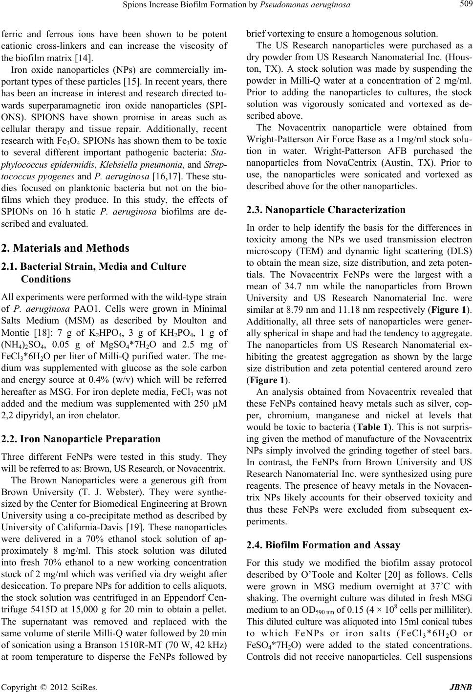 Spions Increase Biofilm Formation by Pseudomonas aeruginosa 509 ferric and ferrous ions have been shown to be potent cationic cross-linkers and can increase the viscosity of the biofilm matrix [14]. Iron oxide nanoparticles (NPs) are commercially im- portant types of these particles [15]. In recent years, there has been an increase in interest and research directed to- wards superparamagnetic iron oxide nanoparticles (SPI- ONS). SPIONS have shown promise in areas such as cellular therapy and tissue repair. Additionally, recent research with Fe3O4 SPIONs has shown them to be toxic to several different important pathogenic bacteria: Sta- phylococcus epidermidis, Klebsiella pneumonia, and Strep- tococcus pyogenes and P. aeruginosa [16,17]. These stu- dies focused on planktonic bacteria but not on the bio- films which they produce. In this study, the effects of SPIONs on 16 h static P. aeruginosa biofilms are de- scribed and evaluated. 2. Materials and Methods 2.1. Bacterial Strain, Media and Culture Conditions All experiments were performed with the wild-type strain of P. aeruginosa PAO1. Cells were grown in Minimal Salts Medium (MSM) as described by Moulton and Montie [18]: 7 g of K2HPO4, 3 g of KH2PO4, 1 g of (NH4)2SO4, 0.05 g of MgSO4*7H2O and 2.5 mg of FeCl3*6H2O per liter of Milli-Q purified water. The me- dium was supplemented with glucose as the sole carbon and energy source at 0.4% (w/v) which will be referred hereafter as MSG. For iron deplete media, FeCl3 was not added and the medium was supplemented with 250 µM 2,2 dipyridyl, an iron chelator. 2.2. Iron Nanoparticle Preparation Three different FeNPs were tested in this study. They will be referred to as: Brown, US Research, or Novacentrix. The Brown Nanoparticles were a generous gift from Brown University (T. J. Webster). They were synthe- sized by the Center for Biomedical Engineering at Brown University using a co-precipitate method as described by University of California-Davis [19]. These nanoparticles were delivered in a 70% ethanol stock solution of ap- proximately 8 mg/ml. This stock solution was diluted into fresh 70% ethanol to a new working concentration stock of 2 mg/ml which was verified via dry weight after desiccation. To prepare NPs for addition to cells aliquots, the stock solution was centrifuged in an Eppendorf Cen- trifuge 5415D at 15,000 g for 20 min to obtain a pellet. The supernatant was removed and replaced with the same volume of sterile Milli-Q water followed by 20 min of sonication using a Branson 1510R-MT (70 W, 42 kHz) at room temperature to disperse the FeNPs followed by brief vortexing to ensure a homogenous solution. The US Research nanoparticles were purchased as a dry powder from US Research Nanomaterial Inc. (Hous- ton, TX). A stock solution was made by suspending the powder in Milli-Q water at a concentration of 2 mg/ml. Prior to adding the nanoparticles to cultures, the stock solution was vigorously sonicated and vortexed as de- scribed above. The Novacentrix nanoparticle were obtained from Wright-Patterson Air Force Base as a 1mg/ml stock solu- tion in water. Wright-Patterson AFB purchased the nanoparticles from NovaCentrix (Austin, TX). Prior to use, the nanoparticles were sonicated and vortexed as described above for the other nanoparticles. 2.3. Nanoparticle Characterization In order to help identify the basis for the differences in toxicity among the NPs we used transmission electron microscopy (TEM) and dynamic light scattering (DLS) to obtain the mean size, size distribution, and zeta poten- tials. The Novacentrix FeNPs were the largest with a mean of 34.7 nm while the nanoparticles from Brown University and US Research Nanomaterial Inc. were similar at 8.79 nm and 11.18 nm respectively (Figure 1). Additionally, all three sets of nanoparticles were gener- ally spherical in shape and had the tendency to aggregate. The nanoparticles from US Research Nanomaterial ex- hibiting the greatest aggregation as shown by the large size distribution and zeta potential centered around zero (Figure 1). An analysis obtained from Novacentrix revealed that these FeNPs contained heavy metals such as silver, cop- per, chromium, manganese and nickel at levels that would be toxic to bacteria (Table 1). This is not surpris- ing given the method of manufacture of the Novacentrix NPs simply involved the grinding together of steel bars. In contrast, the FeNPs from Brown University and US Research Nanomaterial Inc. were synthesized using pure reagents. The presence of heavy metals in the Novacen- trix NPs likely accounts for their observed toxicity and thus these FeNPs were excluded from subsequent ex- periments. 2.4. Biofilm Formation and Assay For this study we modified the biofilm assay protocol described by O’Toole and Kolter [20] as follows. Cells were grown in MSG medium overnight at 37˚C with shaking. The overnight culture was diluted in fresh MSG medium to an OD590 nm of 0.15 (4 × 10⁸ cells per milliliter). This diluted culture was aliquoted into 15ml conical tubes to which FeNPs or iron salts (FeCl3*6H2O or FeSO4*7H2O) were added to the stated concentrations. Controls did not receive nanoparticles. Cell suspensions Copyright © 2012 SciRes. JBNB 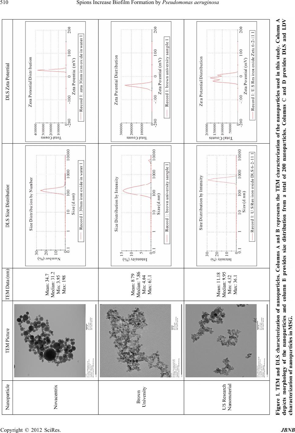 Spions Increase Biofilm Formation by Pseudomonas aeruginosa Copyright © 2012 SciRes. JBNB 510 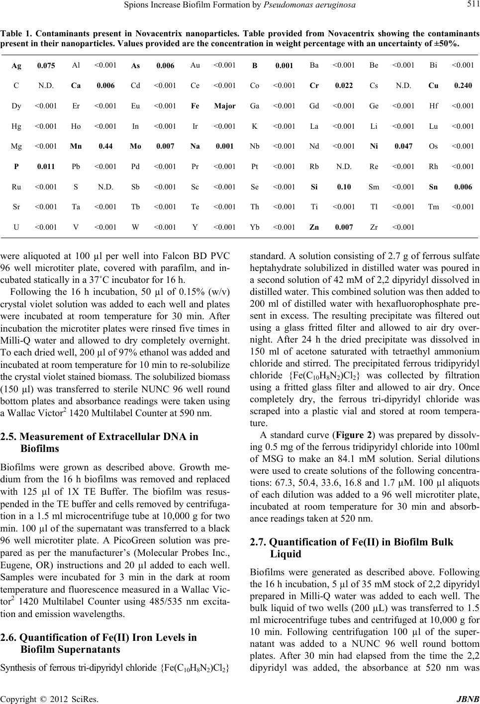 Spions Increase Biofilm Formation by Pseudomonas aeruginosa 511 Table 1. Contaminants present in Novacentrix nanoparticles. Table provided from Novacentrix showing the contaminants present in their nanoparticles. Values provided are the concentration in weight percentage with an uncertainty of ±50%. Ag 0.075 Al <0.001 As 0.006 Au <0.001B 0.001Ba <0.001Be <0.001 Bi <0.001 C N.D. Ca 0.006 Cd <0.001 Ce <0.001Co <0.001Cr 0.022Cs N.D. Cu 0.240 Dy <0.001 Er <0.001 Eu <0.001 Fe MajorGa <0.001 Gd <0.001 Ge <0.001 Hf <0.001 Hg <0.001 Ho <0.001 In <0.001 Ir <0.001K <0.001La <0.001Li <0.001 Lu <0.001 Mg <0.001 Mn 0.44 Mo 0.007 Na 0.001 Nb <0.001 Nd <0.001Ni 0.047 Os <0.001 P 0.011 Pb <0.001 Pd <0.001 Pr <0.001Pt <0.001Rb N.D.Re <0.001 Rh <0.001 Ru <0.001 S N.D. Sb <0.001 Sc <0.001 Se <0.001Si 0.10 Sm <0.001 Sn 0.006 Sr <0.001 Ta <0.001 Tb <0.001 Te <0.001 Th <0.001Ti <0.001Tl <0.001 Tm <0.001 U <0.001 V <0.001 W <0.001 Y <0.001Yb <0.001Zn 0.007Zr <0.001 were aliquoted at 100 µl per well into Falcon BD PVC 96 well microtiter plate, covered with parafilm, and in- cubated statically in a 37˚C incubator for 16 h. Following the 16 h incubation, 50 µl of 0.15% (w/v) crystal violet solution was added to each well and plates were incubated at room temperature for 30 min. After incubation the microtiter plates were rinsed five times in Milli-Q water and allowed to dry completely overnight. To each dried well, 200 µl of 97% ethanol was added and incubated at room temperature for 10 min to re-solubilize the crystal violet stained biomass. The solubilized biomass (150 µl) was transferred to sterile NUNC 96 well round bottom plates and absorbance readings were taken using a Wallac Victor2 1420 Multilabel Counter at 590 nm. 2.5. Measurement of Extracellular DNA in Biofilms Biofilms were grown as described above. Growth me- dium from the 16 h biofilms was removed and replaced with 125 µl of 1X TE Buffer. The biofilm was resus- pended in the TE buffer and cells removed by centrifuga- tion in a 1.5 ml microcentrifuge tube at 10,000 g for two min. 100 µl of the supernatant was transferred to a black 96 well microtiter plate. A PicoGreen solution was pre- pared as per the manufacturer’s (Molecular Probes Inc., Eugene, OR) instructions and 20 µl added to each well. Samples were incubated for 3 min in the dark at room temperature and fluorescence measured in a Wallac Vic- tor2 1420 Multilabel Counter using 485/535 nm excita- tion and emission wavelengths. 2.6. Quantification of Fe(II) Iron Levels in Biofilm Supernatants Synthesis of ferrous tri-dipyridyl chloride {Fe(C10H8N2)Cl2} standard. A solution consisting of 2.7 g of ferrous sulfate heptahydrate solubilized in distilled water was poured in a second solution of 42 mM of 2,2 dipyridyl dissolved in distilled water. This combined solution was then added to 200 ml of distilled water with hexafluorophosphate pre- sent in excess. The resulting precipitate was filtered out using a glass fritted filter and allowed to air dry over- night. After 24 h the dried precipitate was dissolved in 150 ml of acetone saturated with tetraethyl ammonium chloride and stirred. The precipitated ferrous tridipyridyl chloride {Fe(C10H8N2)Cl2} was collected by filtration using a fritted glass filter and allowed to air dry. Once completely dry, the ferrous tri-dipyridyl chloride was scraped into a plastic vial and stored at room tempera- ture. A standard curve (Figure 2) was prepared by dissolv- ing 0.5 mg of the ferrous tridipyridyl chloride into 100ml of MSG to make an 84.1 mM solution. Serial dilutions were used to create solutions of the following concentra- tions: 67.3, 50.4, 33.6, 16.8 and 1.7 µM. 100 µl aliquots of each dilution was added to a 96 well microtiter plate, incubated at room temperature for 30 min and absorb- ance readings taken at 520 nm. 2.7. Quantification of Fe(II) in Biofilm Bulk Liquid Biofilms were generated as described above. Following the 16 h incubation, 5 µl of 35 mM stock of 2,2 dipyridyl prepared in Milli-Q water was added to each well. The bulk liquid of two wells (200 µL) was transferred to 1.5 ml microcentrifuge tubes and centrifuged at 10,000 g for 10 min. Following centrifugation 100 µl of the super- natant was added to a NUNC 96 well round bottom plates. After 30 min had elapsed from the time the 2,2 dipyridyl was added, the absorbance at 520 nm was Copyright © 2012 SciRes. JBNB 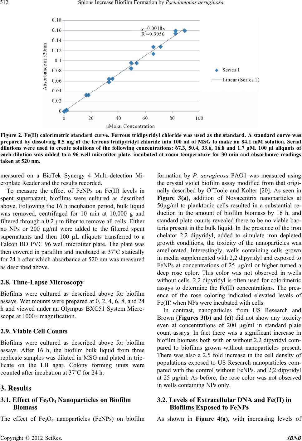 Spions Increase Biofilm Formation by Pseudomonas aeruginosa 512 Figure 2. Fe(II) colorimetric standard curve. Ferrous tridipyridyl chloride was used as the standard. A standard curve was prepared by dissolving 0.5 mg of the ferrous tridipyridyl chloride into 100 ml of MSG to make an 84.1 mM solution. Serial dilutions were used to create solutions of the following concentrations: 67.3, 50.4, 33.6, 16.8 and 1.7 µM. 100 µl aliquots of each dilution was added to a 96 well microtiter plate, incubated at room temperature for 30 min and absorbance readings taken at 520 nm. measured on a BioTek Synergy 4 Multi-detection Mi- croplate Reader and the results recorded. To measure the effect of FeNPs on Fe(II) levels in spent supernatant, biofilms were cultured as described above. Following the 16 h incubation period, bulk liquid was removed, centrifuged for 10 min at 10,000 g and filtered through a 0.2 µm filter to remove all cells. Either no NPs or 200 µg/ml were added to the filtered spent supernatants and then 100 µL aliquots transferred to a Falcon BD PVC 96 well microtiter plate. The plate was then covered in parafilm and incubated at 37˚C statically for 24 h after which absorbance at 520 nm was measured as described above. 2.8. Time-Lapse Microscopy Biofilms were cultured as described above for biofilm assays. Wet mounts were prepared at 0, 2, 4, 6, 8, and 24 h and viewed under an Olympus BXC51 System Micro- scope at 1000× magnification. 2.9. Viable Cell Counts Biofilms were cultured as described above for biofilm assays. After 16 h, the biofilm bulk liquid from three replicate samples was diluted in MSG and plated in trip- licate on the LB agar. Colony forming units were counted after incubation at 37˚C for 24 h. 3. Results 3.1. Effect of Fe3O4 Nanoparticles on Biofilm Biomass The effect of Fe3O4 nanoparticles (FeNPs) on biofilm formation by P. aeruginosa PAO1 was measured using the crystal violet biofilm assay modified from that origi- nally described by O’Toole and Kolter [20]. As seen in Figure 3(a), addition of Novacentrix nanoparticles at 50µg/ml to planktonic cells resulted in a substantial re- duction in the amount of biofilm biomass by 16 h, and standard plate counts revealed there to be no viable bac- teria present in the bulk liquid. In the presence of the iron chelator 2,2 dipyridyl, added to simulate iron depleted growth conditions, the toxicity of the nanoparticles was ameliorated. Interestingly, wells containing cells grown in media supplemented with 2,2 dipyridyl and exposed to FeNPs at concentrations of 25 µg/ml or higher turned a deep rose color. This color was not observed in wells without cells. 2,2 dipyridyl is often used for colorimetric assays to determine the Fe(II) concentrations. The pres- ence of the rose coloring indicated elevated levels of Fe(II) when NPs were incubated with cells. In contrast, nanoparticles from US Research and Brown (Figures 3(b) and (c)) did not show any toxicity even at concentrations of 200 µg/ml in standard plate count assays. In fact there was a significant increase in biofilm biomass both with or without 2,2 dipyridyl com- pared to biofilms grown without nanoparticles present. There was also a 2.5 fold increase in the cell density of populations exposed to US Research nanoparticles com- pared with the control without FeNPs. and 2,2 dipyridyl at 25 µg/ml. As before, the rose color was not observed in wells containing NPs only. 3.2. Levels of Extracellular DNA and Fe(II) in Biofilms Exposed to FeNPs As shown in Figure 4(a), with increasing levels of Copyright © 2012 SciRes. JBNB 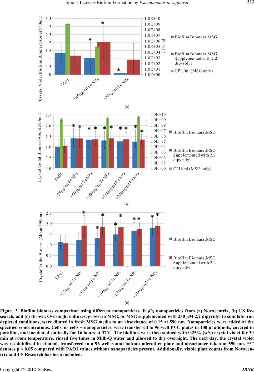 Spions Increase Biofilm Formation by Pseudomonas aeruginosa 513 (a) (b) (c) Figure 3. Biofilm biomass comparison using different nanoparticles. Fe3O4 nanoparticles from (a) Novacentrix, (b) US Re- search, and (c) Brown. Overnight cultures, grown in MSG, or MSG supplemented with 250 µM 2,2 dipyridyl to simulate iron depleted conditions, were diluted in fresh MSG media to an absorbance of 0.15 at 590 nm. Nanoparticles were added at the specified concentrations. Cells, or cells + nanoparticles, were transferred to 96-well PVC plates in 100 µl aliquots, covered in parafilm, and incubated statically for 16 hours at 37˚C. The biofilms were then stained with 0.25% (w/v) crystal violet for 30 min at room temperature, rinsed five times in Milli-Q water and allowed to dry overnight. The next day, the crystal violet was resolubilized in ethanol, transferred to a 96 well round bottom microtiter plate and absorbance taken at 590 nm. “*” denotes p < 0.05 compared with PAO1 values without nanoparticles present. Additionally, viable plate counts from Novacen- trix and US Research has been included. Copyright © 2012 SciRes. JBNB 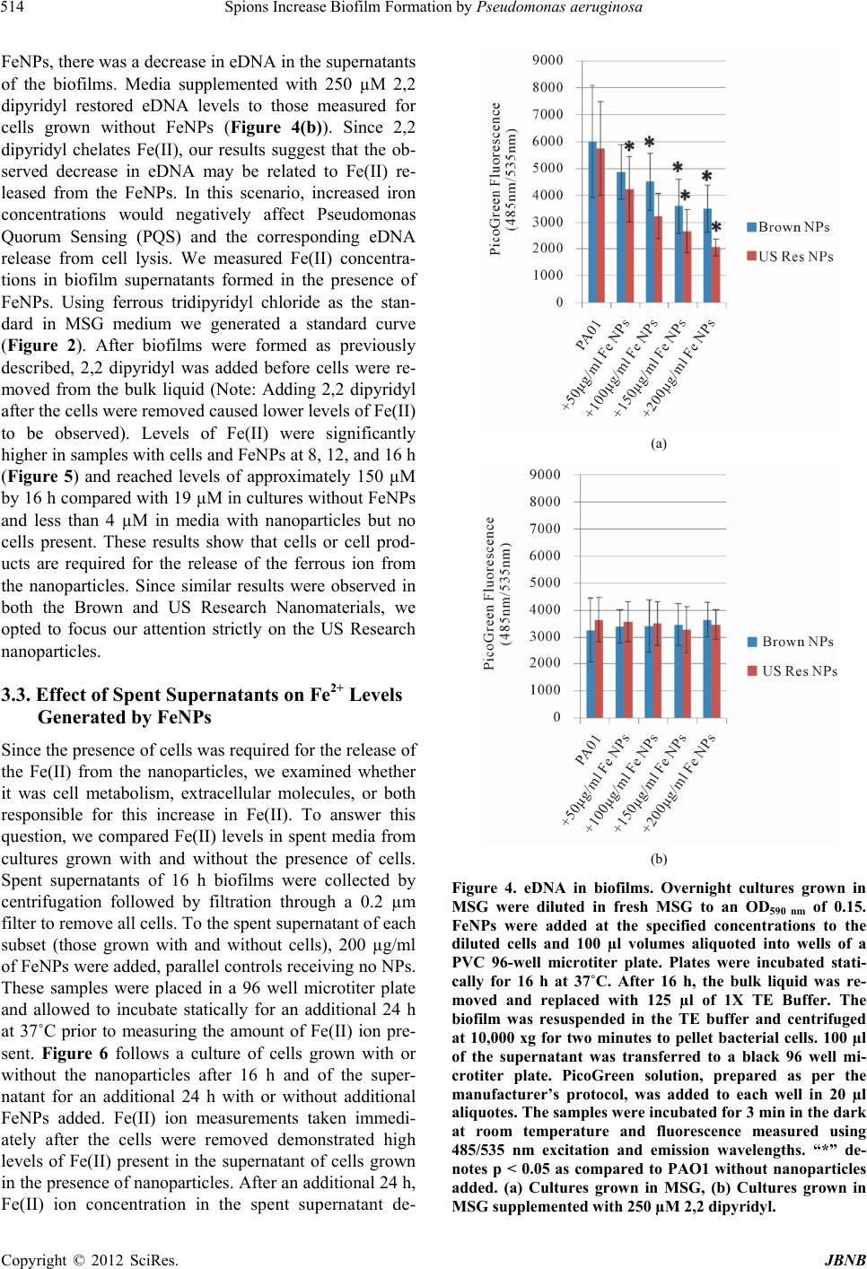 Spions Increase Biofilm Formation by Pseudomonas aeruginosa 514 FeNPs, there was a decrease in eDNA in the supernatants of the biofilms. Media supplemented with 250 µM 2,2 dipyridyl restored eDNA levels to those measured for cells grown without FeNPs (Figure 4(b)). Since 2,2 dipyridyl chelates Fe(II), our results suggest that the ob- served decrease in eDNA may be related to Fe(II) re- leased from the FeNPs. In this scenario, increased iron concentrations would negatively affect Pseudomonas Quorum Sensing (PQS) and the corresponding eDNA release from cell lysis. We measured Fe(II) concentra- tions in biofilm supernatants formed in the presence of FeNPs. Using ferrous tridipyridyl chloride as the stan- dard in MSG medium we generated a standard curve (Figure 2). After biofilms were formed as previously described, 2,2 dipyridyl was added before cells were re- moved from the bulk liquid (Note: Adding 2,2 dipyridyl after the cells were removed caused lower levels of Fe(II) to be observed). Levels of Fe(II) were significantly higher in samples with cells and FeNPs at 8, 12, and 16 h (Figure 5) and reached levels of approximately 150 µM by 16 h compared with 19 µM in cultures without FeNPs and less than 4 µM in media with nanoparticles but no cells present. These results show that cells or cell prod- ucts are required for the release of the ferrous ion from the nanoparticles. Since similar results were observed in both the Brown and US Research Nanomaterials, we opted to focus our attention strictly on the US Research nanoparticles. 3.3. Effect of Spent Supernatants on Fe2+ Levels Generated by FeNPs Since the presence of cells was required for the release of the Fe(II) from the nanoparticles, we examined whether it was cell metabolism, extracellular molecules, or both responsible for this increase in Fe(II). To answer this question, we compared Fe(II) levels in spent media from cultures grown with and without the presence of cells. Spent supernatants of 16 h biofilms were collected by centrifugation followed by filtration through a 0.2 µm filter to remove all cells. To the spent supernatant of each subset (those grown with and without cells), 200 µg/ml of FeNPs were added, parallel controls receiving no NPs. These samples were placed in a 96 well microtiter plate and allowed to incubate statically for an additional 24 h at 37˚C prior to measuring the amount of Fe(II) ion pre- sent. Figure 6 follows a culture of cells grown with or without the nanoparticles after 16 h and of the super- natant for an additional 24 h with or without additional FeNPs added. Fe(II) ion measurements taken immedi- ately after the cells were removed demonstrated high levels of Fe(II) present in the supernatant of cells grown in the presence of nanoparticles. After an additional 24 h, Fe(II) ion concentration in the spent supernatant de- (a) (b) Figure 4. eDNA in biofilms. Overnight cultures grown in MSG were diluted in fresh MSG to an OD590 nm of 0.15. FeNPs were added at the specified concentrations to the diluted cells and 100 µl volumes aliquoted into wells of a PVC 96-well microtiter plate. Plates were incubated stati- cally for 16 h at 37˚C. After 16 h, the bulk liquid was re- moved and replaced with 125 µl of 1X TE Buffer. The biofilm was resuspended in the TE buffer and centrifuged at 10,000 xg for two minutes to pellet bacterial cells. 100 µl of the supernatant was transferred to a black 96 well mi- crotiter plate. PicoGreen solution, prepared as per the manufacturer’s protocol, was added to each well in 20 µl aliquotes. The samples were incubated for 3 min in the dark at room temperature and fluorescence measured using 485/535 nm excitation and emission wavelengths. “*” de- notes p < 0.05 as compared to PAO1 without nanoparticles added. (a) Cultures grown in MSG, (b) Cultures grown in MSG supplemented with 250 µM 2,2 dipyridyl. Copyright © 2012 SciRes. JBNB 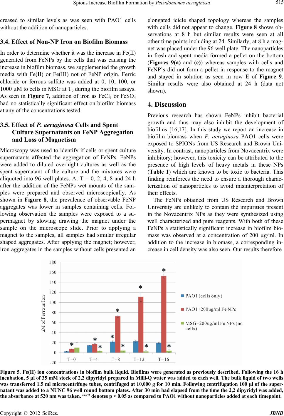 Spions Increase Biofilm Formation by Pseudomonas aeruginosa 515 creased to similar levels as was seen with PAO1 cells without the addition of nanoparticles. 3.4. Effect of Non-NP Iron on Biofilm Biomass In order to determine whether it was the increase in Fe(II) generated from FeNPs by the cells that was causing the increase in biofilm biomass, we supplemented the growth media with Fe(II) or Fe(III) not of FeNP origin. Ferric chloride or ferrous sulfate was added at 0, 10, 100, or 1000 µM to cells in MSG at T0 during the biofilm assays. As seen in Figure 7, addition of iron as FeCl3 or FeSO4 had no statistically significant effect on biofilm biomass at any of the concentrations tested. 3.5. Effect of P. aeruginosa Cells and Spent Culture Supernatants on FeNP Aggregation and Loss of Magnetism Microscopy was used to identify if cells or spent culture supernatants affected the aggregation of FeNPs. FeNPs were added to diluted overnight cultures as well as the spent supernatant of the culture and the mixtures were aliquoted into 96 well plates. At T = 0, 2, 4, 8 and 24 h after the addition of the FeNPs wet mounts of the sam- ples were prepared and observed microscopically. As shown in Figure 8, the prevalence of observable FeNP aggregates was lower in samples containing cells. Fol- lowing observation the samples were exposed to a su- permagnet by slowing drawing the magnet under the sample on the microscope slide. Prior to applying a magnet to the samples, all samples had similar irregular shaped aggregates. After applying the magnet; however, iron aggregates in the samples without cells presented an elongated icicle shaped topology whereas the samples with cells did not appear to change. Figure 8 shows ob- servations at 8 h but similar results were seen at all other time points including at 24. Similarly, at 8 h a mag- net was placed under the 96 well plate. The nanoparticles in fresh and spent media formed a pellet on the bottom (Figures 9(a) and (c)) whereas samples with cells and FeNP’s did not form a pellet in response to the magnet and stayed in solution as seen in row E of Figure 9. Similar results were also obtained at 24 h (data not shown). 4. Discussion Previous research has shown FeNPs inhibit bacterial growth and thus may also inhibit the development of biofilms [16,17]. In this study we report an increase in biofilm biomass when P. aeruginosa PAO1 cells were exposed to SPIONs from US Research and Brown Uni- versity. In contrast, nanoparticles from Novacentrix were inhibitory; however, this toxicity can be attributed to the presence of high levels of heavy metals in these NPs (Table 1) which are known to be toxic to bacteria. This finding reinforces the need to ensure a thorough charac- terization of nanoparticles to avoid misinterpretation of their effects. The FeNPs obtained from US Research and Brown University are unlikely to contain the impurities present in the Novacentrix NPs as they were synthesized using well characterized and pure reagents. With both of these FeNPs a statistically significant increase in biofilm bio- mass was observed at a concentration of 200 µg/ml. In addition to the increase in biomass, a corresponding in- crease in cell density was also seen. Our results therefore Figure 5. Fe(II) ion concentrations in biofilm bulk liquid. Biofilms were generated as previously described. Following the 16 h incubation, 5 µl of 35 mM stock of 2,2 dipyridyl prepared in Milli-Q water was added to each well. The bulk liquid of two wells was transferred 1.5 ml microcentrifuge tubes, centrifuged at 10,000 g for 10 min. Following centrifugation 100 µl of the super- natant was added to a NUNC 96 well round bottom plates. After 30 min had elapsed from the time the 2,2 dipyridyl was added, the absorbance at 520 nm was taken. “*” denotes p < 0.05 as compared to PAO1 without nanoparticles added at each timepoint. Copyright © 2012 SciRes. JBNB 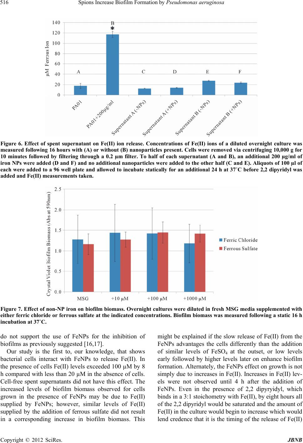 Spions Increase Biofilm Formation by Pseudomonas aeruginosa 516 Figure 6. Effect of spent supernatant on Fe(II) ion release. Concentrations of Fe(II) ions of a diluted overnight culture was measured following 16 hours with (A) or without (B) nanoparticles present. Cells were removed via centrifuging 10,000 g for 10 minutes followed by filtering through a 0.2 µm filter. To half of each supernatant (A and B), an additional 200 µg/ml of iron NPs were added (D and F) and no additional nanoparticles were added to the other half (C and E). Aliquots of 100 µl of each were added to a 96 well plate and allowed to incubate statically for an additional 24 h at 37˚C before 2,2 dipyridyl was added and Fe(II) measurements taken. Figure 7. Effect of non-NP iron on biofilm biomass. Overnight cultures were diluted in fresh MSG media supplemented with either ferric chloride or ferrous sulfate at the indicated concentrations. Biofilm biomass was measured following a static 16 h incubation at 37˚C. do not support the use of FeNPs for the inhibition of biofilms as previously suggested [16,17]. Our study is the first to, our knowledge, that shows bacterial cells interact with FeNPs to release Fe(II). In the presence of cells Fe(II) levels exceeded 100 µM by 8 h compared with less than 20 µM in the absence of cells. Cell-free spent supernatants did not have this effect. The increased levels of biofilm biomass observed for cells grown in the presence of FeNPs may be due to Fe(II) supplied by FeNPs; however, similar levels of Fe(II) supplied by the addition of ferrous sulfate did not result in a corresponding increase in biofilm biomass. This might be explained if the slow release of Fe(II) from the FeNPs advantages the cells differently than the addition of similar levels of FeSO4 at the outset, or low levels early followed by higher levels later on enhance biofilm formation. Alternately, the FeNPs effect on growth is not simply due to increases in Fe(II). Increases in Fe(II) lev- els were not observed until 4 h after the addition of FeNPs. Even in the presence of 2,2 dipyryidyl, which binds in a 3:1 stoichometry with Fe(II), by eight hours all of the 2,2 dipyridyl would be saturated and the amount of Fe(II) in the culture would begin to increase which would lend credence that it is the timing of the release of Fe(II) Copyright © 2012 SciRes. JBNB 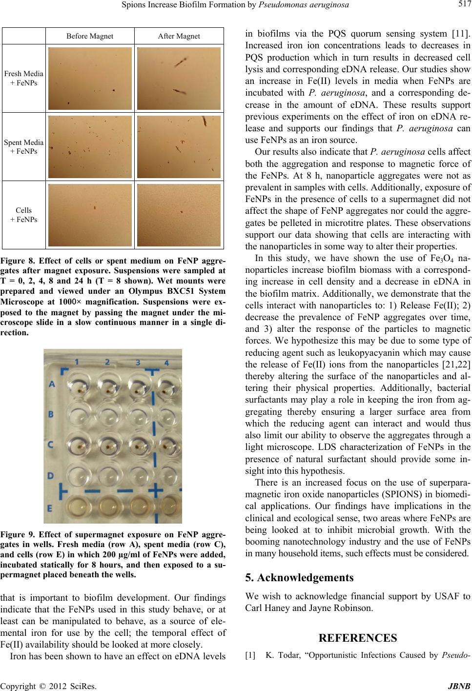 Spions Increase Biofilm Formation by Pseudomonas aeruginosa 517 Before Magnet After Magnet Fresh Media + FeNPs Spent Media + FeNPs Cells + FeNPs Figure 8. Effect of cells or spent medium on FeNP aggre- gates after magnet exposure. Suspensions were sampled at T = 0, 2, 4, 8 and 24 h (T = 8 shown). Wet mounts were prepared and viewed under an Olympus BXC51 System Microscope at 1000× magnification. Suspensions were ex- posed to the magnet by passing the magnet under the mi- croscope slide in a slow continuous manner in a single di- rection. Figure 9. Effect of supermagnet exposure on FeNP aggre- gates in wells. Fresh media (row A), spent media (row C), and cells (row E) in which 200 µg/ml of FeNPs were added, incubated statically for 8 hours, and then exposed to a su- permagnet placed beneath the wells. that is important to biofilm development. Our findings indicate that the FeNPs used in this study behave, or at least can be manipulated to behave, as a source of ele- mental iron for use by the cell; the temporal effect of Fe(II) availability should be looked at more closely. Iron has been shown to have an effect on eDNA levels in biofilms via the PQS quorum sensing system [11]. Increased iron ion concentrations leads to decreases in PQS production which in turn results in decreased cell lysis and corresponding eDNA release. Our studies show an increase in Fe(II) levels in media when FeNPs are incubated with P. aeruginosa, and a corresponding de- crease in the amount of eDNA. These results support previous experiments on the effect of iron on eDNA re- lease and supports our findings that P. aeruginosa can use FeNPs as an iron source. Our results also indicate that P. aeruginosa cells affect both the aggregation and response to magnetic force of the FeNPs. At 8 h, nanoparticle aggregates were not as prevalent in samples with cells. Additionally, exposure of FeNPs in the presence of cells to a supermagnet did not affect the shape of FeNP aggregates nor could the aggre- gates be pelleted in microtitre plates. These observations support our data showing that cells are interacting with the nanoparticles in some way to alter their properties. In this study, we have shown the use of Fe3O4 na- noparticles increase biofilm biomass with a correspond- ing increase in cell density and a decrease in eDNA in the biofilm matrix. Additionally, we demonstrate that the cells interact with nanoparticles to: 1) Release Fe(II); 2) decrease the prevalence of FeNP aggregates over time, and 3) alter the response of the particles to magnetic forces. We hypothesize this may be due to some type of reducing agent such as leukopyacyanin which may cause the release of Fe(II) ions from the nanoparticles [21,22] thereby altering the surface of the nanoparticles and al- tering their physical properties. Additionally, bacterial surfactants may play a role in keeping the iron from ag- gregating thereby ensuring a larger surface area from which the reducing agent can interact and would thus also limit our ability to observe the aggregates through a light microscope. LDS characterization of FeNPs in the presence of natural surfactant should provide some in- sight into this hypothesis. There is an increased focus on the use of superpara- magnetic iron oxide nanoparticles (SPIONS) in biomedi- cal applications. Our findings have implications in the clinical and ecological sense, two areas where FeNPs are being looked at to inhibit microbial growth. With the booming nanotechnology industry and the use of FeNPs in many household items, such effects must be considered. 5. Acknowledgements We wish to acknowledge financial support by USAF to Carl Haney and Jayne Robinson. REFERENCES [1] K. Todar, “Opportunistic Infections Caused by Pseudo- Copyright © 2012 SciRes. JBNB 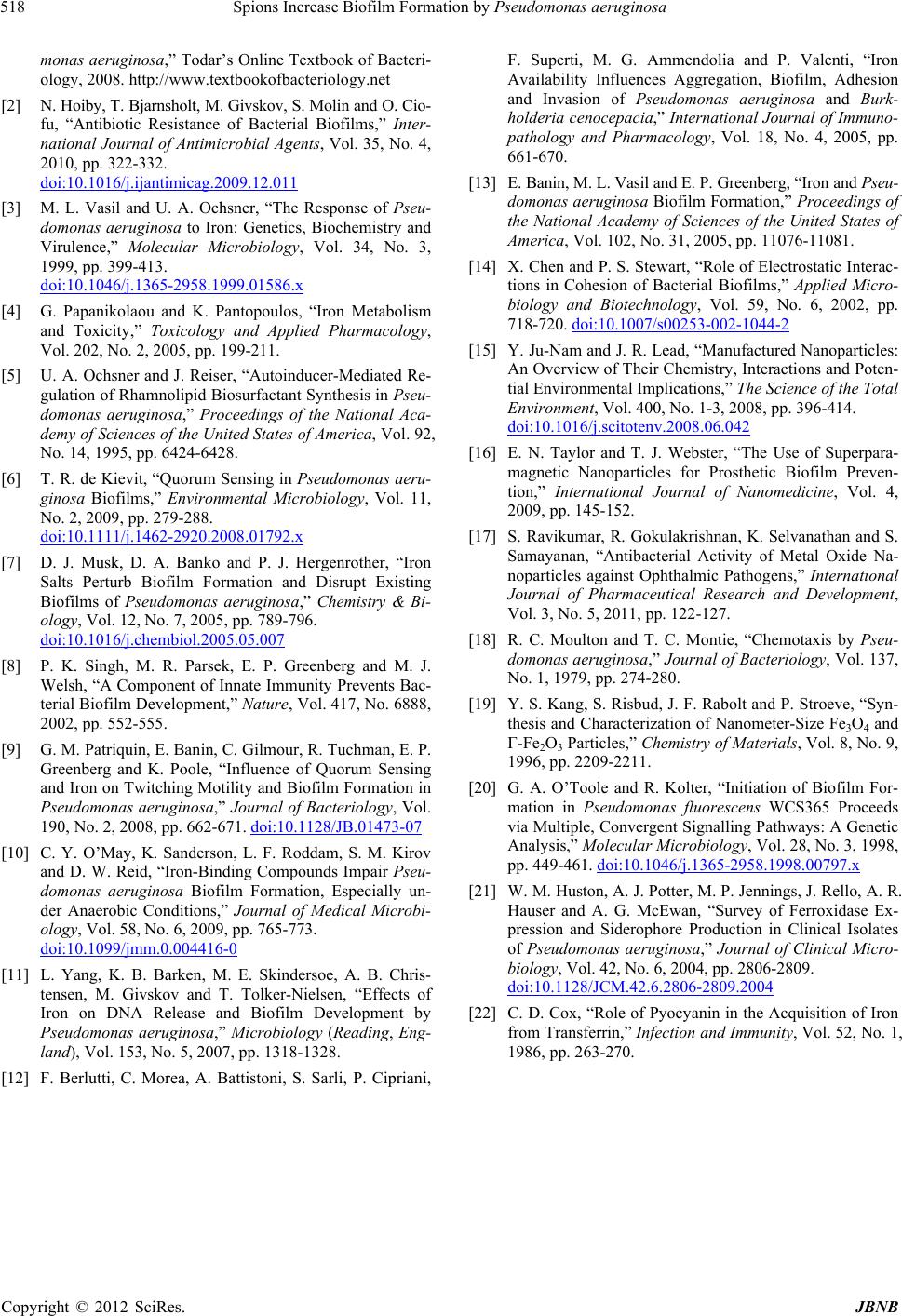 Spions Increase Biofilm Formation by Pseudomonas aeruginosa Copyright © 2012 SciRes. JBNB 518 monas aeruginosa,” Todar’s Online Textbook of Bacteri- ology, 2008. http://www.textbookofbacteriology.net [2] N. Hoiby, T. Bjarnsholt, M. Givskov, S. Molin and O. Cio- fu, “Antibiotic Resistance of Bacterial Biofilms,” Inter- national Journal of Antimicrobial Agents, Vol. 35, No. 4, 2010, pp. 322-332. doi:10.1016/j.ijantimicag.2009.12.011 [3] M. L. Vasil and U. A. Ochsner, “The Response of Pseu- domonas aeruginosa to Iron: Genetics, Biochemistry and Virulence,” Molecular Microbiology, Vol. 34, No. 3, 1999, pp. 399-413. doi:10.1046/j.1365-2958.1999.01586.x [4] G. Papanikolaou and K. Pantopoulos, “Iron Metabolism and Toxicity,” Toxicology and Applied Pharmacology, Vol. 202, No. 2, 2005, pp. 199-211. [5] U. A. Ochsner and J. Reiser, “Autoinducer-Mediated Re- gulation of Rhamnolipid Biosurfactant Synthesis in Pseu- domonas aeruginosa,” Proceedings of the National Aca- demy of Sciences of the United States of America, Vol. 92, No. 14, 1995, pp. 6424-6428. [6] T. R. de Kievit, “Quorum Sensing in Pseudomonas aeru- ginosa Biofilms,” Environmental Microbiology, Vol. 11, No. 2, 2009, pp. 279-288. doi:10.1111/j.1462-2920.2008.01792.x [7] D. J. Musk, D. A. Banko and P. J. Hergenrother, “Iron Salts Perturb Biofilm Formation and Disrupt Existing Biofilms of Pseudomonas aeruginosa,” Chemistry & Bi- ology, Vol. 12, No. 7, 2005, pp. 789-796. doi:10.1016/j.chembiol.2005.05.007 [8] P. K. Singh, M. R. Parsek, E. P. Greenberg and M. J. Welsh, “A Component of Innate Immunity Prevents Bac- terial Biofilm Development,” Nature, Vol. 417, No. 6888, 2002, pp. 552-555. [9] G. M. Patriquin, E. Banin, C. Gilmour, R. Tuchman, E. P. Greenberg and K. Poole, “Influence of Quorum Sensing and Iron on Twitching Motility and Biofilm Formation in Pseudomonas aeruginosa,” Journal of Bacteriology, Vol. 190, No. 2, 2008, pp. 662-671. doi:10.1128/JB.01473-07 [10] C. Y. O’May, K. Sanderson, L. F. Roddam, S. M. Kirov and D. W. Reid, “Iron-Binding Compounds Impair Pseu- domonas aeruginosa Biofilm Formation, Especially un- der Anaerobic Conditions,” Journal of Medical Microbi- ology, Vol. 58, No. 6, 2009, pp. 765-773. doi:10.1099/jmm.0.004416-0 [11] L. Yang, K. B. Barken, M. E. Skindersoe, A. B. Chris- tensen, M. Givskov and T. Tolker-Nielsen, “Effects of Iron on DNA Release and Biofilm Development by Pseudomonas aeruginosa,” Microbiology (Reading, Eng- land), Vol. 153, No. 5, 2007, pp. 1318-1328. [12] F. Berlutti, C. Morea, A. Battistoni, S. Sarli, P. Cipriani, F. Superti, M. G. Ammendolia and P. Valenti, “Iron Availability Influences Aggregation, Biofilm, Adhesion and Invasion of Pseudomonas aeruginosa and Burk- holderia cenocepacia,” International Journal of Immuno- pathology and Pharmacology, Vol. 18, No. 4, 2005, pp. 661-670. [13] E. Banin, M. L. Vasil and E. P. Greenberg, “Iron and Pseu- domonas aeruginosa Biofilm Formation,” Proceedings of the National Academy of Sciences of the United States of America, Vol. 102, No. 31, 2005, pp. 11076-11081. [14] X. Chen and P. S. Stewart, “Role of Electrostatic Interac- tions in Cohesion of Bacterial Biofilms,” Applied Micro- biology and Biotechnology, Vol. 59, No. 6, 2002, pp. 718-720. doi:10.1007/s00253-002-1044-2 [15] Y. Ju-Nam and J. R. Lead, “Manufactured Nanoparticles: An Overview of Their Chemistry, Interactions and Poten- tial Environmental Implications,” The Science of the Total Environment, Vol. 400, No. 1-3, 2008, pp. 396-414. doi:10.1016/j.scitotenv.2008.06.042 [16] E. N. Taylor and T. J. Webster, “The Use of Superpara- magnetic Nanoparticles for Prosthetic Biofilm Preven- tion,” International Journal of Nanomedicine, Vol. 4, 2009, pp. 145-152. [17] S. Ravikumar, R. Gokulakrishnan, K. Selvanathan and S. Samayanan, “Antibacterial Activity of Metal Oxide Na- noparticles against Ophthalmic Pathogens,” International Journal of Pharmaceutical Research and Development, Vol. 3, No. 5, 2011, pp. 122-127. [18] R. C. Moulton and T. C. Montie, “Chemotaxis by Pseu- domonas aeruginosa,” Journal of Bacteriology, Vol. 137, No. 1, 1979, pp. 274-280. [19] Y. S. Kang, S. Risbud, J. F. Rabolt and P. Stroeve, “Syn- thesis and Characterization of Nanometer-Size Fe3O4 and Γ-Fe2O3 Particles,” Chemistry of Materials, Vol. 8, No. 9, 1996, pp. 2209-2211. [20] G. A. O’Toole and R. Kolter, “Initiation of Biofilm For- mation in Pseudomonas fluorescens WCS365 Proceeds via Multiple, Convergent Signalling Pathways: A Genetic Analysis,” Molecular Microbiology, Vol. 28, No. 3, 1998, pp. 449-461. doi:10.1046/j.1365-2958.1998.00797.x [21] W. M. Huston, A. J. Potter, M. P. Jennings, J. Rello, A. R. Hauser and A. G. McEwan, “Survey of Ferroxidase Ex- pression and Siderophore Production in Clinical Isolates of Pseudomonas aeruginosa,” Journal of Clinical Micro- biology, Vol. 42, No. 6, 2004, pp. 2806-2809. doi:10.1128/JCM.42.6.2806-2809.2004 [22] C. D. Cox, “Role of Pyocyanin in the Acquisition of Iron from Transferrin,” Infection and Immunity, Vol. 52, No. 1, 1986, pp. 263-270. |