

Paper Menu >>
Journal Menu >>
 Open Journal of Pathology, 2012, 2, 159-161 Published Online October 2012 (http://www.SciRP.org/journal/ojpathology) http://dx.doi.org/10.4236/ojpathology.2012.24030 Copyright © 2012 SciRes. OJPathology 1 Obstructive Azoospermia in a Patient with VATER Association Zhanyong Bing Department of Pathology and Laboratory Medicine, Hospital of the University of Pennsylvania, Philadelphia, USA. Email: [email protected] Received May 23rd, 2012; revised June 21st, 2012; accepted June 29th, 2012 ABSTRACT VATER association was first described in 1968 by Say and Gerald and is an association of congenital anomalies in- cluding V (vertebral defect), A (anal atresia), TE (tracheoesophegeal fistula), and R (radial dysplasia and renal defects). This report described a 26 year-old man with VATER association presented with infertility. Hormone tests including follicular stimulating hormone, luteinizing hormone and testosterone were normal. Semen analysis showed azoospermia. A testicular biopsy was performed and showed the presence of all of germinative components, germ cell sloughing, and mild hypospermatogenesis. The findings were compatible with obstructive azoospermia. No evidence of intratubular germ cell neoplasia unclassified was identified. Keywords: VATER Association; Obstructive; Azoospermia 1. Introduction Infertility can be attributed to men in 30% - 40% of in- fertile couples, which may be du e to endocrin e disruption of gonadal development during early pregnancy, environ- ment pollution and genetic factors. Azoospermia can be secondary to obstructive or non-obstructive etiologies. Testicular biopsy plays a definitive role in confirming the obstructive azoo spermia, which in turn leads to appropri- ate management. VATER association was first described in 1968 by Say and Gerald [1] and is an association of congenital anomalies including V ( vertebral defect), A (anal atresia), TE (tracheoesophegeal fistula), and R (radial dysplasia and renal defects) [2,3]. Since the introduction of the acronym, various expansions in an effort to further define the scope of the VATER association have been suggested, such as V in VATER was expanded to include vascular anomalies [4]. It has been suggested to expand the acro- nym to VACTERL with the C denoting cardiovascular anomalies and the L denoting limb anomalies [5]. About half of the patients with VATER association also show the urologic anomalies [6,7]. No obstructive azoospermia has been reported in the patients with VATER associa- tion. In this report a men with VATER association with an obstructive azoospermia was describ ed. 2. Clinical Histor y The patient was a 26 year old man who was born with an imperforate anus and hypospadias. He had one functional kidney. He had a colostomy from 1986 to 1997 and then was reversed. He had fecal incontinence. Colorectal evaluation showed minimal tone and no appreciable functional sphincter. He developed hydrocele in 2005 and it was enlarged since then. He had no issues with erection or orgasm. In 2 009 he had a semen analysis that showed azoospermia. Hormone tests were done in 2011 and showed that follicular stimulating hormone, lu- teinizing hormone and total testosterone were within normal limits. He reported occasional dysuria and had to strain to void at times. He had in intermittent stream. He denied hematuria or urolithiasis. A testicular biopsy and resection of hydrocele sac was performed. 3. Pathology The testicular biopsy showed the presence of all germi- native elements (Figures 1(a) and (b)). The majority of the tubules showed sloughing of the immature germina- tive elements. The modal Johnsen score was 9. The mean spermatid count per tubules was 10. The tubular base- ment membranes showed mild thickening. No Leydig cell hyperplasia was seen. The vessels were unremark- able. No morphological evidence for intratubular germ cell neoplasia unspecificed was identified. Immunohis- tochemical stains for OCT3/4 and PLAP were performed and were negative. 4. Discussion VATER association describes a non-random group of 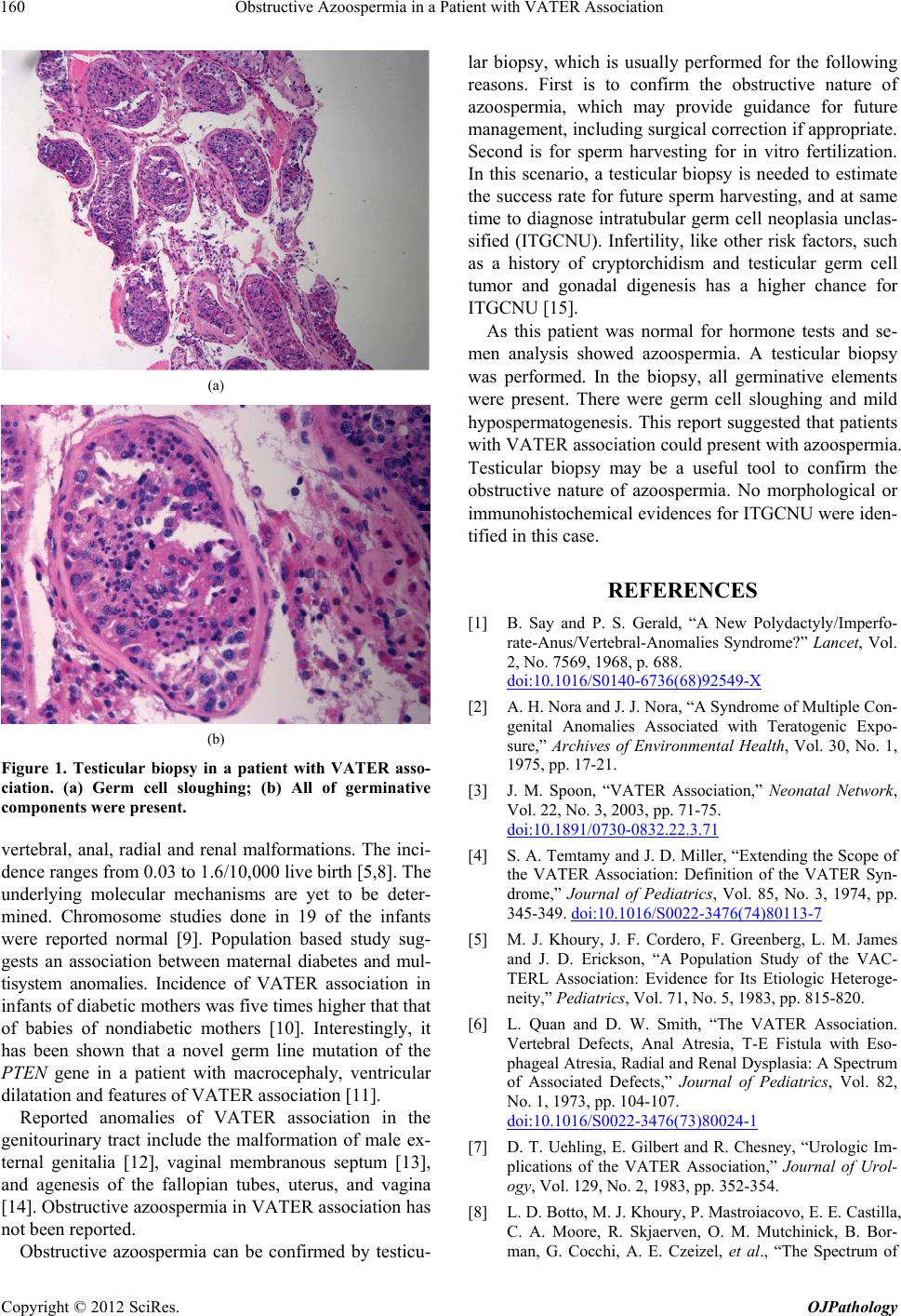 Obstructive Azoospermia in a Patient with VATER Association 160 (a) (b) Figure 1. Testicular biopsy in a patient with VATER asso- ciation. (a) Germ cell sloughing; (b) All of germinative components were present. vertebral, anal, radial and renal malformations. The inci- dence ranges from 0.03 to 1.6/10,000 live birth [5,8]. The underlying molecular mechanisms are yet to be deter- mined. Chromosome studies done in 19 of the infants were reported normal [9]. Population based study sug- gests an association between maternal diabetes and mul- tisystem anomalies. Incidence of VATER association in infants of diabetic mothers was five times high er that that of babies of nondiabetic mothers [10]. Interestingly, it has been shown that a novel germ line mutation of the PTEN gene in a patient with macrocephaly, ventricular dilatation and features of VATER association [11]. Reported anomalies of VATER association in the genitourinary tract include the malformation of male ex- ternal genitalia [12], vaginal membranous septum [13], and agenesis of the fallopian tubes, uterus, and vagina [14]. Obstructive azoospermia in VATER association has not been reported. Obstructive azoospermia can be confirmed by testicu- lar biopsy, which is usually performed for the following reasons. First is to confirm the obstructive nature of azoospermia, which may provide guidance for future management, including surgical correction if appropriate. Second is for sperm harvesting for in vitro fertilization. In this scenario, a testicular biopsy is needed to estimate the success rate for future sperm harvesting, and at same time to diagnose intratubular germ cell neoplasia unclas- sified (ITGCNU). Infertility, like other risk factors, such as a history of cryptorchidism and testicular germ cell tumor and gonadal digenesis has a higher chance for ITGCNU [15]. As this patient was normal for hormone tests and se- men analysis showed azoospermia. A testicular biopsy was performed. In the biopsy, all germinative elements were present. There were germ cell sloughing and mild hypospermatogenesis. This report suggested that patients with VATER association could present with azoospermia. Testicular biopsy may be a useful tool to confirm the obstructive nature of azoospermia. No morphological or immunohistochemical evidences for ITGCNU were iden- tified in this case. REFERENCES [1] B. Say and P. S. Gerald, “A New Polydactyly/Imperfo- rate-Anus/Vertebral-Anomalies Syndrome?” Lancet, Vol. 2, No. 7569, 1968, p. 688. doi:10.1016/S0140-6736(68)92549-X [2] A. H. Nora and J. J. Nora, “A Syndrome of Multiple Con- genital Anomalies Associated with Teratogenic Expo- sure,” Archives of Environmental Health, Vol. 30, No. 1, 1975, pp. 17-21. [3] J. M. Spoon, “VATER Association,” Neonatal Network, Vol. 22, No. 3, 2003, pp. 71-75. doi:10.1891/0730-0832.22.3.71 [4] S. A. Temtamy and J. D. Miller, “Extending the Scope of the VATER Association: Definition of the VATER Syn- drome,” Journal of Pediatrics, Vol. 85, No. 3, 1974, pp. 345-349. doi:10.1016/S0022-3476(74)80113-7 [5] M. J. Khoury, J. F. Cordero, F. Greenberg, L. M. James and J. D. Erickson, “A Population Study of the VAC- TERL Association: Evidence for Its Etiologic Heteroge- neity,” Pediatrics, Vol. 71, No. 5, 1983, pp. 815-820. [6] L. Quan and D. W. Smith, “The VATER Association. Vertebral Defects, Anal Atresia, T-E Fistula with Eso- phageal Atresia, Radial and Renal Dysplasia: A Spectrum of Associated Defects,” Journal of Pediatrics, Vol. 82, No. 1, 1973, pp. 104-107. doi:10.1016/S0022-3476(73)80024-1 [7] D. T. Uehling, E. Gilbert and R. Chesney, “Urologic Im- plications of the VATER Association,” Journal of Urol- ogy, Vol. 129, No. 2, 1983, pp. 352-354. [8] L. D. Botto, M. J. Khoury, P. Mastroiacovo, E. E. Castilla, C. A. Moore, R. Skjaerven, O. M. Mutchinick, B. Bor- man, G. Cocchi, A. E. Czeizel, et al., “The Spectrum of Copyright © 2012 SciRes. OJPathology  Obstructive Azoospermia in a Patient with VATER Association Copyright © 2012 SciRes. OJPathology 161 Congenital Anomalies of the VATER Association: An International Study,” American Journal of Medical Ge- netics, Vol. 71, No. 1, 1997, pp. 8-15. doi:10.1002/(SICI)1096-8628(19970711)71:1<8::AID-AJ MG2>3.0.CO;2-V [9] D. D. Weaver, C. L. Mapstone and P. L. Yu, “The VATER Association. Analysis of 46 Patients,” American Journal of Diseases of Children, Vol. 140, No. 3, 1986, pp. 225-229. [10] M. J. Bull, C. Q. Bryson, J. Grosfeld and R. L. Schreiner, “VATER Association: Analysis of Growth and Devel- opment,” American Journal of Perinatology, Vol. 2, No. 1, 1985, pp. 35-38. doi:10.1055/s-2007-999908 [11] W. Reardon, X. P. Zhou and C. Eng, “A Novel Germline Mutation of the PTEN Gene in a Patient with Macro- cephaly, Ventricular Dilatation, and Features of VATER Association,” Journal of Medical Genetics, Vol. 38, No. 12, 2001, pp. 820-823. doi:10.1136/jmg.38.12.820 [12] J. Apold, E. Dahl and D. Aarskog, “The Vater Associa- tion: Malformations of the Male External Genitalia,” Acta Paediatrica Scandinavica, Vol. 65, No. 2, 1976, pp. 150- 152. doi:10.1111/j.1651-2227.1976.tb16528.x [13] B. Say, N. J. Carpenter and E. I. Smith, “Genital Malfor- mations in a Child with VATER Association,” American Journal of Diseases of Children, Vol. 133, No. 4, 1979, pp. 438-439. [14] S. L. King, R. L. Ladda and S. J. Shochat, “Monozygotic Twins Concordant for Tracheo-Esophageal Fistula and Discordant for the VATER Association,” Acta Paedi- atrica Scandinavica, Vol. 66, No. 6, 1977, pp. 783-785. doi:10.1111/j.1651-2227.1977.tb07989.x [15] G. R. Dohle, S. Elzanaty and N. J. van Casteren, “Tes- ticular Biopsy: Clinical Practice and Interpretation,” Asian Journal of Andrology, Vol. 14, No. 1, 2012, pp. 88-93. doi:10.1038/aja.2011.57 |