

Paper Menu >>
Journal Menu >>
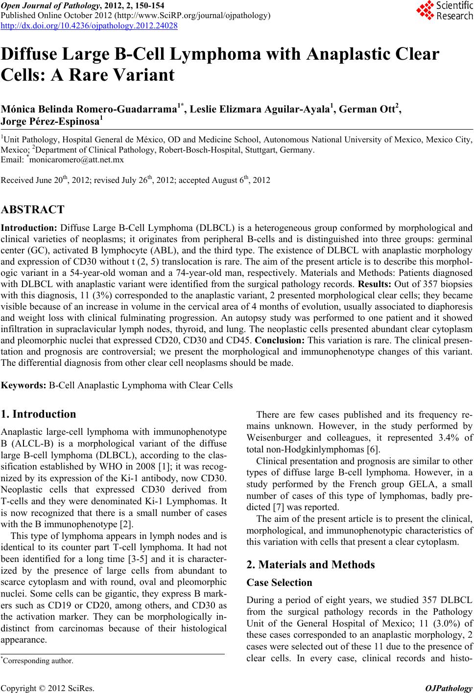 Open Journal of Pathology, 2012, 2, 150-154 Published Online October 2012 (http://www.SciRP.org/journal/ojpathology) http://dx.doi.org/10.4236/ojpathology.2012.24028 Copyright © 2012 SciRes. OJPathology 1 Diffuse Large B-Cell Lymphoma with Anaplastic Clear Cells: A Rare Variant Mónica Belinda Romero-Guadarrama1*, Leslie Elizmara Aguilar-Ayala1, German Ott2, Jorge Pérez-Espinosa1 1Unit Pathology, Hospital General de México, OD and Medicine School, Autonomous National University of Mexico, Mexico City, Mexico; 2Department of Clinical Pathology, Robert-Bosch-Hospital, Stuttgart, Germany. Email: *[email protected] Received June 20th, 2012; revised July 26th, 2012; accepted August 6th, 2012 ABSTRACT Introduction: Diffuse Large B-Cell Lymphoma (DLBCL) is a heterogeneous group conformed by morphological and clinical varieties of neoplasms; it originates from peripheral B-cells and is distinguished into three groups: germinal center (GC), activated B lymphocyte (ABL), and the third type. The existence of DLBCL with anaplastic morphology and expression of CD30 without t (2, 5) translocation is rare. The aim of the present article is to describe this morphol- ogic variant in a 54-year-old woman and a 74-year-old man, respectively. Materials and Methods: Patients diagnosed with DLBCL with anaplastic variant were identified from the surgical pathology records. Results: Out of 357 biopsies with this diagnosis, 11 (3%) corresponded to the anaplastic variant, 2 presented morphological clear cells; they became visible because of an increase in volume in the cervical area of 4 months of evolution, usually associated to diaphoresis and weight loss with clinical fulminating progression. An autopsy study was performed to one patient and it showed infiltration in supraclavicular lymph nodes, thyroid, and lung. The neoplastic cells presented abundant clear cytoplasm and pleomorphic nuclei that expressed CD20, CD30 and CD45. Conclusion: This variation is rare. The clinical presen- tation and prognosis are controversial; we present the morphological and immunophenotype changes of this variant. The differential diagnosis from other clear cell neoplasms should be made. Keywords: B-Cell Anaplastic Lymphoma with Clear Cells 1. Introduction Anaplastic large-cell lymphoma with immunophenotype B (ALCL-B) is a morphological variant of the diffuse large B-cell lymphoma (DLBCL), according to the clas- sification established by WHO in 2008 [1]; it was recog- nized by its expression of the Ki-1 antibody, now CD30. Neoplastic cells that expressed CD30 derived from T-cells and they were denominated Ki-1 Lymphomas. It is now recognized that there is a small number of cases with the B immunophenotype [2]. This type of lymphoma appears in lymph nodes and is identical to its counter part T-cell lymphoma. It had not been identified for a long time [3-5] and it is character- ized by the presence of large cells from abundant to scarce cytoplasm and with round, oval and pleomorphic nuclei. Some cells can be gigantic, they express B mark- ers such as CD19 or CD20, among others, and CD30 as the activation marker. They can be morphologically in- distinct from carcinomas because of their histological appearance. There are few cases published and its frequency re- mains unknown. However, in the study performed by Weisenburger and colleagues, it represented 3.4% of total non-Hodgkinlymphomas [6]. Clinical presentation and prognosis are similar to other types of diffuse large B-cell lymphoma. However, in a study performed by the French group GELA, a small number of cases of this type of lymphomas, badly pre- dicted [7] was reported. The aim of the present article is to present the clinical, morphological, and immunophenotypic characteristics of this variation with cells that present a clear cytoplasm. 2. Materials and Methods Case Selection During a period of eight years, we studied 357 DLBCL from the surgical pathology records in the Pathology Unit of the General Hospital of Mexico; 11 (3.0%) of these cases corresponded to an anaplastic morphology, 2 cases were selected out of these 11 due to the presence of clear cells. In every case, clinical records and histo- *Corresponding author. 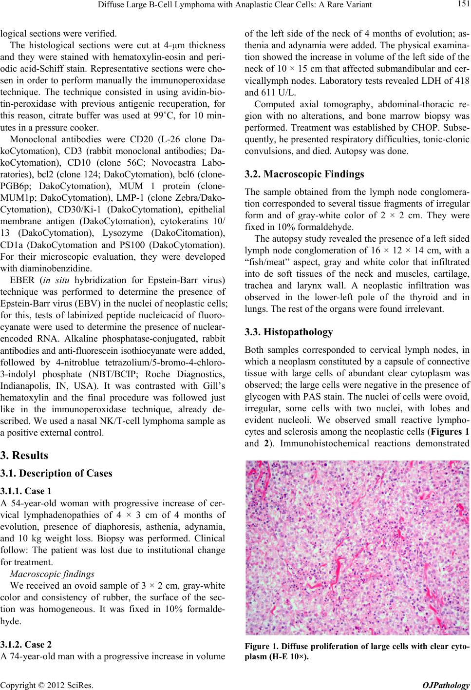 Diffuse Large B-Cell Lymphoma with Anaplastic Clear Cells: A Rare Variant 151 logical sections were verified. The histological sections were cut at 4-μm thickness and they were stained with hematoxylin-eosin and peri- odic acid-Schiff stain. Representative sections were cho- sen in order to perform manually the immunoperoxidase technique. The technique consisted in using avidin-bio- tin-peroxidase with previous antigenic recuperation, for this reason, citrate buffer was used at 99˚C, for 10 min- utes in a pressure cooker. Monoclonal antibodies were CD20 (L-26 clone Da- koCytomation), CD3 (rabbit monoclonal antibodies; Da- koCytomation), CD10 (clone 56C; Novocastra Labo- ratories), bcl2 (clone 124; DakoCytomation), bcl6 (clone- PGB6p; DakoCytomation), MUM 1 protein (clone- MUM1p; DakoCytomation), LMP-1 (clone Zebra/Dako- Cytomation), CD30/Ki-1 (DakoCytomation), epithelial membrane antigen (DakoCytomation), cytokeratins 10/ 13 (DakoCytomation), Lysozyme (DakoCitomation), CD1a (DakoCytomation and PS100 (DakoCytomation). For their microscopic evaluation, they were developed with diaminobenzidine. EBER (in situ hybridization for Epstein-Barr virus) technique was performed to determine the presence of Epstein-Barr virus (EBV) in the nuclei of neoplastic cells; for this, tests of labinized peptide nucleicacid of fluoro- cyanate were used to determine the presence of nuclear- encoded RNA. Alkaline phosphatase-conjugated, rabbit antibodies and anti-fluorescein isothiocyanate were added, followed by 4-nitroblue tetrazolium/5-bromo-4-chloro- 3-indolyl phosphate (NBT/BCIP; Roche Diagnostics, Indianapolis, IN, USA). It was contrasted with Gill’s hematoxylin and the final procedure was followed just like in the immunoperoxidase technique, already de- scribed. We used a nasal NK/T-cell lymphoma sample as a positive external control. 3. Results 3.1. Description of Cases 3.1.1. C as e 1 A 54-year-old woman with progressive increase of cer- vical lymphadenopathies of 4 × 3 cm of 4 months of evolution, presence of diaphoresis, asthenia, adynamia, and 10 kg weight loss. Biopsy was performed. Clinical follow: The patient was lost due to institutional change for treatment. Macroscopic findings We received an ovoid sample of 3 × 2 cm, gray-white color and consistency of rubber, the surface of the sec- tion was homogeneous. It was fixed in 10% formalde- hyde. 3.1.2. C as e 2 A 74-year-old man with a progressive increase in volume of the left side of the neck of 4 months of evolution; as- thenia and adynamia were added. The physical examina- tion showed the increase in volume of the left side of the neck of 10 × 15 cm that affected submandibular and cer- vicallymph nodes. Laboratory tests revealed LDH of 418 and 611 U/L. Computed axial tomography, abdominal-thoracic re- gion with no alterations, and bone marrow biopsy was performed. Treatment was established by CHOP. Subse- quently, he presented respiratory difficulties, tonic-clonic convulsions, and died. Autopsy was done. 3.2. Macroscopic Findings The sample obtained from the lymph node conglomera- tion corresponded to several tissue fragments of irregular form and of gray-white color of 2 × 2 cm. They were fixed in 10% formaldehyde. The autopsy study revealed the presence of a left sided lymph node conglomeration of 16 × 12 × 14 cm, with a “fish/meat” aspect, gray and white color that infiltrated into de soft tissues of the neck and muscles, cartilage, trachea and larynx wall. A neoplastic infiltration was observed in the lower-left pole of the thyroid and in lungs. The rest of the organs were found irrelevant. 3.3. Histopathology Both samples corresponded to cervical lymph nodes, in which a neoplasm constituted by a capsule of connective tissue with large cells of abundant clear cytoplasm was observed; the large cells were negative in the presence of glycogen with PAS stain. The nuclei of cells were ovoid, irregular, some cells with two nuclei, with lobes and evident nucleoli. We observed small reactive lympho- cytes and sclerosis among the neoplastic cells (Figures 1 and 2). Immunohistochemical reactions demonstrated Figure 1. Diffuse proliferation of large cells with clear cyto- plasm (H-E 10×). Copyright © 2012 SciRes. OJPathology 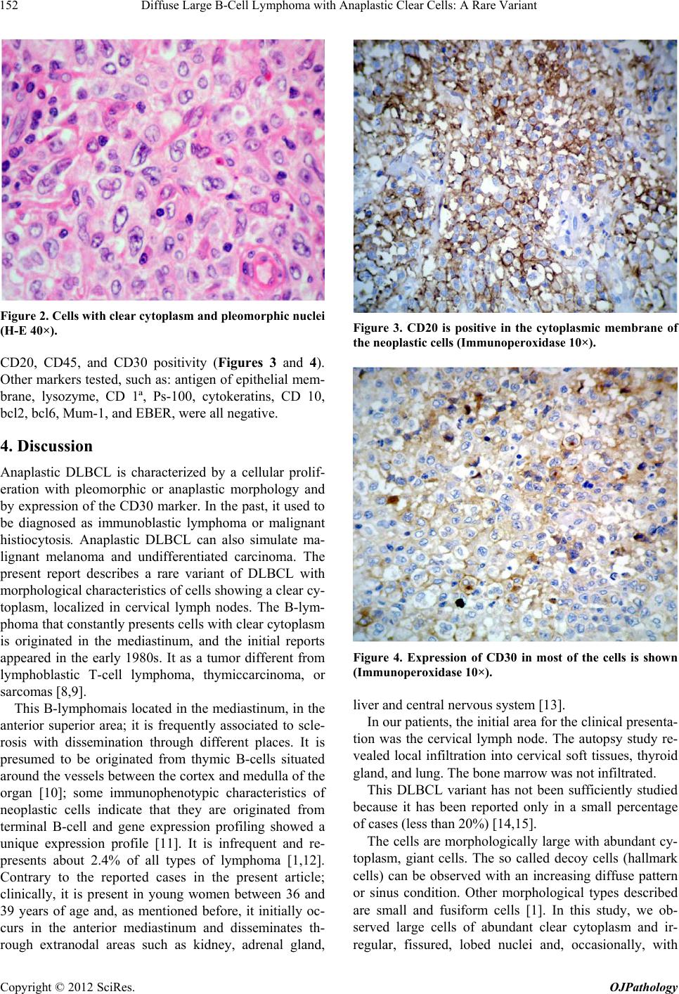 Diffuse Large B-Cell Lymphoma with Anaplastic Clear Cells: A Rare Variant 152 Figure 2. Cells with clear cytoplasm and pleomorphic nuclei (H-E 40×). CD20, CD45, and CD30 positivity (Figures 3 and 4). Other markers tested, such as: antigen of epithelial mem- brane, lysozyme, CD 1ª, Ps-100, cytokeratins, CD 10, bcl2, bcl6, Mum-1, and EBER, were all negative. 4. Discussion Anaplastic DLBCL is characterized by a cellular prolif- eration with pleomorphic or anaplastic morphology and by expression of the CD30 marker. In the past, it used to be diagnosed as immunoblastic lymphoma or malignant histiocytosis. Anaplastic DLBCL can also simulate ma- lignant melanoma and undifferentiated carcinoma. The present report describes a rare variant of DLBCL with morphological characteristics of cells showing a clear cy- toplasm, localized in cervical lymph nodes. The B-lym- phoma that constantly presents cells with clear cytoplasm is originated in the mediastinum, and the initial reports appeared in the early 1980s. It as a tumor different from lymphoblastic T-cell lymphoma, thymiccarcinoma, or sarcomas [8,9]. This B-lymphomais located in the mediastinum, in the anterior superior area; it is frequently associated to scle- rosis with dissemination through different places. It is presumed to be originated from thymic B-cells situated around the vessels between the cortex and medulla of the organ [10]; some immunophenotypic characteristics of neoplastic cells indicate that they are originated from terminal B-cell and gene expression profiling showed a unique expression profile [11]. It is infrequent and re- presents about 2.4% of all types of lymphoma [1,12]. Contrary to the reported cases in the present article; clinically, it is present in young women between 36 and 39 years of age and, as mentioned before, it initially oc- curs in the anterior mediastinum and disseminates th- rough extranodal areas such as kidney, adrenal gland, Figure 3. CD20 is positive in the cytoplasmic membrane of the neoplastic cells (Immunoperoxidase 10×). Figure 4. Expression of CD30 in most of the cells is shown (Immunoperoxidase 10×). liver and central nervous system [13]. In our patients, the initial area for the clinical presenta- tion was the cervical lymph node. The autopsy study re- vealed local infiltration into cervical soft tissues, thyroid gland, and lung. The bone marrow was not infiltrated. This DLBCL variant has not been sufficiently studied because it has been reported only in a small percentage of cases (less than 20%) [14,15]. The cells are morphologically large with abundant cy- toplasm, giant cells. The so called decoy cells (hallmark cells) can be observed with an increasing diffuse pattern or sinus condition. Other morphological types described are small and fusiform cells [1]. In this study, we ob- served large cells of abundant clear cytoplasm and ir- regular, fissured, lobed nuclei and, occasionally, with Copyright © 2012 SciRes. OJPathology 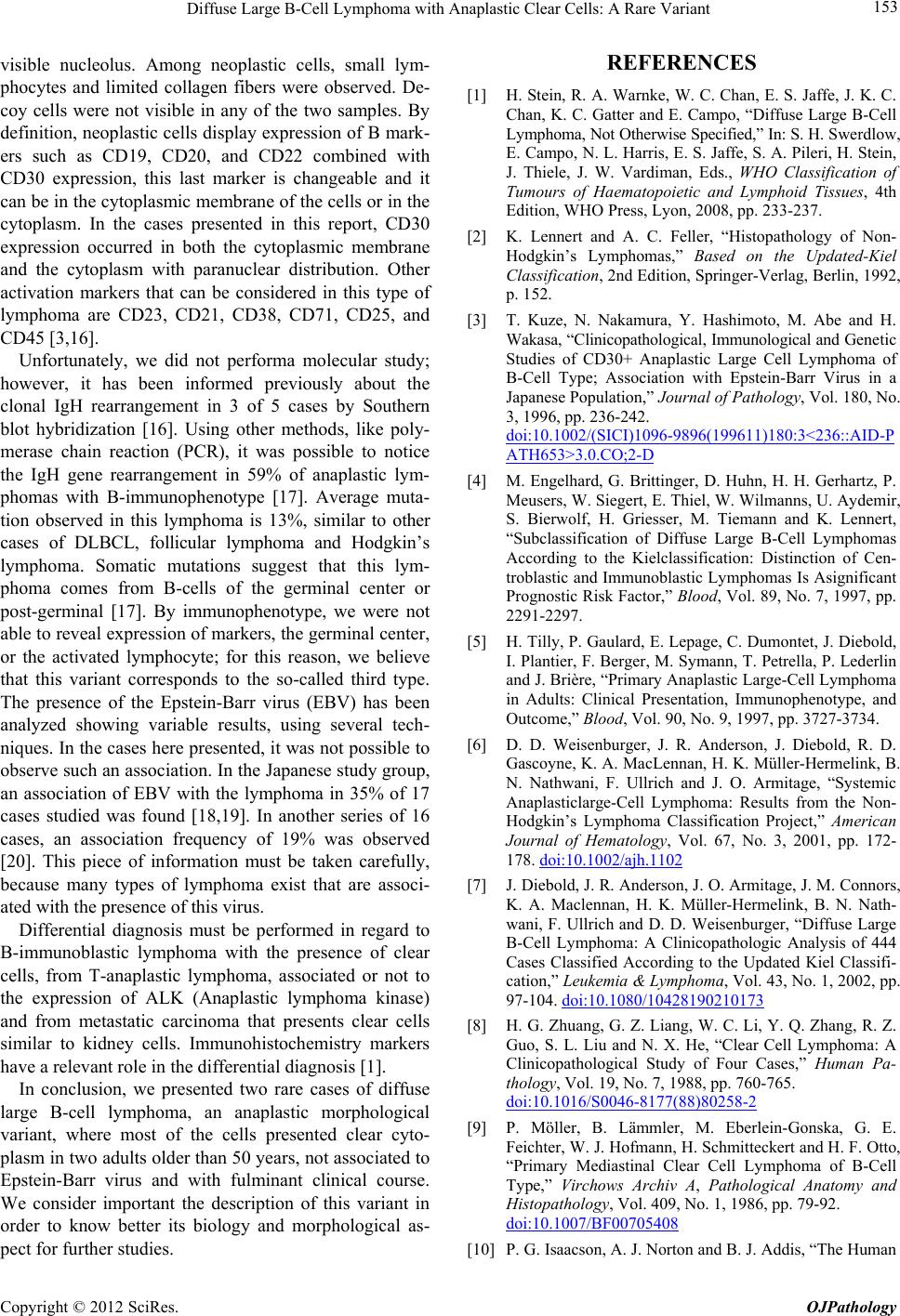 Diffuse Large B-Cell Lymphoma with Anaplastic Clear Cells: A Rare Variant 153 visible nucleolus. Among neoplastic cells, small lym- phocytes and limited collagen fibers were observed. De- coy cells were not visible in any of the two samples. By definition, neoplastic cells display expression of B mark- ers such as CD19, CD20, and CD22 combined with CD30 expression, this last marker is changeable and it can be in the cytoplasmic membrane of the cells or in the cytoplasm. In the cases presented in this report, CD30 expression occurred in both the cytoplasmic membrane and the cytoplasm with paranuclear distribution. Other activation markers that can be considered in this type of lymphoma are CD23, CD21, CD38, CD71, CD25, and CD45 [3,16]. Unfortunately, we did not performa molecular study; however, it has been informed previously about the clonal IgH rearrangement in 3 of 5 cases by Southern blot hybridization [16]. Using other methods, like poly- merase chain reaction (PCR), it was possible to notice the IgH gene rearrangement in 59% of anaplastic lym- phomas with B-immunophenotype [17]. Average muta- tion observed in this lymphoma is 13%, similar to other cases of DLBCL, follicular lymphoma and Hodgkin’s lymphoma. Somatic mutations suggest that this lym- phoma comes from B-cells of the germinal center or post-germinal [17]. By immunophenotype, we were not able to reveal expression of markers, the germinal center, or the activated lymphocyte; for this reason, we believe that this variant corresponds to the so-called third type. The presence of the Epstein-Barr virus (EBV) has been analyzed showing variable results, using several tech- niques. In the cases here presented, it was not possible to observe such an association. In the Japanese study group, an association of EBV with the lymphoma in 35% of 17 cases studied was found [18,19]. In another series of 16 cases, an association frequency of 19% was observed [20]. This piece of information must be taken carefully, because many types of lymphoma exist that are associ- ated with the presence of this virus. Differential diagnosis must be performed in regard to B-immunoblastic lymphoma with the presence of clear cells, from T-anaplastic lymphoma, associated or not to the expression of ALK (Anaplastic lymphoma kinase) and from metastatic carcinoma that presents clear cells similar to kidney cells. Immunohistochemistry markers have a relevant role in the differential diagnosis [1]. In conclusion, we presented two rare cases of diffuse large B-cell lymphoma, an anaplastic morphological variant, where most of the cells presented clear cyto- plasm in two adults older than 50 years, not associated to Epstein-Barr virus and with fulminant clinical course. We consider important the description of this variant in order to know better its biology and morphological as- pect for further studies. REFERENCES [1] H. Stein, R. A. Warnke, W. C. Chan, E. S. Jaffe, J. K. C. Chan, K. C. Gatter and E. Campo, “Diffuse Large B-Cell Lymphoma, Not Otherwise Specified,” In: S. H. Swerdlow, E. Campo, N. L. Harris, E. S. Jaffe, S. A. Pileri, H. Stein, J. Thiele, J. W. Vardiman, Eds., WHO Classification of Tumours of Haematopoietic and Lymphoid Tissues, 4th Edition, WHO Press, Lyon, 2008, pp. 233-237. [2] K. Lennert and A. C. Feller, “Histopathology of Non- Hodgkin’s Lymphomas,” Based on the Updated-Kiel Classification, 2nd Edition, Springer-Verlag, Berlin, 1992, p. 152. [3] T. Kuze, N. Nakamura, Y. Hashimoto, M. Abe and H. Wakasa, “Clinicopathological, Immunological and Genetic Studies of CD30+ Anaplastic Large Cell Lymphoma of B-Cell Type; Association with Epstein-Barr Virus in a Japanese Population,” Journal of Pathology, Vol. 180, No. 3, 1996, pp. 236-242. doi:10.1002/(SICI)1096-9896(199611)180:3<236::AID-P ATH653>3.0.CO;2-D [4] M. Engelhard, G. Brittinger, D. Huhn, H. H. Gerhartz, P. Meusers, W. Siegert, E. Thiel, W. Wilmanns, U. Aydemir, S. Bierwolf, H. Griesser, M. Tiemann and K. Lennert, “Subclassification of Diffuse Large B-Cell Lymphomas According to the Kielclassification: Distinction of Cen- troblastic and Immunoblastic Lymphomas Is Asignificant Prognostic Risk Factor,” Blood, Vol. 89, No. 7, 1997, pp. 2291-2297. [5] H. Tilly, P. Gaulard, E. Lepage, C. Dumontet, J. Diebold, I. Plantier, F. Berger, M. Symann, T. Petrella, P. Lederlin and J. Brière, “Primary Anaplastic Large-Cell Lymphoma in Adults: Clinical Presentation, Immunophenotype, and Outcome,” Blood, Vol. 90, No. 9, 1997, pp. 3727-3734. [6] D. D. Weisenburger, J. R. Anderson, J. Diebold, R. D. Gascoyne, K. A. MacLennan, H. K. Müller-Hermelink, B. N. Nathwani, F. Ullrich and J. O. Armitage, “Systemic Anaplasticlarge-Cell Lymphoma: Results from the Non- Hodgkin’s Lymphoma Classification Project,” American Journal of Hematology, Vol. 67, No. 3, 2001, pp. 172- 178. doi:10.1002/ajh.1102 [7] J. Diebold, J. R. Anderson, J. O. Armitage, J. M. Connors, K. A. Maclennan, H. K. Müller-Hermelink, B. N. Nath- wani, F. Ullrich and D. D. Weisenburger, “Diffuse Large B-Cell Lymphoma: A Clinicopathologic Analysis of 444 Cases Classified According to the Updated Kiel Classifi- cation,” Leukemia & Lymphoma, Vol. 43, No. 1, 2002, pp. 97-104. doi:10.1080/10428190210173 [8] H. G. Zhuang, G. Z. Liang, W. C. Li, Y. Q. Zhang, R. Z. Guo, S. L. Liu and N. X. He, “Clear Cell Lymphoma: A Clinicopathological Study of Four Cases,” Human Pa- thology, Vol. 19, No. 7, 1988, pp. 760-765. doi:10.1016/S0046-8177(88)80258-2 [9] P. Möller, B. Lämmler, M. Eberlein-Gonska, G. E. Feichter, W. J. Hofmann, H. Schmitteckert and H. F. Otto, “Primary Mediastinal Clear Cell Lymphoma of B-Cell Type,” Virchows Archiv A, Pathological Anatomy and Histopathology, Vol. 409, No. 1, 1986, pp. 79-92. doi:10.1007/BF00705408 [10] P. G. Isaacson, A. J. Norton and B. J. Addis, “The Human Copyright © 2012 SciRes. OJPathology  Diffuse Large B-Cell Lymphoma with Anaplastic Clear Cells: A Rare Variant Copyright © 2012 SciRes. OJPathology 154 Thymus Contains a Novel Population of B Lympho- cytes,” Lancet, Vol. 2, No. 8574, 1987, pp. 1488-1491. doi:10.1016/S0140-6736(87)92622-5 [11] P. Möller, G. Moldenhauer, F. Momburg, B. Lämmler, M. Eberlein-Gonska, S. Kiesel and B. Dörken, “Mediastinal Lymphoma of Clear Cell Type Is a Tumor Corresponding Toterminal Steps of B Cell Differentiation,” Blood, Vol. 69, No. 4, 1987, pp. 1087-1095. [12] J. O. Armitage and D. D. Weisenburger, “New Approach to Classifying Non-Hodgkin’s Lymphomas: Clinical Fea- tures of the Major Histologic Subtypes, Non-Hodgkin’s Lymphoma Classification Project,” Journal of Clinical Oncology, Vol. 16, No. 8, 1998, pp. 2780-2795. [13] P. C. Bishop, W. H. Wilson, D. Pearson, J. Janik, E. S. Jaffe and P. C. Elwood, “CNS Involvement in Primary Mediastinal Large B-Cell Lymphoma,” Journal of Clini- cal Oncology, Vol. 17, No. 8, 1999, pp. 2479-2485. [14] H. Stein, D. Y. Mason, J. Gerdes, N. O’Connor, J. Wain- scoat, G. Pallesen, K. Gatter, B. Falini, G. Delsol, H. Lemke, et al., “The Expression of the Hodgkin’s Disease Associated Antigen Ki-1 in Reactive and Neoplastic Lymphoid Tissue: Evidence That Reed-Sternberg Cells and Histiocytic Malignancies Are Derived from Activated Lymphoid Cells,” Blood, Vol. 66, No. 4, 1985, pp. 848- 858. [15] J. K. Chan, C. S. Ng, P. K. Hui, T. W. Leung, E. S. Lo, W. H. Lau and L. J. McGuire, “Anaplastic Large Cell Ki-1 Lymphoma. Delineation of Two Morphological Types,” Histopathology, Vol. 15, No. 1, 1989, pp. 11-34. doi:10.1111/j.1365-2559.1989.tb03038.x [16] H. Herbst, G. Tippelmann, I. Anagnostopoulos, J. Gerdes, R. Schwarting, T. Boehm, S. Pileri, D. B. Jones and H. Stein, “Immunoglobulin and T-Cell Receptor Gene Rear- rangements in Hodgkin’s Disease and Ki-1-Positive Anaplastic Large Cell Lymphoma: Dissociation between Phenotype and Genotype,” Leukemia Research, Vol. 13, No. 2, 1989, pp. 103-116. doi:10.1016/0145-2126(89)90134-3 [17] T. Kuze, N. Nakamura, Y. Hashimoto and M. Abe, “Most of CD30+ Anaplastic Large Cell Lymphoma of B Cell Type Show a Somatic Mutation in the IgH V Region Genes,” Leukemia, Vol. 12, No. 5, 1998, pp. 753-757. [18] I. Anagnostopoulos, H. Herbst, G. Niedobitek and H. Stein, “Demonstration of Monoclonal EBV Genomes in Hodgkin’s Disease and Ki-1-Positive Anaplastic Large Cell Lymphoma by Combined Southern Blot and in Situ Hybridization,” Blood, Vol. 74, No. 2, 1989, pp. 810-816. [19] H. Herbst, F. Dallenbach, M. Hummel, G. Niedobitek, T. Finn, L. S. Young, M. Rowe, N. Müller-Lantzsch and H. Stein, “Epstein-Barr Virus DNA and Latent Gene Prod- ucts in Ki-1 (CD30)-Positive Anaplastic Large Cell Lym- phomas,” Blood, Vol. 78, No. 10, 1991, pp. 2666-2673. [20] M. Hummel, I. Anagnostopoulos, P. Korbjuhn and H. Stein, “Epstein-Barr Virus in B-Cell Non-Hodgkin’s Lym- phomas: Unexpected Infection Patterns and Different In- fection Incidence in Low- and High-Grade Types,” Jour- nal of Pathology, Vol. 175, No. 3, 1995, pp. 263-271. Abreviations DLBCL = Diffuse Large B Cell- Lymphoma GC = Germinal Center ALCL-B = Anaplastic Large-Cell Lymphoma with Im- munophenotype B WHO = World Health Organization GELA = French Group EBER = In Situ Hybridization for Epstein-Barr Virus PCR = Polymerase Chain Reaction |