

Paper Menu >>
Journal Menu >>
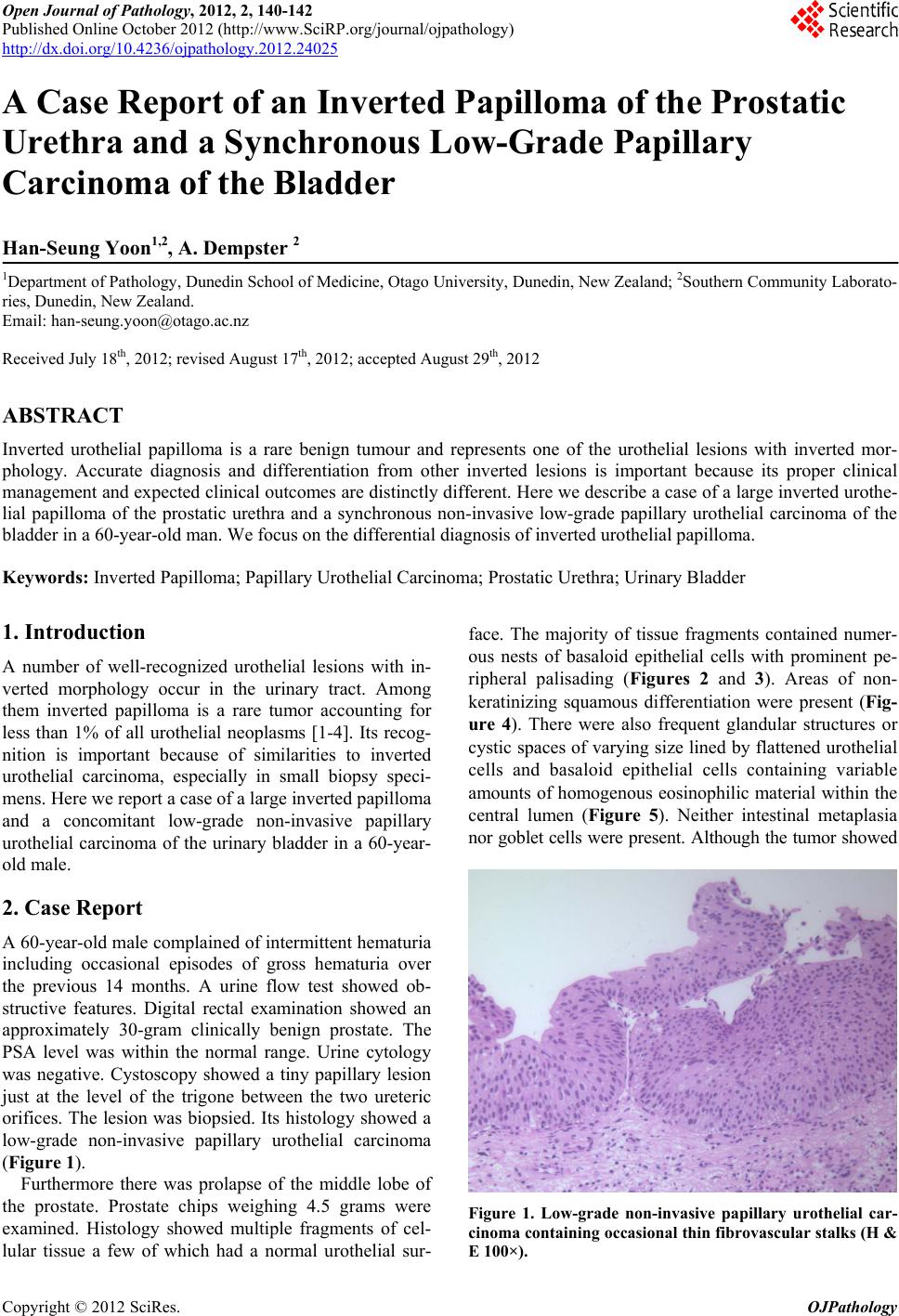 Open Journal of Pathology, 2012, 2, 140-142 Published Online October 2012 (http://www.SciRP.org/journal/ojpathology) http://dx.doi.org/10.4236/ojpathology.2012.24025 Copyright © 2012 SciRes. OJPathology 1 A Case Report of an Inverted Papilloma of the Prostatic Urethra and a Synchronous Low-Grade Papillary Carcinoma of the Bladder Han-Seung Yoon1,2, A. Dempster 2 1Department of Pathology, Dunedin School of Medicine, Otago University, Dunedin, New Zealand; 2Southern Community Laborato- ries, Dunedin, New Zealand. Email: [email protected] Received July 18th, 2012; revised August 17th, 2012; accepted August 29th, 2012 ABSTRACT Inverted urothelial papilloma is a rare benign tumour and represents one of the urothelial lesions with inverted mor- phology. Accurate diagnosis and differentiation from other inverted lesions is important because its proper clinical management and expected clinical outcomes are distinctly different. Here we describe a case of a large inverted urothe- lial papilloma of the prostatic urethra and a synchronous non-invasive low-grade papillary urothelial carcinoma of the bladder in a 60-year-old man. We focus on the differential diagnosis of inverted urothelial papilloma. Keywords: Inverted Papilloma; Papillary Urothelial Carcino ma; Prostatic Urethra; Urinary Bladder 1. Introduction A number of well-recognized urothelial lesions with in- verted morphology occur in the urinary tract. Among them inverted papilloma is a rare tumor accounting for less than 1% of all urothelial neoplasms [1-4]. Its recog- nition is important because of similarities to inverted urothelial carcinoma, especially in small biopsy speci- mens. Here we report a case of a large inverted papilloma and a concomitant low-grade non-invasive papillary urothelial carcinoma of the urinary bladder in a 60-year- old male. 2. Case Report A 60-year-old male complained o f intermittent hematuria including occasional episodes of gross hematuria over the previous 14 months. A urine flow test showed ob- structive features. Digital rectal examination showed an approximately 30-gram clinically benign prostate. The PSA level was within the normal range. Urine cytology was negative. Cystoscopy showed a tiny papillary lesion just at the level of the trigone between the two ureteric orifices. The lesion was biopsied. Its histology showed a low-grade non-invasive papillary urothelial carcinoma (Figure 1). Furthermore there was prolapse of the middle lobe of the prostate. Prostate chips weighing 4.5 grams were examined. Histology showed multiple fragments of cel- lular tissue a few of which had a normal urothelial sur- face. The majority of tissue fragments contained numer- ous nests of basaloid epithelial cells with prominent pe- ripheral palisading (Figures 2 and 3). Areas of non- keratinizing squamous differentiation were present (Fig- ure 4). There were also frequent glandular structures or cystic spaces of varying size lined by flattened urothelial cells and basaloid epithelial cells containing variable amounts of homogenous eosinophilic material within the central lumen (Figure 5). Neither intestinal metaplasia nor goblet cells were present. Although the tumor showed Figure 1. Low-grade non-invasive papillary urothelial car- cinoma containing occasional thin fibrovascular stalks (H & E 100×). 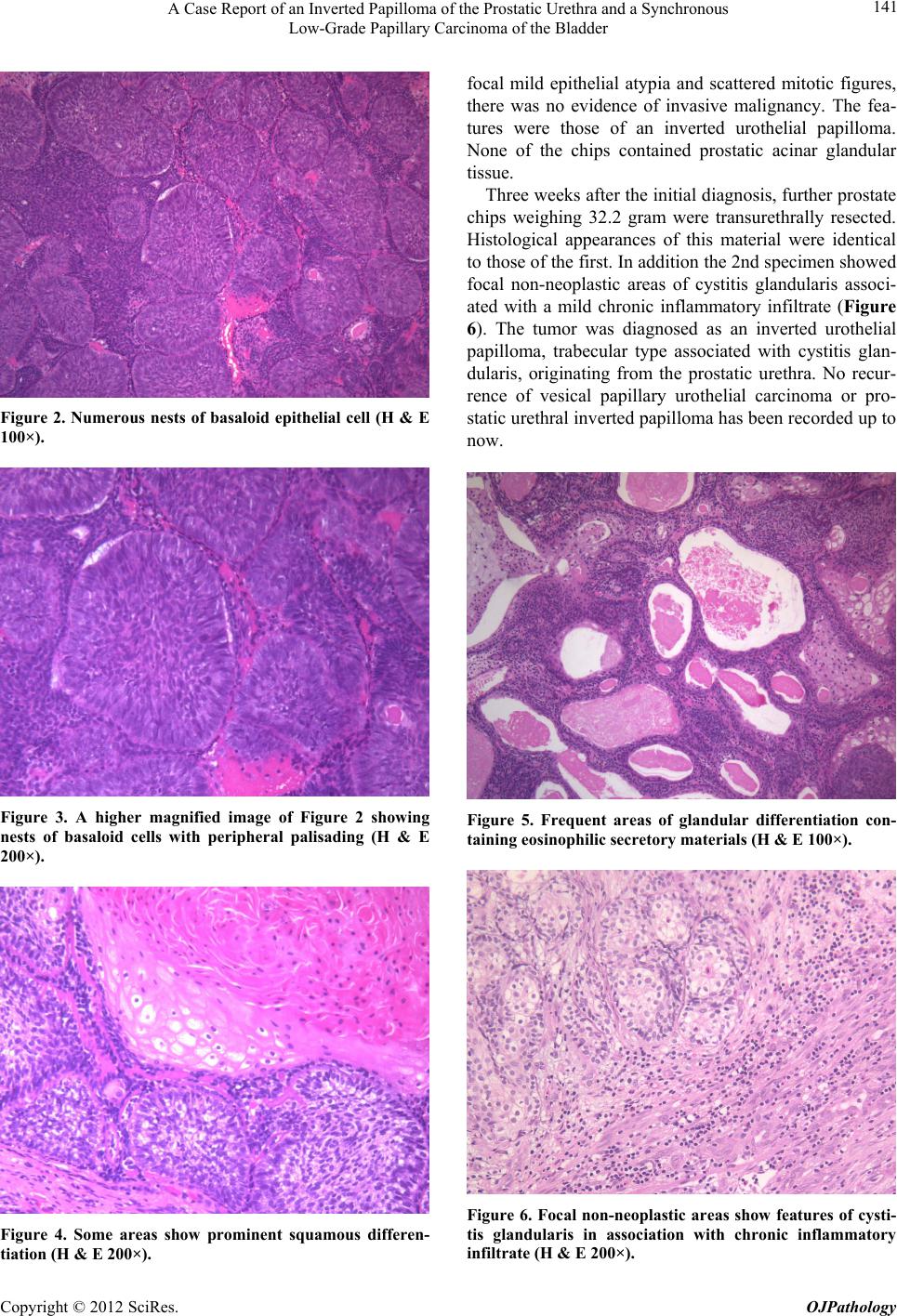 A Case Report of an Inverted Papilloma of the Prostatic Urethra and a Synchronous Low-Grade Papillary Carcinoma of the Bladder 141 Figure 2. Numerous nests of basaloid epithelial cell (H & E 100×). Figure 3. A higher magnified image of Figure 2 showing nests of basaloid cells with peripheral palisading (H & E 200×). Figure 4. Some areas show prominent squamous differen- tiation (H & E 200×). focal mild epithelial atypia and scattered mitotic figures, there was no evidence of invasive malignancy. The fea- tures were those of an inverted urothelial papilloma. None of the chips contained prostatic acinar glandular tissue. Three weeks after the initial d iagnosis, further prostate chips weighing 32.2 gram were transurethrally resected. Histological appearances of this material were identical to those of the first. In add ition th e 2nd sp ecimen sho w ed focal non-neoplastic areas of cystitis glandularis associ- ated with a mild chronic inflammatory infiltrate (Figure 6). The tumor was diagnosed as an inverted urothelial papilloma, trabecular type associated with cystitis glan- dularis, originating from the prostatic urethra. No recur- rence of vesical papillary urothelial carcinoma or pro- static urethral inverted papilloma has b een record ed up to now. Figure 5. Frequent areas of glandular differentiation con- taining eosinophilic secretory materials (H & E 100×). Figure 6. Focal non-neoplastic areas show features of cysti- tis glandularis in association with chronic inflammatory infiltrate (H & E 200×). Copyright © 2012 SciRes. OJPathology  A Case Report of an Inverted Papilloma of the Prostatic Urethra and a Synchronous Low-Grade Papillary Carcinoma of the Bladder Copyright © 2012 SciRes. OJPathology 142 3. Discussion Inverted papilloma of the urinary tract is a rare benign tumor most commonly diagnosed in older men present- ing with hematuria or symptoms of lower urinary tract obstruction [3,4]. It is most frequently identified in the bladder neck or trigone as a polypoid growth with a smooth surface. These lesions are usually small (<3 cm) but can be large. Most are solitary although 1% - 4% may be multifocal. Histologically there are two main subtypes of inverted papillomas; trabecular and glandular [1]. The former is characterized by widely branched, anastomosing cords of urothelial cells originating directly from the overlying transitional epithelium. The latter is characterized by multiple round to oval islands of proliferating urothelial cells together with pseudoglandular and true glandular structures, which are often connected with the surface urothelium. Although inverted papilloma is generally regarded to be a benign neoplasm, sporadic cases of inverted papil- loma with malignant features have been reported raising concern that inverted papilloma may be a precursor le- sion of utorhelial malignancy [5-8]. However most of these patients had a history of previous or concurrent urothelial carcinoma. Inverted papilloma is associated with a low risk of recurrence (5%), in marked contrast to the high recurrence rates of papillary urothelial carci- noma [3]. Sung et al. [2] reported only one recurrence among 75 patients with inverted papilloma (1.3%) during a mean follow-up of 68 months after treatment. The au- thors recommend that complete transurethral resection appears to be adequate surgical therapy for inverted papilloma [2]. The present case showed an inverted papilloma of the prostatic urethra and a synchronous low-grade non-inva- sive papillary urothelial carcinoma of the bladder. Spo- radic cases with inverted papilloma have been reported to show metachronous or synchronou s urothelial carcinoma. Brown and Cohen in a series of 41 cases of inverted papilloma reported 2 patients (4.9%) had a history of urothelial carcinoma and one of the 2 (2.4%) was diag- nosed with concomitant urothelial carcinoma [4]. The authors recommend ed postoperativ e surveillance because 2 patients developed urothelial carcinoma among 25 pa- tients during a 9 - 25 months cystoscopic follow-up for non-recurrent inverted papilloma. Differential diagnosis of inverted papilloma from other inverted urothelial lesions rests primarily on morpho- logical criteria. Recognition of the possibility o f inverted papilloma in the differential diagnosis remains the best safeguard against incorrect diagnosis [9]. The differential diagnosis includes non-neoplastic lesions such as von Brunn’s nest, florid von Brunn’s nest proliferation, cysti- tis cystica and cystitis glandularis as well as neoplastic lesions such as inverted urothelial carcinoma, nested variant of urothelial carcinoma and verrucous squamous cell carcinoma. REFERENCES [1] E. Kunze, A. Schauer and M. Schmitt, “Histology and Histogenesis of Two Different Types of Inverted Uro- thelial Pa pillomas,” Cancer, Vol. 51, 1983, pp. 348-358. doi:10.1002/1097-0142(19830115)51:2<348::AID-CNCR 2820510231>3.0.CO;2-O [2] M. T. Sung, G. T. MacLennan, A. Lopez-Beltran, R. Montironi and L. Cheng, “Natural History of Urothelial Inverted Papilloma,” Cancer, Vol. 107, No. 11, 2006, pp. 2622-2627. doi:10.1002/cncr.22311 [3] T. D. Jones, S. Zhang, A. Lopez-Beltran, J. N. Eble, M.-T., Sung, G. T. MacLennan, R. Montironi, P.-H. Tan, S. Zheng, L. A. Baldridge and L. Cheng, “Urothelial Car- cinoma with an Invereted Growth Pattern Can Be Dis- tinguished from Inverted Papilloma by Fluorescence in Situ Hybridization, Immunohistochemistry, and Mor- phologic Analysis,” American Journal of Surgical Patho- logy, Vol. 31, No. 12, 2007, pp. 1861-1867. [4] K. B. Hodges, A. Lopez-Beltran, G. T. MacLennan, R. Montironi and L. Cheng, “Urothel ial Lesions with In v e rt e d Growth Patterns: Histogenesis, Molecular Genetic Findings, Differential Diagnosis and Clinical Management,” BJU International, Vol. 107, No. 4, 2010, pp. 532-537. doi:10.1111/j.1464-410X.2010.09853.x [5] K. Asano, J. Miki, S. Maeda, T. Naruoka, H. Takahasi and Y. Oishi, “Clinical Studies on Inverted Papilloma of the Urinary Tract: Report of 48 Cases and Review of the Literature,” Journal of Urology, Vol. 170, No. 4, 2003, pp. 1209-1212. doi:10.1097/01.ju.0000085342.15918.d7 [6] S. Picozzi, S. Casellato, G. Bozzini, D. Ratti, A. Macchi, B. Rubino, G. Pace and L. Carmignani, “Inverted Papil- loma of the Bladder: A Review and an Analysis of the Recent Literature of 365 Patients,” Urologic Oncology, 2012, in Press. doi:10.1016/j.urolonc.2012.03.009 [7] M. Eiber, J. M. van Oers, E. C. Zwarthoff, et al., “Low Frequency of Molecular Changes and Tumor Recurrence in Inverted Papillomas of the Urinary Tract,” American Journal of Surgical Pathology, Vol. 31, No. 6, 2007, pp. 938-946. doi:10.1097/01.pas.0000249448.13466.75 [8] L. F. Altaffer III, S. Y. Wilkerson, G. H. Jordan and D. F. Lynch, “Malignant Inverted Papilloma and Carcinoma in Situ of the Bladder,” Journal of Urology, Vol. 128, No. 4, 1982, pp. 816-822. [9] A. L. Brown and R. J. Cohen, “Inverted Papilloma of the Urinaru Bladder,” BJU International, Vol. 107, Suppl. 3, 2011, pp. 24-26. doi:10.1111/j.1464-410X.2011.10046.x |