

Paper Menu >>
Journal Menu >>
 Journal of Biomaterials and Nanobiotechnology, 2012, 3, 413-420 http://dx.doi.org/10.4236/jbnb.2012.34041 Published Online October 2012 (http://www.SciRP.org/journal/jbnb) 413 Formation of Calcium Carbonate Polymorphs Induced by Living Microalgae Giulia Santomauro*, Johannes Baier, Wanjing Huang, Stefan Pezold, Joachim Bill Institute for Materials Science, University of Stuttgart, Stuttgart, Germany. Email: *[email protected] Received August 7th, 2012; revised September 17th, 2012; accepted September 26th, 2012 ABSTRACT Calcium carbonate (CaCO3) occurs in the three polymorphs calcite, aragonite and vaterite. The formation of these crys- tals in inorganic solutions is influenced by parameters like pH, temperature or impurities. Living freshwater microalgae can also induce the formation of CaCO3 when they live in a suitable environment containing saturated amounts of Ca2+. Through this biologically induced biomineralization only the formation of the polymorph calcite has been reported yet. We investigated the precipitates which have been formed in solutions containing the freshwater microalgae Scenedes- mus obliquus and different zinc amounts (0, 3.27 and 6.53 mg Zn2+/l) by XRD and SEM. As references precipitates from the same solutions but without algae were investigated. We could show that the presence of living microalgae has a great influence on the precipitation of calcium carbonate crystals. In algae-containing media without or with a low zinc amount always calcite and aragonite are formed. In the corresponding medium with 6.53 mg Zn2+/l pure aragonite crystals were built. In contrast, in the inorganic, algae-free solutions without zinc, pure calcite is precipitated. Both in- organic solutions with zinc show major calcite precipitation and weak aragonite precipitation. Thus the algae cells ad- vance significantly the formation of aragonite, which is enhanced by the presence of zinc cations in the media. Possible mechanisms are discussed. Keywords: CaCO3 Polymorphs; Biomineralization; Zinc; Microalgae; XRD 1. Introduction The formation of the three calcium carbonate (CaCO3) polymorphs calcite, aragonite and vaterite in inorganic solutions has been the topic of many studies [1-4]. It was proposed that first an unstable amorphous phase is built which transforms to the metastable phase vaterite or ara- gonite, followed by the transformation to the stable phase calcite [1]. The precipitation of the polymorphs can be affected by different factors like temperature, pH of the medium, concentration ratio of individual components, supersaturation, ionic strength or impurities [3]. The in- vestigations of Ogino et al. (1987) [1] revealed that ara- gonite formation is favored at higher temperatures (>40˚C) in aqueous solution, which has also been sup- ported in other experiments [4]. At room temperature, the pH of the solution in a constant-composition environ- ment is the most important factor for the formation of the polymorphs [3]. At 24˚C, vaterite is the major product in the pH range between 8.5 and 10. Aragonite preferably formed at pH 11; when the pH is higher than 12, calcite is the dominant product. The precipitation of the different polymorphs can also be modified by impurities in the solution. When the ionic radius of the impurities is smaller than that of the Ca2+ cations, what is the case for Zn2+ cations [5], aragonite is deposited [6]. This influ- ence is explained by adsorption phenomena on the crys- talline faces of the nuclei. Kitano et al. (1976) [7] exam- ined specifically the adsorption behavior of Zn2+ on CaCO3 polymorphs in aqueous solutions. They showed that Zn2+ is strongly adsorbed on aragonite crystals in comparison to other polymorphs. It was assumed that the zinc ions inhibit the transformation of aragonite to calcite [1,7]. The Zn2+ adsorption on the calcite surface occurs via an exchange of Ca2+ in a surface-adsorbed layer [8]. Also organic additives show an effect on the minerali- zation of calcium carbonate (reviews in: Meldrum, 2003 [9]; Ren et al., 2011 [10]). Amino acids like glycine, aspartic acid and glutamic acid or polysaccharides like cellulose influence the precipitation and morphology of calcite and vaterite [11-13]. Living organisms like photosynthetic microalgae can induce the precipitation of CaCO3 through biominerali- zation. It is known that many freshwater algae build cal- cite crystals when they live in a supersaturated environ- ment regarding calcium [14,15]. This is a result of the *Corresponding author. Copyright © 2012 SciRes. JBNB 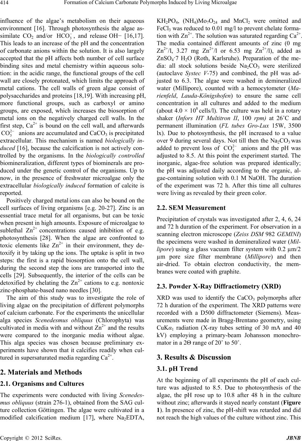 Formation of Calcium Carbonate Polymorphs Induced by Living Microalgae 414 influence of the algae’s metabolism on their aqueous environment [16]. Through photosynthesis the algae as- similate CO2 and/or 3, and release OH− [16,17]. This leads to an increase of the pH and the concentration of carbonate anions within the solution. It is also largely accepted that the pH affects both number of cell surface binding sites and metal chemistry within aqueous solu- tion: in the acidic range, the functional groups of the cell wall are closely protonated, which limits the approach of metal cations. The cell walls of green algae consist of polysaccharides and proteins [18,19]. With increasing pH, more functional groups, such as carboxyl or amino groups, are exposed, which increases the biosorption of metal ions on the negatively charged cell walls. In the first step, Ca2+ is bound on the cell wall, and afterwards anions are accumulated and CaCO3 is precipitated extracellular. This mechanism is named biologically in- duced [16], because the calcification is not actively con- trolled by the organisms. In the biologically controlled biomineralization, different types of biominerals are pro- duced under the genetic control of the organisms. Up to now, in the presence of freshwater microalgae only the extracellular biologically induced formation of calcite is reported. HCO 2 3 CO Positively charged metal ions can also be bound on the cell surfaces of living organisms [e.g. 20-27]. Zinc is an essential trace metal for all organisms, but can be toxic when present in high amounts. Exposure of microalgae to sublethal Zn2+ concentrations caused inhibition of e.g. photosynthesis [28]. When the algae are confronted to toxic elements like Zn2+ in their environment, they de- toxify it by taking up the ions. The uptake is split in two steps: the first is a rapid biosorption onto the cell wall, during the second step the ions are transported into the cells [29]. Subsequently, the interior of the cells can be detoxified by chelating the Zn2+ cations to e.g. nontoxic zinc-phosphate-based nano needles [30]. The aim of this study was to investigate the role of living algae on the precipitation of different polymorphs of calcium carbonate. For the experiments the unicellular alga species Scenedesmus obliquus (Chlorophyta) was cultivated in media with and without Zn2+ and the results were compared to the inorganic media without algae. This alga species was chosen because preliminary ex- periments have shown that it calcifies readily when cul- tured in supersaturated media regarding Ca2+. 2. Materials and Methods 2.1. Organisms and Cultures The experiments were conducted with living Scenedes- mus obliquus (strain 276-1), obtained from the SAG cul- ture collection Göttingen. The algae were cultivated in a modified calcification medium [17], where Na2EDTA, KH2PO4, (NH4)Mo7O24 and MnCl2 were omitted and FeCl3 was reduced to 0.01 mg/l to prevent chelate forma- tion with Zn2+. The solution was saturated regarding Ca2+. The media contained different amounts of zinc (0 mg Zn2+/l, 3.27 mg Zn2+/l or 6.53 mg Zn2+/l), added as ZnSO4·7 H2O (Roth, Karlsruhe). Preparation of the me- dia: all stock solutions beside Na2CO3 were sterilized (autoclave Systec V-75) and combined, the pH was ad- justed to 6.3. The algae were washed in demineralized water (Millipore), counted with a hemocytometer (Ma- rienfeld, Lauda-Königshofen) to ensure the same cell concentration in all cultures and added to the medium (about 4.0 × 10⁸ cells/l). The culture was held in a rotary shaker (Infors HT Multitron II, 100 rpm) at 26˚C and permanent illumination (FL tubes Gro-Lux 15W, 3500 lx). Due to photosynthesis, the pH increased to a value over 9 during several days. Not till then the Na2CO3 was added to prevent loss of anions and the pH was adjusted to 8.5. At this point the experiment started. The inorganic, algae-free solution was prepared identically; the pH was adjusted daily according to the organic, al- gae-containing solution with 0.1 M NaOH. The duration of the experiment was 72 h. After this time all cultures were living as revealed by their green color. 2 3 CO 2.2. SEM Measurement Precipitation of crystals was investigated after 2, 4, 6, 24 and 72 h duration of the experiment. For observation in a scanning electron microscope (Zeiss DSM 982 GEMINI) the specimens were washed in demineralized water (Mil- lipore) using a glass vacuum filter system with 0.2 µm/2 µm pore size filter membrane (Millipore) and then air-dried. To obtain electron conductivity, the mem- branes were coated with graphite. 2.3. Powder X-Ray Diffractiometry (XRD) XRD was used to identify the CaCO3 polymorphs after 72 h duration of the experiment. The XRD patterns were recorded with a D500 diffractometer (Siemens). Meas- urements were made in Bragg-Brentano geometry, using CuKα1 radiation (X-ray tubes setting of 30 mA and 40 kV) employing a primary-beam Johansson monochro- mator in a 2Θ range of 20˚ to 50˚. 3. Results & Discussion 3.1. pH Trend At the beginning of all experiments the pH of each cul- ture was adjusted to 8.5. Due to photosynthesis of the algae, the pH rose up to 10.8 after 48 h in the culture without zinc; afterwards it stayed nearly constant (Figure 1). In presence of zinc, the pH-shift was retarded and did not reach the high values of the culture without zinc. This Copyright © 2012 SciRes. JBNB 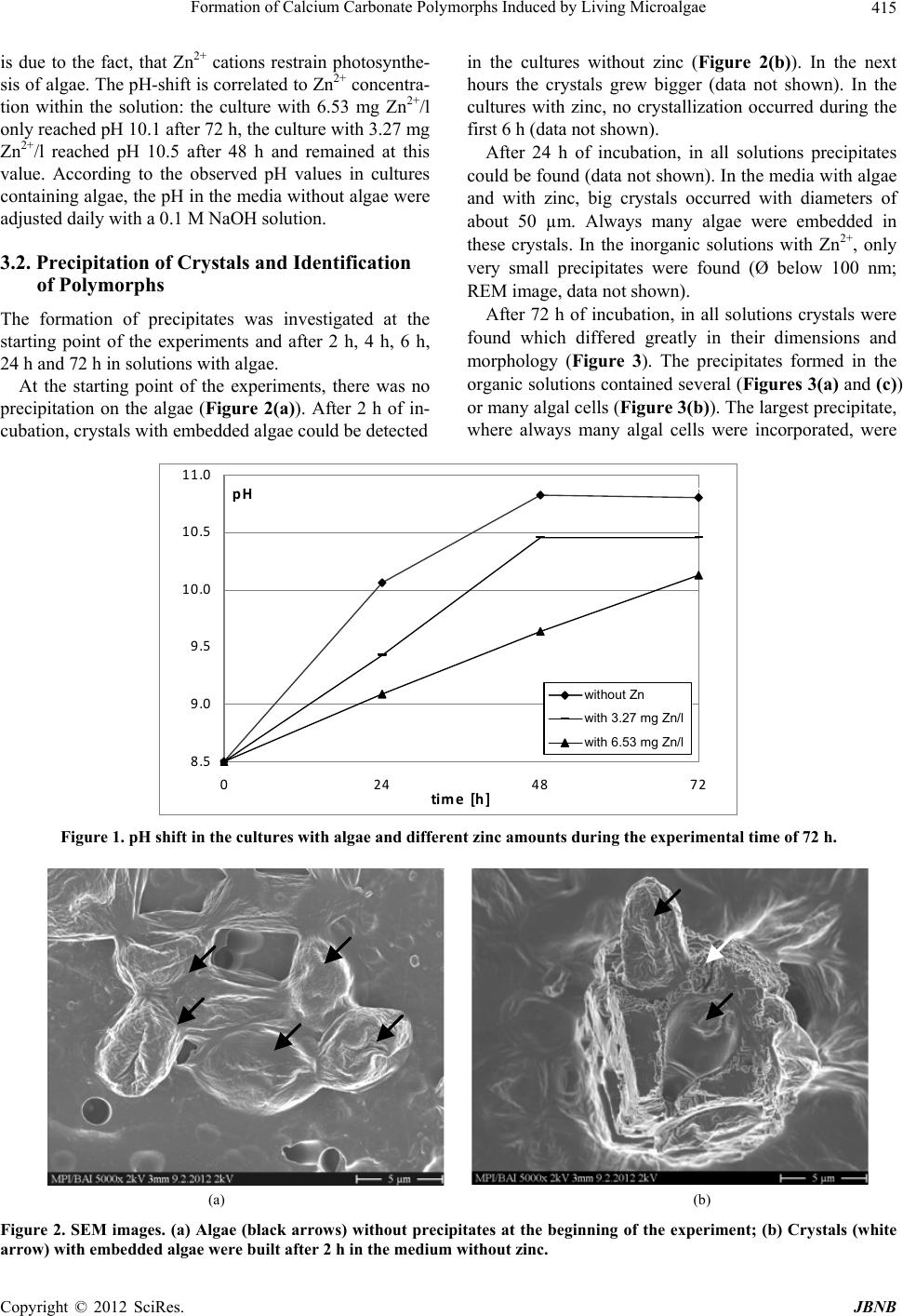 Formation of Calcium Carbonate Polymorphs Induced by Living Microalgae Copyright © 2012 SciRes. JBNB 415 is due to the fact, that Zn2+ cations restrain photosynthe- sis of algae. The pH-shift is correlated to Zn2+ concentra- tion within the solution: the culture with 6.53 mg Zn2+/l only reached pH 10.1 after 72 h, the culture with 3.27 mg Zn2+/l reached pH 10.5 after 48 h and remained at this value. According to the observed pH values in cultures containing algae, the pH in the media without algae were adjusted daily with a 0.1 M NaOH solution. in the cultures without zinc (Figure 2(b)). In the next hours the crystals grew bigger (data not shown). In the cultures with zinc, no crystallization occurred during the first 6 h (data not shown). After 24 h of incubation, in all solutions precipitates could be found (data not shown). In the media with algae and with zinc, big crystals occurred with diameters of about 50 µm. Always many algae were embedded in these crystals. In the inorganic solutions with Zn2+, only very small precipitates were found (Ø below 100 nm; REM image, data not shown). 3.2. Precipitation of Crystals and Identification of Polymorphs After 72 h of incubation, in all solutions crystals were found which differed greatly in their dimensions and morphology (Figure 3). The precipitates formed in the organic solutions contained several (Figures 3(a) and (c)) or many algal cells (Figure 3(b)). The largest precipitate, where always many algal cells were incorporated, were The formation of precipitates was investigated at the starting point of the experiments and after 2 h, 4 h, 6 h, 24 h and 72 h in solutions with algae. At the starting point of the experiments, there was no precipitation on the algae (Figure 2(a)). After 2 h of in- cubation, crystals with embedded algae could be detected 8.5 9.0 9.5 10.0 10.5 11.0 0 24487 time [h] pH 2 w ithout Zn with 3.27 mg Zn/l with 6.53 mg Zn/l wit h ou t Zn with 3.27 mg Zn/l with 6.53 mg Zn/l Figure 1. pH shift in the cultures with algae and different zinc amounts during the experimental time of 72 h. (a) (b) Figure 2. SEM images. (a) Algae (black arrows) without precipitates at the beginning of the experiment; (b) Crystals (white arrow) with embedded algae were built afte r 2 h in the medium without zinc. 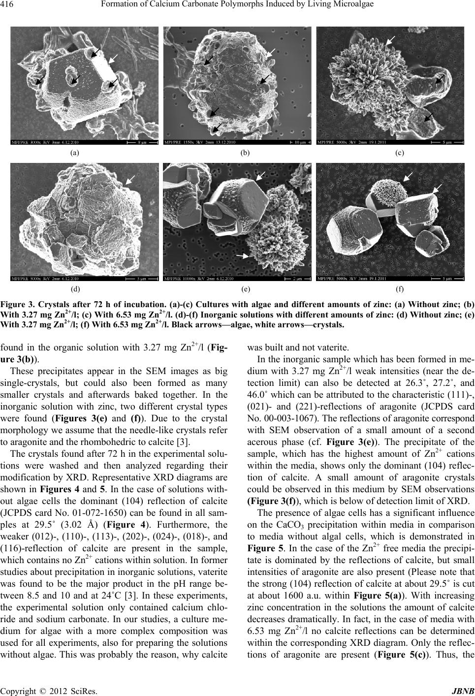 Formation of Calcium Carbonate Polymorphs Induced by Living Microalgae 416 (a) (b) (c) (d) (e) (f) Figure 3. Crystals after 72 h of incubation. (a)-(c) Cultures with algae and different amounts of zinc: (a) Without zinc; (b) With 3.27 mg Zn2+/l; (c) With 6.53 mg Zn2+/l. (d)-(f) Inorganic solutions with different amounts of zinc: (d) Without zinc; (e) With 3.27 mg Zn2+/l; (f) With 6.53 mg Zn2+/l. Black arrows—algae, white arrows—crystals. found in the organic solution with 3.27 mg Zn2+/l (Fig- ure 3(b)). These precipitates appear in the SEM images as big single-crystals, but could also been formed as many smaller crystals and afterwards baked together. In the inorganic solution with zinc, two different crystal types were found (Figures 3(e) and (f)). Due to the crystal morphology we assume that the needle-like crystals refer to aragonite and the rhombohedric to calcite [3]. The crystals found after 72 h in the experimental solu- tions were washed and then analyzed regarding their modification by XRD. Representative XRD diagrams are shown in Figures 4 and 5. In the case of solutions with- out algae cells the dominant (104) reflection of calcite (JCPDS card No. 01-072-1650) can be found in all sam- ples at 29.5˚ (3.02 Å) (Figure 4). Furthermore, the weaker (012)-, (110)-, (113)-, (202)-, (024)-, (018)-, and (116)-reflection of calcite are present in the sample, which contains no Zn2+ cations within solution. In former studies about precipitation in inorganic solutions, vaterite was found to be the major product in the pH range be- tween 8.5 and 10 and at 24˚C [3]. In these experiments, the experimental solution only contained calcium chlo- ride and sodium carbonate. In our studies, a culture me- dium for algae with a more complex composition was used for all experiments, also for preparing the solutions without algae. This was probably the reason, why calcite was built and not vaterite. In the inorganic sample which has been formed in me- dium with 3.27 mg Zn2+/l weak intensities (near the de- tection limit) can also be detected at 26.3˚, 27.2˚, and 46.0˚ which can be attributed to the characteristic (111)-, (021)- and (221)-reflections of aragonite (JCPDS card No. 00-003-1067). The reflections of aragonite correspond with SEM observation of a small amount of a second acerous phase (cf. Figure 3(e)). The precipitate of the sample, which has the highest amount of Zn2+ cations within the media, shows only the dominant (104) reflec- tion of calcite. A small amount of aragonite crystals could be observed in this medium by SEM observations (Figure 3(f)), which is below of detection limit of XRD. The presence of algae cells has a significant influence on the CaCO3 precipitation within media in comparison to media without algal cells, which is demonstrated in Figure 5. In the case of the Zn2+ free media the precipi- tate is dominated by the reflections of calcite, but small intensities of aragonite are also present (Please note that the strong (104) reflection of calcite at about 29.5˚ is cut at about 1600 a.u. within Figure 5(a)). With increasing zinc concentration in the solutions the amount of calcite decreases dramatically. In fact, in the case of media with 6.53 mg Zn2+/l no calcite reflections can be determined within the corresponding XRD diagram. Only the reflec- tions of aragonite are presen(Figure 5(c)). Thus, the t Copyright © 2012 SciRes. JBNB 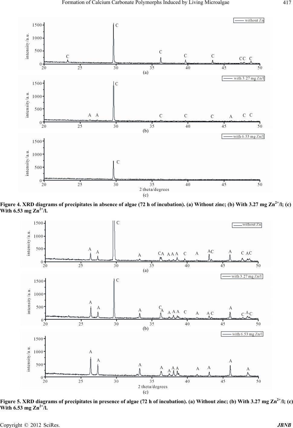 Formation of Calcium Carbonate Polymorphs Induced by Living Microalgae 417 (a) (b) (c) Figure 4. XRD diagrams of precipitates in absence of algae (72 h of incubation). (a) Without zinc; (b) With 3.27 mg Zn2+/l; (c) With 6.53 mg Zn2+/l. (a) (b) (c) Figure 5. XRD diagrams of precipitates in presence of algae (72 h of incubation). (a) Without zinc; (b) With 3.27 mg Zn2+/l; (c) With 6.53 mg Zn2+/l. Copyright © 2012 SciRes. JBNB 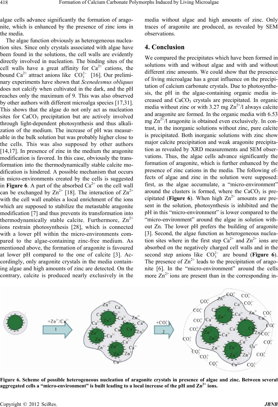 Formation of Calcium Carbonate Polymorphs Induced by Living Microalgae 418 algae cells advance significantly the formation of arago- nite, which is enhanced by the presence of zinc ions in the media. The algae function obviously as heterogeneous nuclea- tion sites. Since only crystals associated with algae have been found in the solutions, the cell walls are evidently directly involved in nucleation. The binding sites of the cell walls have a great affinity for Ca2+ cations, the bound Ca2+ attract anions like [16]. Our prelimi- nary experiments have shown that Scenedesmus obliquus does not calcify when cultivated in the dark, and the pH reaches only the maximum of 9. This was also observed by other authors with different microalga species [17,31]. This shows that the algae do not only act as nucleation sites for CaCO3 precipitation but are actively involved through light-dependent photosynthesis and thus alkali- zation of the medium. The increase of pH was measur- able in the bulk solution but was probably higher close to the cells. This was also supposed by other authors [14,17]. In presence of zinc in the medium the aragonite modification is favored. In this case, obviously the trans- formation into the thermodynamically stable calcite mo- dification is hindered. A possible mechanism that occurs in micro-environments created by the cells is suggested in Figure 6. A part of the absorbed Ca2+ on the cell wall can be exchanged by Zn2+ [18]. The interaction of Zn2+ with the cell wall enables a local enrichment of the ions which are supposed to stabilize the metastable aragonite modification [7] and thus prevents its transformation into thermodynamically stable calcite. Furthermore, Zn2+ ions restrain photosynthesis [28], which is connected with a lower pH within the micro-environments com- pared to the algae-containing zinc-free medium. As mentioned above, the formation of aragonite is favoured at lower pH compared to the one of calcite [3]. Ac- cordingly, only aragonite crystals in the media contain- ing algae and high amounts of zinc are detected. On the contrary, calcite is produced nearly exclusively in the 2 3 CO media without algae and high amounts of zinc. Only traces of aragonite are produced, as revealed by SEM observations. 4. Conclusion We compared the precipitates which have been formed in solutions with and without algae and with and without different zinc amounts. We could show that the presence of living microalgae has a great influence on the precipi- tation of calcium carbonate crystals. Due to photosynthe- sis, the pH in the algae-containing organic media in- creased and CaCO3 crystals are precipitated. In organic media without zinc or with 3.27 mg Zn2+/l always calcite and aragonite are formed. In the organic media with 6.53 mg Zn2+/l aragonite is obtained even exclusively. In con- trast, in the inorganic solutions without zinc, pure calcite is precipitated. Both inorganic solutions with zinc show major calcite precipitation and weak aragonite precipita- tion as revealed by XRD measurements and SEM obser- vations. Thus, the algae cells advance significantly the formation of aragonite, which is further enhanced by the presence of zinc cations in the media. The following ef- fects of algae and zinc in the solution were supposed: first, as the algae accumulate, a “micro-environment” around the clusters is formed, where the CaCO3 is pre- cipitated (Figure 6). When high Zn2+ amounts are pre- sent in the solution, photosynthesis is inhibited and the pH in this “micro-environment” is lower compared to the “micro-environment” around the algae in solution with- out Zn. The lower pH prefers the building of aragonite [3]. Second, the algae function as heterogeneous nuclea- tion sites where in the first step Ca2+ and Zn2+ ions are absorbed on the negatively charged cell walls and in the second step anions like are bound (Figure 6). The presence of Zn2+ leads to the precipitation of arago- nite [6]. In the “micro-environment” around the cells more Zn2+ ions are present than in the corresponding in- 2 3 CO Figure 6. Scheme of possible heterogeneous nucleation of aragonite crystals in presence of algae and zinc. Between several ggregated cells a “micro-environment” is built leading to a local increase of the pH and Zn2+ ions. a Copyright © 2012 SciRes. JBNB 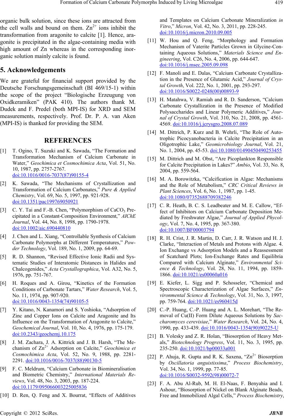 Formation of Calcium Carbonate Polymorphs Induced by Living Microalgae 419 organic bulk solution, since these ions are attracted from the cell walls and bound on them. Zn2+ ions inhibit the transformation from aragonite to calcite [1]. Hence, ara- gonite is precipitated in the algae-containing media with high amount of Zn whereas in the corresponding inor- ganic solution mainly calcite is found. 5. Acknowledgements We are grateful for financial support provided by the Deutsche Forschungsgemeinschaft (BI 469/15-1) within the scope of the project “Biologische Erzeugung von Oxidkeramiken” (PAK 410). The authors thank M. Dudek and F. Predel (both MPI-IS) for XRD and SEM measurements, respectively. Prof. Dr. P. A. van Aken (MPI-IS) is thanked for providing the SEM. REFERENCES [1] T. Ogino, T. Suzuki and K. Sawada, “The Formation and Transformation Mechanism of Calcium Carbonate in Water,” Geochimica et Cosmochimica Acta, Vol. 51, No. 10, 1987, pp. 2757-2767. doi:10.1016/0016-7037(87)90155-4 [2] K. Sawada, “The Mechanisms of Crystallization and Transformation of Calcium Carbonates,” Pure & Applied Chemistry, Vol. 69, No. 5, 1997, pp. 921-928. doi:10.1351/pac199769050921 [3] C. Y. Tai and F.-B. Chen, “Polymorphism of CaCO3 Pre- cipitated in a Constant-Composition Environment,” AIChE Journal, Vol. 44, No. 8, 1998, pp. 1790-1978. doi:10.1002/aic.690440810 [4] J. Chen and L. Xiang, “Controllable Synthesis of Calcium Carbonate Polymorphs at Different Temperatures,” Pow- der Technology, Vol. 189, No. 1, 2009, pp. 64-69. [5] R. D. Shannon, “Revised Effective Ionic Radii and Sys- tematic Studies of Interatomic Distances in Halides and Chalcogenides,” Acta Crystallographica, Vol. A32, No. 5, 1976, pp. 751-767. [6] H. Roques and A. Girou, “Kinetics of the Formation Conditions of Carbonate Tartars,” Water Research, Vol. 5, No. 11, 1974, pp. 907-920. doi:10.1016/0043-1354(74)90105-5 [7] Y. Kitano, N. Kanamori and S. Yoshioka, “Adsorption of Zinc and Copper Ions on Calcite and Aragonite and Its Influence on the Transformation of Aragonite to Calcite,” Geochemical Journal, Vol. 10, No. 4, 1976, pp. 175-179. doi:10.2343/geochemj.10.175 [8] J. M. Zachara, J. A. Kittrick and J. B. Harsh, “The Me- chanism of Zn2+ Adsorption on Calcite,” Geochimica et Cosmochimica Acta, Vol. 52, No. 9, 1988, pp. 2281- 2291. doi:10.1016/0016-7037(88)90130-5 [9] F. C. Meldrum, “Calcium Carbonate in Biomineralisation and Biometric Chemistry,” International Materials Re- views, Vol. 48, No. 3, 2003, pp. 187-224. doi:10.1179/095066003225005836 [10] D. Ren, Q. Feng and X. Bourrat, “Effects of Additives and Templates on Calcium Carbonate Mineralization in Vitro,” Micron, Vol. 42, No. 3, 2011, pp. 228-245. doi:10.1016/j.micron.2010.09.005 [11] W. Hou and Q. Feng, “Morphology and Formation Mechanism of Vaterite Particles Grown in Glycine-Con- taining Aqueous Solutions,” Materials Science and En- gineering, Vol. C26, No. 4, 2006, pp. 644-647. doi:10.1016/j.msec.2005.09.098 [12] F. Manoli and E. Dalas, “Calcium Carbonate Crystalliza- tion in the Presence of Glutamic Acid,” Journal of Crys- tal Growth, Vol. 222, No. 1, 2001, pp. 293-297. doi:10.1016/S0022-0248(00)00893-9 [13] H. Matahwa, V. Ramiah and R. D. Sanderson, “Calcium Carbonate Crystallization in the Presence of Modified Polysaccharides and Linear Polymeric Additives,” Jour- nal of Crystal Growth, Vol. 310, No. 21, 2008, pp. 4561- 4569. doi:10.1016/j.jcrysgro.2008.07.089 [14] M. Dittrich, P. Kurz and B. Wehrli, “The Role of Auto- trophic Picocyanobacteria in Calcite Precipitation in an Oligotrophic Lake,” Geomicrobiology Journal, Vol. 21, No. 1, 2004, pp. 45-53. doi:10.1080/01490450490253455 [15] M. Dittrich and M. Obst, “Are Picoplankton Responsible for Calcite Precipitation in Lakes?” Ambio, Vol. 33, No. 8, 2004, pp. 559-564. [16] M. A. Borowitzka, “Calcification in Algae: Mechanisms and the Role of Metabolism,” CRC Critical Reviews in Plant Sciences, Vol. 6, No. 1, 1987, pp. 1-45. doi:10.1080/07352688709382246 [17] C. R. Heath, B. C. S. Leadbeater and M. E. Callow, “Ef- fect of Inhibitors on Calcium Carbonate Deposition Me- diated by Freshwater Algae,” Journal of Applied Phycol- ogy, Vol. 7, No. 4, 1995, pp. 367-380. doi:10.1007/BF00003794 [18] R. H. Crist, J. R. Martin, D. Carr, J. R. Watson and H. J. Clarke, “Interaction of Metals and Protons with Algae. 4. Ion Exchange vs Adsorption Models and a Reassessment of Scatchard Plots; Ion-Exchange Rates and Equilibria Compared with Calcium Alginate,” Environmental Sci- ence & Technology, Vol. 28, No. 11, 1994, pp. 1859- 1866. doi:10.1021/es00060a016 [19] E. Kiefer, L. Sigg and P. Schosseler, “Chemical and Spectroscopic Characterization of Algae Surfaces,” En- vironmental Science & Technology, Vol. 31, No. 3, 1997, pp. 759-764. doi:10.1021/es960415d [20] C.-P. Huang, C.-P. Huang and A. L. Morehart, “The Re- moval of Cu(II) Form Dilute Aqueous Solutions by Sac- charomyces cerevisiae,” Water Research, Vol. 24, No. 4, 1990, pp. 433-439. doi:10.1016/0043-1354(90)90225-U [21] B. Volesky and Z. R. Holan, “Biosorption of Heavy Met- als,” Biotechnology Progress, Vol. 11, No. 3, 1995, pp. 235-250. doi:10.1021/bp00033a001 [22] P. Ahuja, R. Gupta and R. K. Saxena, “Zn2+ Biosorption by Oscillatoria anguistissima,” Process Biochemistry, Vol. 34, No. 1, 1999, pp. 77-85. doi:10.1016/S0032-9592(98)00072-7 [23] F. A. Abu Al-Rub, M. H. El-Naas, F. Benyahia and I. Ashour, “Biosorption of Nickel on Blank Alginate Beads, Free and Immobilized Algal Cells,” Process Biochemistry, Copyright © 2012 SciRes. JBNB  Formation of Calcium Carbonate Polymorphs Induced by Living Microalgae 420 Vol. 39, No. 11, 2004, pp. 1767-1773. doi:10.1016/j.procbio.2003.08.002 [24] K. Chojnacka, A. Chojnacki and H. Górecka, “Biosorp- tion of Cr3+, Cd2+ and Cu2+ Ions by Blue-Green Algae Spirulina sp.: Kinetics, Equilibrium and the Mechanism of the Process,” Chemosphere, Vol. 59, No. 1, 2005, pp. 75-84. doi:10.1016/j.chemosphere.2004.10.005 [25] R. Gong, Y. Ding, H. Liu, Q. Chen and Z. Liu, “Lead Biosorption and Desorption by Intact and Pretreated Spirulina Maxima Biomass,” Chemosphere, Vol. 58, No. 1, 2005, pp. 125-130. doi:10.1016/j.chemosphere.2004.08.055 [26] L. Deng, Y. Su, H. Su, X. Wang and X. Zhu, “Sorption and Desorption of Lead (II) from Wastewater by Green Algae Cladophora fascicularis,” Journal of Hazardous Materials, Vol. 143, No. 1-2, 2007, pp. 220-225. doi:10.1016/j.jhazmat.2006.09.009 [27] C. M. Monteiro, A. P. G. C. Marques, P. M. L. Castro and F. X. Malcata, “Characterization of Desmodesmus pleio- morphus Isolated from a Heavy Metal-Comtamined Site: Biosorption of Zinc,” Biodegradation, Vol. 20, No. 5, 2009, pp. 629-641. doi:10.1007/s10532-009-9250-6 [28] B. N. Tripathi and J. P. Gaur, “Physilogical Behavior of Scenedesmus sp. during Exposure to Elevated Levels of Cu and Zn and after Withdrawal of Metal Stress,” Proto- plasma, Vol. 229, No. 1, 2006, pp. 1-9. doi:10.1007/s00709-006-0196-9 [29] Y. P. Ting, F. Lawson and I. G. Prince, “Uptake of Cad- mium and Zinc by the Alga Chlorella vulgaris: Part 1. Individual Ion Species,” Biotechnology and Bioengineer- ing, Vol. 34, No. 7, 1989, pp. 990-999. doi:10.1002/bit.260340713 [30] G. Santomauro, V. Srot, B. Bussmann, P. A. van Aken, F. Brümmer, H. Strunk and J. Bill, “Biomineralization of Zinc-Phosphate-Based Nano Needles by Living Microal- gae,” Journal of Biomaterials and Nanobiotechnology, Vol. 3, No. 3, 2012, pp. 362-370. doi:10.4236/jbnb.2012.33034 [31] A. M. Hartley, W. A. House, M. E. Callow and B. S. C. Leadbeater, “The Role of a Green Alga in the Precipita- tion of Calcite and the Coprecipitation of Phosphate in Freshwater,” Internationale Revue gesamten Hydrobiolo- gie, Vol. 80, No. 3, 1995, pp. 385-401. doi:10.1002/iroh.19950800302 Copyright © 2012 SciRes. JBNB |