

Paper Menu >>
Journal Menu >>
 Modern Plastic Surgery, 2012, 2, 87-90 http://dx.doi.org/10.4236/mps.2012.24021 Published Online October 2012 (http://www.SciRP.org/journal/mps) 87 Usefulness of Free Nipple-Areola Complex Graft for Nipple Malposition after Nipple Sparing Mastectomy Kyoichi Matsuzaki1,2 1Department of Plastic and Reconstructive Surgery, Kawasaki Municipal Tama Hospital, Kawasaki, Japan; 2Department of Plastic and Reconstructive Surgery, St. Marianna University School of Medicine, Kawasaki, Japan. Email: [email protected] Received July 9th, 2012; revised August 6th, 2012; accepted September 7th, 2012 ABSTRACT Purpose: This article identifies the advantage and disadvantage of a free nipple areola complex graft (FNACG) for nip- ple malposition which resulted from tissue-expander insertion and subsequently replaced with an implant after nipple sparing mastectomy (NSM). Methods: The subjects were three such patients treated using FNACG and who were fol- lowed up for at least one year postoperatively. The surgical outcome was assessed for symmetry of nipple-areola posi- tion, graft take, depigmentation, and shrinkage. Results: In all patients, the graft was accurately transferred to a po sition to achiev e symmetry with the unaffected breast, and there was complete graft take in the areola by simple surgical de- sign and techniques. No depigmentation of the areola was observed. The size of the areola was almost unchanged after grafting in two patien ts, but areolar shrinkage occu rred in one other patient. There was complete graft take in th e nipple in one patient and no depigmentation of the nipple was observed. Necrosis occurred at the tip of the nipple in two other patients. These patients had depigmentation, and the height of nipples decreased in proportion to the level of necrosis. Conclusion: FNACG can be a useful method if its advantages and disadvantages are well considered. Keywords: High-Riding Nipple; Nipple Areola Complex Graft; Nipple Sparing Mastectomy; Nipple Malpositio n; Nipple Transposition 1. Introduction Many women who undergo breast cancer surgery not only face anxiety and fear toward cancer but also are affected by the sense of loss of their breasts. Thus, nipple sparing mastectomy (NSM) tends to be selected in pa- tients who want to preserve their nipples and areolas and for whom this treatment is indicated [1]. In NSM, the re- sected breast tissue needs to be replaced with autologous tissue in a single-stage surgery. Otherwise, nipple-areola complex malposition occurs due to contraction of the skin and changes to the breast volume. Patients are placed in an unexpected situation of facing breast cancer, and some of them are unprepared to decide at the stage of breast cancer surgery whether they want to use autologous tissue to recon struct their breasts. Thus, a tissue expander is often inserted at the site of the breast tissue resection in NSM. Postoperatively, the patients can take their time to decide on breast reconstruction when they are in a state of mind to make such a decision. There have been many reports on surgical methods to correct nipple-areola complex malposition. This article identifies the adv antage and disadvantage of a free nipple areola complex graft (FNACG) [2]. 2. Patients and Methods 2.1. Patients The subjects were selected among patients in whom a tissue expander (Integra® Tissue Expander Model 3612- 06, PMT® Corporation, USA) was inserted after NSM and it was subsequently replaced with an implant (Mc- Ghan Style 410, INAMED Corporation, Ireland). The subjects were three such patients whose nipple sensation was lost, nipple-areola complex malposition was cor- rected using FNACG, and who were followed up for at least one year postoperatively (Table 1). 2.2. Operative Procedures The tissue expander inserted after NSM was inflated so that the affected breast was approximately 20% more than the volume of the unaffected breast (Figures 1(a) and (b)). One year after NSM, the tissue expander was re- placed with an implant (Figure 1(c)). After replacement with an implant and six months postoperatively, FNACG was performed with local anesthesia. The nipple-areola complex was resected in a spindle shape. The resection was performed in the same drection as the laxity of sk in i Copyright © 2012 SciRes. MPS 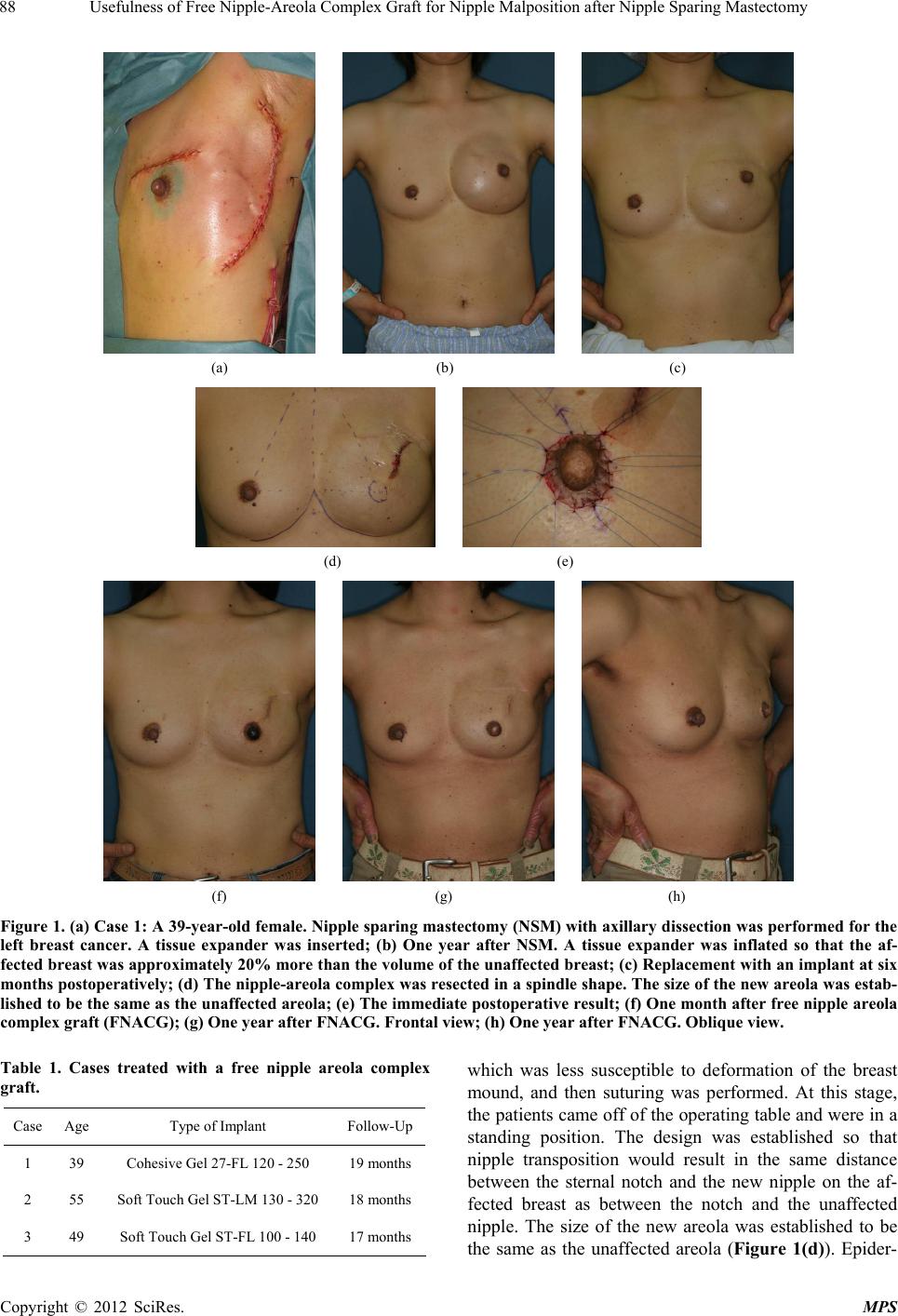 Usefulness of Free Nipple-Areola Complex Graft for Nipple Malposition after Nipple Sparing Mastectomy 88 (a) (b) (c) (d) (e) (f) (g) (h) Figure 1. (a) Case 1: A 39-year-old female. Nipple sparing mastectomy (NSM) with axillary dissection was performed for the left breast cancer. A tissue expander was inserted; (b) One year after NSM. A tissue expander was inflated so that the af- fected breast was approximately 20% more than the volume of the unaffected breast; (c) Replacement with an implant at six months postoperatively; (d) The nipple-areola complex was resected in a spindle shape. The size of the new areola was estab- lished to be the same as the unaffected areola; (e) The immediate postoperative result; (f) One month after free nipple areola complex graft (FNACG); (g) One year after FNACG. Frontal view; (h) One year after FNACG. Oblique view. Table 1. Cases treated with a free nipple areola complex graft. Case Age Type of Implant Follow-Up 1 39 Cohesive Gel 27-FL 1 2 0 - 250 19 months 2 55 Soft Touch Gel ST-LM 130 - 320 18 months 3 49 Soft Touch Gel ST-FL 100 - 140 17 months which was less susceptible to deformation of the breast mound, and then suturing was performed. At this stage, the patients came off of the operating table and were in a standing position. The design was established so that nipple transposition would result in the same distance between the sternal notch and the new nipple on the af- fected breast as between the notch and the unaffected nipple. The size of the new areola was established to be the same as the unaffected areola (Figure 1(d)). Epider- Copyright © 2012 SciRes. MPS  Usefulness of Free Nipple-Areola Complex Graft for Nipple Malposition after Nipple Sparing Mastectomy 89 mal resection was performed in the area where the areola was to be created. A full-thickness graft was used to re- construct the nipple-areola complex and fixed with a tie-over dressing (Figure 1(e)). Seven days postopera- tively, the tie-over dressing was removed. Subsequently, petroleum jelly was applied followed by a dressing until complete epithelialization occurred in the nipple-areola area. In the six-month postoperative period, the nipple was protected by a sponge with the center cut out to cre- ate a doughnut shape. This sponge was used to prevent pressure on the nipple by underwear or while sleeping. 2.3. Assessment of Surgical Outcome At least one year postoperatively, the results were as- sessed for symmetry of nipple-areola p osition, graft tak e, depigmentation, and shrinkage. 3. Results In all patients, the graft was accurately transferred to a position to achieve symmetry with the unaffected breast, and there was complete graft take in the areola. No de- pigmentation of the areola was observed. The size of the areola was almost unchanged after grafting in two pa- tients, but areolar shrinkage occurred in one other patient (Figures 1( f) -(h)). There was complete graft take in the nipple in one pa- tient. No depigmentation of the nipple was observed (Figure 2). Necrosis occurred at the tip of the nipple in two other patients. These patients had depigmentation, and the height of their nipples decreased in proportion to the level of necrosis (Figures 1(f)-(h)). In one patient without necrosis, there was slight flattening of the nipple (Figure 2 and Table 2). 4. Discussion There have been many reports on surgical methods to correct nipple-areola complex malposition (Table 3). There is a method in which a tissue expander is inserted and the skin on the cranial aspect is inflated to lower the high-riding nipple-areola complex [3]. The advantage of this method is that no new breast scar is created if the (a) (b) (c) Figure 2. (a) Case 2: A 55-year-old female. Before free nip- ple areola complex graft (FNACG); (b) The immediate postoperative result; (c) One and a half years after FNACG. Table 2. Results of a free nipple areola complex graft for 3 cases. No. of Areolae No. of Nipples Symmetry of nipple-areola position 3 3 Complete graft take 3 1 Depigmentation 0 2 Shrinkage 1 Not applicable Table 3. Transposition of the nipple areola complex. Tissue expander Periareolar skin excision Myocutaneous flap Skin flap (Z-plasty flap technique) Island flap (Subcutaneous pedicle) Free nipple areola complex graft tissue expander is inserted from the preexisting scar from NSM. However, its procedure can become complicated, the treatment period can be prolonged, and the stretched skin can revert back to its original state. The method of periareolar skin resection [4] has the advantage of a camouflaged breast scar because the areolar margin ob- scures the scar. If there is insufficient skin laxity of the breast, it is difficult to su ccessfully achieve nipple-areola complex transposition. A defect resulting from the trans- position can be treated using a myocutaneous flap [5,6]. This method enables successful nipple-areola complex transposition. However, there is morbid ity of the myocu- taneous flap donor site and prominent patchworklike scars of flaps. A method using a Z-plasty flap technique [7] can result in prominent geometric scars with an un- natural appearance. Another method involves an island flap in which the nipple-areola complex is transferred as a subcutaneous pedicle flap [8]. In this method, the transfer can be achieved without the interruption of the blood flow. Therefore, compared with the FNACG me- thod, this method is less susceptible to problems such as areolar shrinkage, necrosis of the tip of the nipple, and depigmentation. However, the skin of the breast is thin, and caution is required due to potential flap necrosis if blood flow of the subcutaneous pedicle is unstable. In addition, sufficient breast skin laxity is important to pre- vent impeded blood flow due to pressure on the subcuta- neous pedicle in the subcutaneous tunnel in the breast. In FNACG, suturing also cannot be performed at the donor site of the nipple-areola complex if there is insufficient skin laxity of the breast. Thus, a tissue expander is in- serted after NSM to adequately stretch the skin of the Copyright © 2012 SciRes. MPS  Usefulness of Free Nipple-Areola Complex Graft for Nipple Malposition after Nipple Sparing Mastectomy Copyright © 2012 SciRes. MPS 90 breast. The disadvantages of FNACG are that areolar sh- rinkage can occur, the height of the nipple can decrease due to necrosis if the nipple is high, and depigmentation can occur. Ahmed et al. [9] reported on temporary banking of the nipple-areola complex in cases of skin sparing mastec- tomy. The complex was temporarily banked in areas such as the groin region. The banked nipple-areola complex was replanted onto the reconstructed breast mound in a second surgery. Their results of the first transplantation showed that 88% of the cases had nipple-areola complex graft survival rates of at least 80%, and that 51% of the cases maintained at least 50% nipple projection. In their study, the color change was graded at 3 levels of good, moderate, and poor. Pig mentation was defined as good if the color of the nipple areola complex had not changed. Loss of pigment was considered a moderate result in cases where the nipple-areola complex was still clearly darker than the surrounding skin of the breast mound. The result was poor if there was no distinction between the color of the nipple areola complex and surrounding skin. Their results of the first transplantation were good in 48% of the cases, moderate in 51% of the cases, and poor in 1% of the cases. In this study, there was areolar shrinkage in one patient. Since areolar shrinkage was not included as an item examined by Ahmed et al., there could be racial differences in the results of the FNACG just as there are racial differences in scar contracture and keloid formation. Further studies will be necessary to examine the usefulness of this method in Asians, who are more susceptible to conspicuous scar formation than Caucasians [10,11]. The advantage of FNACG is that a graft can be trans- ferred to a site far from the donor site. Treatment using flaps can result in th e position o f a nipp le-areola co mplex that might be slightly different from the preoperatively planned position because of the state of the subcutaneous scar and the thickness and texture of the skin of the breast. In FNACG, the position of the nipple-areo la com- plex is determined after the donor site of the complex is sutured. Th erefo r e, th e graf t can be accurately transferred to a position to achieve symmetry with the unaffected breast. The method of FNACG involves simple surgical design and techniques, and is not greatly affected by the technical skills of surgeons. Thus, the method should be considered as one surgical treatment option. In conclusion, the disadvantages of FNACG are that areolar shrinkage can occur, the height of the nipple can decrease due to necrosis of the tip if the nipple is high, and depigmentation can occur. The advantage of FN- ACG is that a graft can be transferred to a site far from the donor site. It involves simple surgical design and techniques and is not greatly affected by the technical skills of surgeons. Therefore, the method can be useful if its advantages and disadvantages are well considered. REFERENCES [1] J. Y. Petit, U. Veronesi, A. Luini, R. Orecchia, P. C. Rey, S. Martella, F. Didier, F. De Lorenzi, M. Rietjens, C. Ga- rusi, A. Sonzogni, V. Galimberti, E. Leida, R. Lazzari and A. Giraldo, “When Mastectomy Becomes Inevitable: The Nipple-Sparing Approach,” Breast, Vol. 14, No. 6, 2005, pp. 527-531. doi:10.1016/j.breast.2005.08.028 [2] S. R. Colen, “Breast Reduction with Use of the Free Nip- ple Graft Technique,” Aesthetic Surgery Journal, Vol. 21, No. 3, 2001, pp. 261-271. doi:10.1067/maj.2001.116439 [3] A. S. Colwell, J. W. May Jr. and S. A. Slavin, “Lowering the Postoperative High-Riding Nipple,” Plastic and Re- constructive Surgery, Vol. 120, No. 3, 2007, pp. 596-599. doi:10.1097/01.prs.0000270283.64971.dd [4] R. P. Gruber and H. W. Jones Jr., “The ‘Donut’ Mas- topexy: Indications and Complications,” Plastic and Re- constructive Surgery, Vol. 65, No. 1, 1980, pp. 34-38. doi:10.1097/00006534-198001000-00007 [5] P. Berrino, E. Campora and P. Santi, “Postquadrantec- tomy Breast Deformities: Classification and Techniques of Surgical Correction,” Plastic and Reconstructive Sur- gery, Vol. 79, No. 4, 1987, pp. 567-572. doi:10.1097/00006534-198704000-00010 [6] S. A. Slavin, S. M. Love and N. L. Sadowsky, “Recon- struction of the Radiated Partial Mastectomy Defect with Autogenous Tissues,” Plastic and Reconstructive Surgery, Vol. 90, No. 5, 1992, pp. 854-865. doi:10.1097/00006534-199211000-00022 [7] B. A. Frenkiel, M. D. Pacifico, M. Ritz and G. Southwick, “A Solution to the High-Riding Nipple-Areola Complex,” Aesthetic Plastic Surgery, Vol. 34, No. 4, 2010, pp. 525- 527. doi:10.1007/s00266-009-9421-1 [8] H. Mohmand and A. Naasan, “Double U-Plasty for Cor- rection of Geometric Malposition of the Nipple-Areola Complex,” Plastic and Reconstructive Surgery, Vol. 109, No. 6, 2002, pp. 2019-2022. doi:10.1097/00006534-200205000-00036 [9] A. K. Ahmed, D. E. Hahn, J. J. Hage, E. M. Bleiker and L. A. Woerdeman, “Temporary Banking of the Nipple-Are- ola Complex in 97 Skin-Sparing Mastectomies,” Plastic and Reconstructive Surgery, Vol. 127, No. 2, 2011, pp. 531-539. doi:10.1097/PRS.0b013e3181fed578 [10] H. P. Dustan, “Does Keloid Pathogenesis Hold the Key to Understanding Black/White Differences in Hypertension Severity?” Hypertension, Vol. 26, No. 6, 1995, pp. 858- 862. [11] D. D. Datubo-Brown, “Keloids: A Review of the Litera- ture,” British Journal of Plastic Surgery, Vol. 43, No. 1, 1990, pp. 70-77. doi:10.1016/0007-1226(90)90047-4 |