

Paper Menu >>
Journal Menu >>
 Modern Plastic Surgery, 2012, 2, 80-82 http://dx.doi.org/10.4236/mps.2012.24019 Published Online October 2012 (http://www.SciRP.org/journal/mps) A New Method of Limb Salvage and Functional Upper Limb Reconstruction Ruka Shimizu1, Kazuo Kishi1, Hideo Morioka2, Hiroo Yabe2 1Department of Plastic and Reconstructive Surgery, Keio University School of Medicine, Tokyo, Japan; 2Department of Orthopaedic Surgery, Keio University School of Medicine, Tokyo, Japan. Email: [email protected] Received July 18th, 2012; revised August 17th, 2012; accepted September 17th, 2012 ABSTRACT Amputation is frequently used to treat malignant tumors invading into the shoulder joint. When the vessels and major nerves of the upper arm are preserved, however, the limb may be salvaged with a combined osseous, myocutaneous flap. We reconstructed large defects, which included the shoulder joint, using a vascularized rib-latissmus dorsi combined flap. Ribs were connected via the ninth or tenth posterior intercostal artery. Several slits were cut in the ribs, and the rib was glass stick fractured to straighten it. The thoracodorsal nerve was preserved and the latissmus dorsi muscle was transferred to the defect previously occupied by the biceps humerus. The ribs were connected to the residual clavicle via a Leeds-Keio artificial ligament. The flaps completely engrafted . The patient was able to flex h er elbow relatively early postoperatively. The function of the hand was preserved intact. In conclusion, the combined vascularized rib-latissmus dorsi flap is useful for patients who require functional or bony reconstruction of large defects involving the shoulder joint. Keywords: Shoulder; Vascularized Rib; Latissmus Dorsi; Elbow Flexion; Limb Salvage 1. Introduction Amputation is frequently used to treat malignant tumors invading into the shoulder joint. When the vessels and major nerves of the upper arm are preserved, however, the limb may be salvaged with a combined osseous, myocutaneous flap. In cases where limb salvage is cho- sen, various tissues including muscle, skin, and bone may be required depen di n g o n the extent o f the defect . In such cases, flaps based on the thoracodorsal artery will fulfill most of the closure requirements. Large flaps may be created around this artery, which supplies the latissmus dorsi (LD) muscle, serratus anterior muscle, and ribs. Flaps from this area may be structured to in- corporate varying amounts of bone and muscle as indi- cated by the defect. The ribs are primarily supplied by the intercostal artery, which anastomoses with the thora- codorsal artery in the LD muscle [1,2]. The option to incorporate bone and soft tissue in a single pedicle has made flaps from the thoracodorsal system popular in reconstruction of the maxillofacial region [3,4]. The LD is also frequently used to functionally reconstruct the elbow joint [5- 10] . In the present report, we describe a patient in whom a large defect involving most of the upper arm and gleno- humeral joint was reconstructed with the combined vas- cularized rib-latissmus dorsi flap, resulting in successful limb salvage. 2. Case Report A 33-year-old woman with a right proximal humeral chondrosarcoma underwent a radical tumor resection that included the proximal half of the humerus, the shoulder, the lateral one third of the clavicle, the deltoid muscle, the triceps brachii muscle, the proximal two thirds of the biceps brachii muscle, and the overlying skin (Figure 1(a)). We planned the reconstruction using a vascularized rib-latissmus dorsi flap based on the thoracod orsal artery system (Figure 1(b)). The ninth and tenth ribs were raised with the ninth intercostal artery (Figure 1(c)). The ribs were partially cut with a bone saw to straighten them and placed facing each other, then fixed with surgical wire. They were then fixed to the residual humerus with a titanium plate and sutured to an artificial ligament (Leeds-Keio ligament). The other side of the artificial ligament was fixed to the remnant of the clavicle (Fig- ures 1(d) and (e)). The insertion of the LD was sutured to the remnant of the biceps brachii, and the origin of the LD was sutured to the dissected shoulder with 3 - 0 po- lyglactin 910 (Vycryl, Ethicon Inc., Somerville, NJ) Copyright © 2012 SciRes. MPS 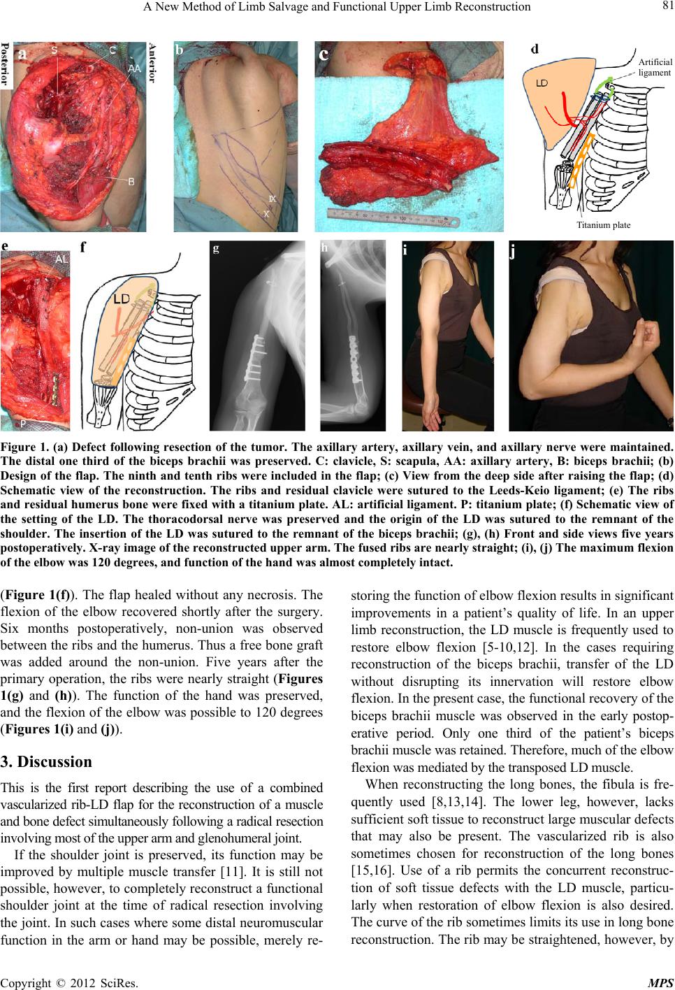 A New Method of Limb Salvage and Functional Upper Limb Reconstruction 81 Artificial ligame nt Titanium plate Figure 1. (a) Defect following resection of the tumor. The axillary artery, axillary vein, and axillary nerve were maintained. The distal one third of the biceps brachii was preserved. C: clavicle, S: scapula, AA: axillary artery, B: biceps brachii; (b) Design of the flap. The ninth and tenth ri bs were included in the flap; (c) View from the deep side after raising the flap; (d) Schematic view of the reconstruction. The ribs and residual clavicle were sutured to the Leeds-Keio ligament; (e) The ribs and residual humerus bone were fixed with a titanium plate. AL: artificial ligament. P: titanium plate; (f) Schematic view of the setting of the LD. The thoracodorsal nerve was preserved and the origin of the LD was sutured to the remnant of the shoulder. The insertion of the LD was sutured to the remnant of the biceps brachii; (g), (h) Front and side views five years postoperatively. X-ray image of the reconstructed upper arm. The fused ribs are nearly straight; (i), (j) The maximum flexion of the elbow was 120 degrees, and function of the hand was almost completely intact. (Figure 1(f)). The flap healed without any necrosis. The flexion of the elbow recovered shortly after the surgery. Six months postoperatively, non-union was observed between the ribs and the humerus. Thus a free bone graft was added around the non-union. Five years after the primary operation, the ribs were nearly straight (Figures 1(g) and (h)). The function of the hand was preserved, and the flexion of the elbow was possible to 120 degrees (Figures 1(i) and (j)). 3. Discussion This is the first report describing the use of a combined vascularized rib-LD flap for the reconstruction of a muscle and bone defect simultaneously following a radical resection involving most of the upper arm and glenohumeral joint. If the shoulder joint is preserved, its function may be improved by multiple muscle transfer [11]. It is still not possible, however, to completely reconstruct a functional shoulder joint at the time of radical resection involving the joint. In such cases where some distal neuromuscular function in the arm or hand may be possible, merely re- storing the function of elbow flexion results in significant improvements in a patient’s quality of life. In an upper limb reconstruction, the LD muscle is frequently used to restore elbow flexion [5-10,12]. In the cases requiring reconstruction of the biceps brachii, transfer of the LD without disrupting its innervation will restore elbow flexion. In the present case, the functional recovery of the biceps brachii muscle was observed in the early postop- erative period. Only one third of the patient’s biceps brachii muscle was retained. Therefore, much of the elbow flexion was mediated by the transposed LD muscle. When reconstructing the long bones, the fibula is fre- quently used [8,13,14]. The lower leg, however, lacks sufficient soft tissue to reconstruct large muscular defects that may also be present. The vascularized rib is also sometimes chosen for reconstruction of the long bones [15,16]. Use of a rib permits the concurrent reconstruc- tion of soft tissue defects with the LD muscle, particu- larly when restoration of elbow flexion is also desired. The curve of the rib sometimes limits its use in long bone reconstruction. The rib may be straightened, however, by Copyright © 2012 SciRes. MPS  A New Method of Limb Salvage and Functional Upper Limb Reconstruction 82 creating a glass stick fracture and suturing the rib to- gether in a fashion that straightens it but does not com- promise its strength. The ability of the reconstructed shoulder joint to sup- port the weight of the arm was another concern in our case. The Leed-Keio ligament is made of polyester woven into an open mesh-like structure [17]. After implantation, longitudinally aligned collagen-fiber bundles with spin- dle-shaped nuclei form over the polyester matrix [18,19]. Use of this material permitted the strength of the artificial joint to be preserved even after five years in our case. Our report illustrates that the combined vascularized rib-LD flap is useful in the reconstruction of large soft tissue and bony defects that involve the upper limb and the glenohumeral joint. REFERENCES [1] K. Kawamura, H. Yajima, Y. Tomita, Y. Kobata, K. Shigematsu and Y. Takakura, “Restoratio n of Elbow Fu nc- tion with Pedicled Latissimus Dorsi Myocutaneous Flap Transfer,” Journal of Shoulder and Elbow Surgery, Vol. 16, No. 1, 2007, pp. 84-90. doi:10.1016/j.jse.2006.03.006 [2] K. Onishi and Y. Maruyama, “Compound Rib-Latissimus Dorsi Osteomusculocutaneous Flap in Reconstruction of the Upper Arm,” Annals of Plastic Surgery, Vol. 36, 1996, pp. 191-194. doi:10.1097/00000637-199608000-00014 [3] C. H. Lin, F. C. Wei, L, S, Levin, J. Su, K. F. Fan, W. L. Yeh and D. T. Hsu, “Free Composite Serratus Anterior and Rib Flaps for Tibial Composite Bone and Soft-Tissue Defect,” Plastic and Reconstructive Surgery, Vol. 99, No. 6, 1997, pp. 1656-1665. doi:10.1097/00006534-199705000-00028 [4] M. D. Vekris, A. E. Beris, M. G. Lykissas, A. V. Ko- rompilias, A. D. Vekris and P. N. Soucacos, “Restoration of Elbow Function in Severe Brachial Plexus Paralysis via Muscle Transfers,” Injury, Vol. 39, No. 3, 2008, pp. S15-S22. doi:10.1016/j.injury.2008.06.008 [5] L. De Smet, “The Latissimus Dorsi Flap for Reconstruc- tion of a Paralysed Deltoid,” Acta Chirurgica Belgica, Vol. 104, 2004, pp. 328-329. [6] K. Fujikawa, F. Iseki and B. B. Seedhom, “Arthroscopy after Anterior Cruciate Reconstruction with the Leeds- Keio Ligament,” Journal of Bone & Joint Surgery, Brit- ish Volume, Vol. 71, No. 4, 1989, pp. 566-570. [7] Y. Maruyama, K. Onishi, Y. Iwahira, Y. Okajima and M. Motegi, “Free Compound Rib-Latissimus Dorsi Osteo- musculocutaneous Flap in Reconstruction of the Leg,” Journal of Reconstructive Microsurgery, Vol. 3, No. 1, 1986, pp. 13-18. doi:10.1055/s-2007-1007032 [8] T. Schoeller, G. Wechselberger, H. Hussl and G. M. Huemer, “Functional Transposition of the Latissimus Dorsi Muscle for Biceps Reconstruction after Upper Arm Replantation,” Journal of Plastic, Reconstructive & Aes- thetic Surgery, Vol. 60, No. 7, 2007, pp. 755-759. doi:10.1016/j.bjps.2006.11.032 [9] D. C. Sundaresh, D. Gopalakrishnan and N. Shetty, “Vascularised Rib Graft Defects of the Diaphysis of the Humerus in Children. A Report of Two Cases,” Journal of Bone & Joint Surgery, British Volume, Vol. 82, 2000, pp. 28-32. doi:10.1302/0301-620X.82B1.9678 [10] S. Yazar, C. H. Lin and F. C. Wei, “One-Stage Recon- struction of Composite Bone and Soft-Tissue Defects in Traumatic Lower Extremities,” Plastic and Reconstruc- tive Surgery, Vol. 114, No. 6, 2004, pp. 1457-1466. doi:10.1097/01.PRS.0000138811.88807.65 [11] J. E. Aviv, M. L. Urke n and C. Vickery, H. Weinberg, D. Buchbinder and H. F. Biller, “The combined latissimus dorsi-scapular free flap in head and neck reconstruction,” Archives of Otolaryngology—Head & Neck Surgery, Vol. 117, No. 11, 1991, No. 1242-1250. doi:10.1001/archotol.1991.01870230058008 [12] S. O’Ceallaigh, K. S. A. Mehboob Ali and T. P. F. O’ Co n - nor, “Functional Latissimus Dorsi Muscle Transfer to Re- store Elbow Flexion in Extensive Electrical Burns,” Burns, Vol. 31, No. 1, 2005, pp. 113-115. doi:10.1016/j.burns.2004.06.017 [13] E. Nomura, M. Inoue and H. Sugiura, “Histological Evaluation of Medial Patellofemoral Ligament Recon- structed Using the Leeds-Keio Ligament Prosthesis,” Biomaterials, Vol. 26, No. 15, 2005, pp. 2663-2670. doi:10.1016/j.biomaterials.2004.07.053 [14] C. H. Lin, F. C. Wei, H. C. Chen and D. C. Chuang, “Outcome Comparison in Traumatic Lower-Extremity Reconstruction by Using Various Composite Vascula- rized Bone Transplantation,” Plastic and Reconstructive Surgery, Vol. 104, No. 4, 1999, pp. 984-992. doi:10.1097/00006534-199909040-00013 [15] T. D. Pierce and M. M. Tomaino, “Use of the Pedicled Latissimus Muscle Flap for Upper-Extremity Reconstruc- tion,” Journal of the American Academy of Orthopaedic Surgeons, Vol. 8, No. 5, 2000, pp. 324-331. [16] Y. K. Tu, C. Y. Yen, W. L. Yeh, I. C. Wang, K. C. Wang and W. N. Ueng, “Reconstruction of Posttraumatic Long Bone Defect with Free Vascularized Bone Graft: Good Outcome in 48 Patients with 6 Years’ Follow-Up,” Acta Orthopaedica Scandinavica, Vol. 72, No. 4, 2001, pp. 359-364. doi:10.1080/000164701753542014 [17] P. D. Kim and K. E. Blackwell, “Latissimus-Serratus-Rib Free Flap for Oromandibular and Maxillary Reconstruc- tion,” Archives of Otolaryngology—Head and Neck Sur- gery, Vol. 133, No. 8, 2007, pp. 791-795. doi:10.1001/archotol.133.8.791 [18] P. J. Stern and J. P. Carey, “The Latissimus Dorsi Flap for Reconstruction of the Brachium and Shoulder,” The Journal of Bone & Joint Surgery, Vol. 70, No. 4, 1988, pp. 526-535. [19] S. Zaffagnini, G. M. Marcheggiani Muccioli, V. Chatrath, A. Bondi, V. De Pasquale, D. Martini, B. Bacchelli and M. Marcacci, “Histological and Ultrastructural Evaluation of Leeds-Keio Ligament 20 Years after Implant: A Case Report,” Knee Surgery, Sports Traumatology, Arthro- scopy, Vol. 16, No. 11, 2008, pp. 1026-1029. doi:10.1007/s00167-008-0608-9 Copyright © 2012 SciRes. MPS |