

Paper Menu >>
Journal Menu >>
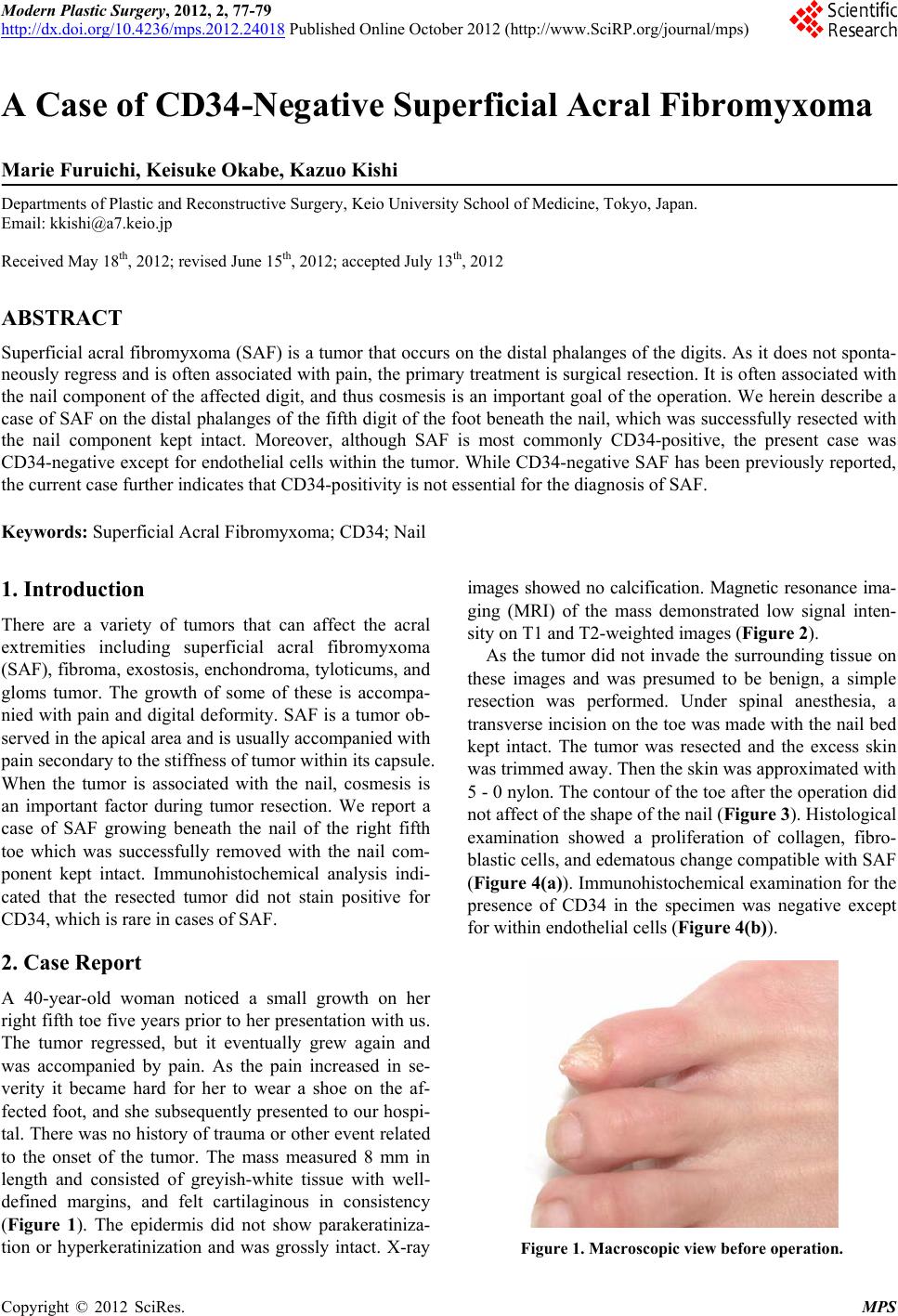 Modern Plastic Surgery, 2012, 2, 77-79 http://dx.doi.org/10.4236/mps.2012.24018 Published Online October 2012 (http://www.SciRP.org/journal/mps) 77 A Case of CD34-Negative Superficial Acral Fibromyxoma Marie Furuichi, Keisuke Okabe, Kazuo Kishi Departments of Plastic and Reconstructive Surgery, Keio University School of Medicine, Tokyo, Japan. Email: [email protected] Received May 18th, 2012; revised June 15th, 2012; accepted July 13th, 2012 ABSTRACT Superficial acral fibromyxoma (SAF) is a tumor that occurs on the distal phalanges of the digits. As it does not sponta- neously regress and is often associated with pain, the primary treatment is surgical resection. It is often associated with the nail component of the affected digit, and thus cosmesis is an important goal of the operation. We herein describe a case of SAF on the distal phalanges of the fifth digit of the foot beneath the nail, which was successfully resected with the nail component kept intact. Moreover, although SAF is most commonly CD34-positive, the present case was CD34-negative ex cept for endothelial cells within the tumor. While CD34-n egative SAF has been previously reported, the current case further indicates that CD34-positivity is not essential for the diagnosis of SAF. Keywords: Superficial Acral Fibromyxoma; CD34; Nail 1. Introduction There are a variety of tumors that can affect the acral extremities including superficial acral fibromyxoma (SAF), fibroma, exostosis, enchondroma, tyloticums, and gloms tumor. The growth of some of these is accompa- nied with pain and digital deformity. SAF is a tumor ob- served in the apical area and is usually accompanied with pain secondary to the stiffness of tumor within its capsule. When the tumor is associated with the nail, cosmesis is an important factor during tumor resection. We report a case of SAF growing beneath the nail of the right fifth toe which was successfully removed with the nail com- ponent kept intact. Immunohistochemical analysis indi- cated that the resected tumor did not stain positive for CD34, which is rare in cases of SAF. 2. Case Report A 40-year-old woman noticed a small growth on her right fifth toe five years prior to her presentation with us. The tumor regressed, but it eventually grew again and was accompanied by pain. As the pain increased in se- verity it became hard for her to wear a shoe on the af- fected foot, and she subsequently presented to our hospi- tal. There was no history of trauma or other event related to the onset of the tumor. The mass measured 8 mm in length and consisted of greyish-white tissue with well- defined margins, and felt cartilaginous in consistency (Figure 1). The epidermis did not show parakeratiniza- tion or hyperkeratinization and was grossly intact. X-ray images showed no calcification. Magnetic resonance ima- ging (MRI) of the mass demonstrated low signal inten- sity on T1 and T2-weighted images (Figure 2). As the tumor did not invade the surrounding tissue on these images and was presumed to be benign, a simple resection was performed. Under spinal anesthesia, a transverse incision on the to e was made with the nail bed kept intact. The tumor was resected and the excess skin was trimmed away. Then the skin was approximated with 5 - 0 nylon. The contour of the toe after the operation did not affect of the shape of the nail (Figure 3). Histological examination showed a proliferation of collagen, fibro- blastic cells, and edematous change compatible with SAF (Figure 4(a)). Immunohistochemical examination for the presence of CD34 in the specimen was negative except for within endothelial cells (Figure 4(b)). Figure 1. Macroscopic view before operation. Copyright © 2012 SciRes. MPS 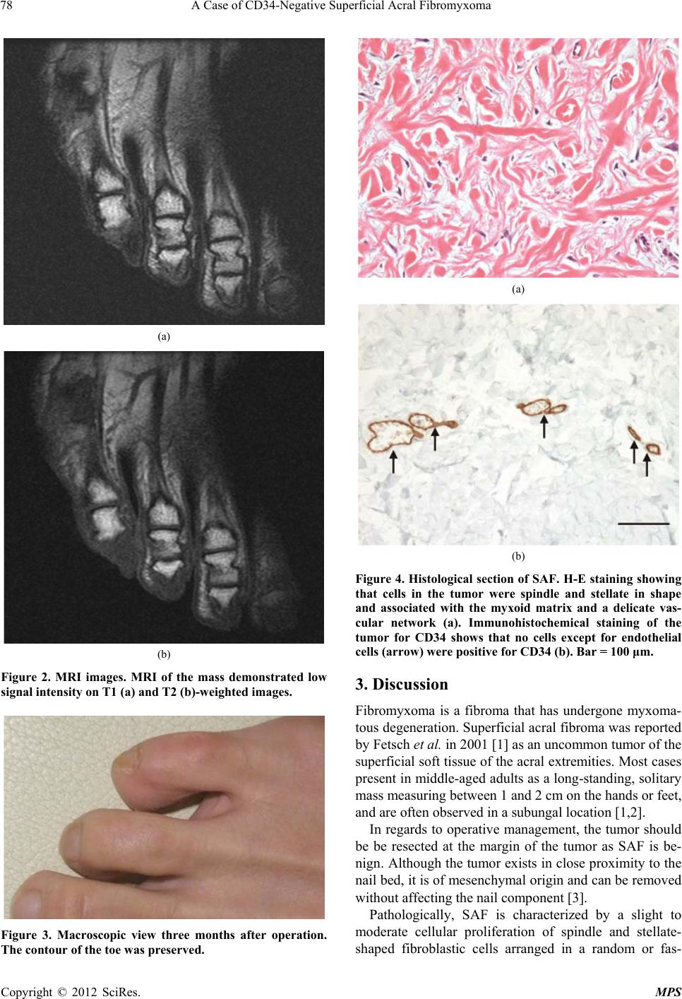 A Case of CD34-Negative Superficial Acral Fibromyxoma 78 (a) (b) Figure 2. MRI images. MRI of the mass demonstrated low signal intensity on T1 (a) and T2 (b)-weighted images. Figure 3. Macroscopic view three months after operation. The contour of the toe was preserved. (a) (b) Figure 4. Histological section of SAF. H-E staining showing that cells in the tumor were spindle and stellate in shape and associated with the myxoid matrix and a delicate vas- cular network (a). Immunohistochemical staining of the tumor for CD34 shows that no cells except for endothelial cells (arrow) were positive for CD34 (b). Bar = 100 μm. 3. Discussion Fibromyxoma is a fibroma that has undergone myxoma- tous degeneration. Superficial acral fibroma was reported by Fetsch et al. in 2001 [1] as an uncommon tumor of the superficial soft tissue of the acral extremities. Most cases present in middle-ag ed adults as a long-standing, so litary mass measuring between 1 and 2 cm on the hands or feet, and are often observed in a subungal location [1,2]. In regards to operative management, the tumor should be be resected at the margin of the tumor as SAF is be- nign. Although the tumor exists in close proximity to the nail bed, it is of mesenchymal origin and can be removed without affecting the nail component [3]. Pathologically, SAF is characterized by a slight to moderate cellular proliferation of spindle and stellate- shaped fibroblastic cells arranged in a random or fas- Copyright © 2012 SciRes. MPS 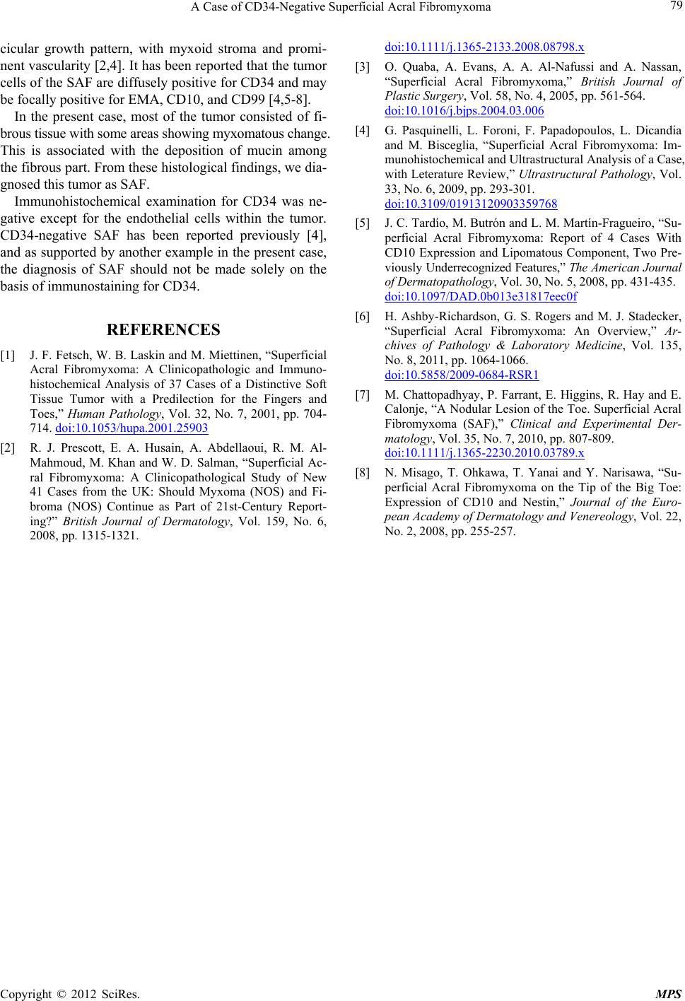 A Case of CD34-Negative Superficial Acral Fibromyxoma Copyright © 2012 SciRes. MPS 79 cicular growth pattern, with myxoid stroma and promi- nent vascularity [2,4]. It has been reported that the tumor cells of the SAF are diffusely positiv e for CD34 and may be focally positive for EMA, CD10, and CD99 [4,5-8]. In the present case, most of the tumor consisted of fi- brous tissue with some areas showing m y xomatous change. This is associated with the deposition of mucin among the fibrous part. From these histological findings, we dia- gnosed this tumor as SAF. Immunohistochemical examination for CD34 was ne- gative except for the endothelial cells within the tumor. CD34-negative SAF has been reported previously [4], and as supported by another example in the present case, the diagnosis of SAF should not be made solely on the basis of immunostaining for CD34. REFERENCES [1] J. F. Fetsch, W. B. Laskin and M. Miettinen, “Superficial Acral Fibromyxoma: A Clinicopathologic and Immuno- histochemical Analysis of 37 Cases of a Distinctive Soft Tissue Tumor with a Predilection for the Fingers and Toes,” Human Pathology, Vol. 32, No. 7, 2001, pp. 704- 714. doi:10.1053/hupa.2001.25903 [2] R. J. Prescott, E. A. Husain, A. Abdellaoui, R. M. Al- Mahmoud, M. Khan and W. D. Salman, “Superficial Ac- ral Fibromyxoma: A Clinicopathological Study of New 41 Cases from the UK: Should Myxoma (NOS) and Fi- broma (NOS) Continue as Part of 21st-Century Report- ing?” British Journal of Dermatology, Vol. 159, No. 6, 2008, pp. 1315-1321. doi:10.1111/j.1365-2133.2008.08798.x [3] O. Quaba, A. Evans, A. A. Al-Nafussi and A. Nassan, “Superficial Acral Fibromyxoma,” British Journal of Plastic Surgery, Vol. 58, No. 4, 2005, pp. 561-564. doi:10.1016/j.bjps.2004.03.006 [4] G. Pasquinelli, L. Foroni, F. Papadopoulos, L. Dicandia and M. Bisceglia, “Superficial Acral Fibromyxoma: Im- munohistochemical and Ultrastructural Analysis of a Case, with Leterature Review,” Ultrastructural Pathology, Vol. 33, No. 6, 2009, pp. 293-301. doi:10.3109/01913120903359768 [5] J. C. Tardío, M. Butrón and L. M. Martín-Fragueiro, “Su- perficial Acral Fibromyxoma: Report of 4 Cases With CD10 Expression and Lipomatous Component, Two Pre- vio usly Underrecognized Features,” The American Journal of Dermatopathology, Vol. 30, No. 5, 2008, pp. 431-435. doi:10.1097/DAD.0b013e31817eec0f [6] H. Ashby-Richardson, G. S. Rogers and M. J. Stadecker, “Superficial Acral Fibromyxoma: An Overview,” Ar- chives of Pathology & Laboratory Medicine, Vol. 135, No. 8, 2011, pp. 1064-1066. doi:10.5858/2009-0684-RSR1 [7] M. Chattopadhyay, P. Farrant, E. Higgins, R. Hay and E. Calonje, “A Nodular Lesion of the Toe. Superficial Acral Fibromyxoma (SAF),” Clinical and Experimental Der- matology, Vol. 35, No. 7, 2010, pp. 807-809. doi:10.1111/j.1365-2230.2010.03789.x [8] N. Misago, T. Ohkawa, T. Yanai and Y. Narisawa, “Su- perficial Acral Fibromyxoma on the Tip of the Big Toe: Expression of CD10 and Nestin,” Journal of the Euro- pean Academy of Dermatology and Venereology, Vol. 22, No. 2, 2008, pp. 255-257. |