

Paper Menu >>
Journal Menu >>
 Journal of Biomaterials and Nanobiotechnology, 2012, 3, 480-485 http://dx.doi.org/10.4236/jbnb.2012.34049 Published Online October 2012 (http://www.SciRP.org/journal/jbnb) Comparative Study on Bactericidal Effect of Silver Nanoparticles, Synthesized Using Green Technology, in Combination with Antibiotics on Salmonella typhi Shweta Rajawat, Mohammad Shums Qureshi Department of Physics, Maulana Azad National Institute of Technology, Bhopal, India. Email: [email protected], [email protected] Received March 12th, 2012; revised June 25th, 2012; accepted July 24th, 2012 ABSTRACT In this work bactericidal study of silver nanoparticles was taken up in combination with two standard antibiotics, am- picillin and gentamycin, for Salmonella typhi. The antibacterial activities of antibiotics were increased in the presence of AgNPs against test strains. The higher enhancing effect was observed for ampicillin in comparison to gentamicin against test strains. Silver nanoparticles were synthesized elctrolytically using silver wire of 99% purity as anode and carbon rod wrapped with LDPE as cathode. Silver nitrate [of Merck] of 0.01N is used as an electrolyte. Here tea extract is added as capping and mild reducing agent. The polyphenols theaflavins and thearubigins, present in tea perform the role of stabilizing or capping agents due to their bulky and steric nature. A brown coloured colloidal solution of silver nanoparticles is obtained. The as-synthesized silver nanoparticles were characterized using XRD, TEM and UV-Vis spectroscopy. Keywords: Silver Nanoparticles; Salmonella typhi; Tea Extract; Ampicillin; Gentamicin 1. Introduction Historically, silver has been extensively used for both hygienic and healing purposes (Cheng and Schluesener, 2008). With time, the use of silver has reduced as an anti-infection agent due to the advent of antibiotics and other disinfectants and the poorly understood mecha- nisms of their toxic effects. However, resistance of bac- teria to bactericides and antibiotics has increased in re- cent years. Some antimicrobial agents are extremely irri- tant and toxic. Hence there is a need to find ways to for- mulate safe and cost-effective Biocidal materials. Previ- ous studies show that antimicrobial formulations of silver in the form of nanoparticles could be used as effective bactericidal materials. Highly reactive metal oxide nano- particles exhibit excellent biocidal action against Gram- positive and Gram-negative bacteria [1]. Thus, the syn- theses, characterization, functionalization of nanosized particles open the possibility of formulation of a new generation of bactericidal materials. Researchers have found that nanosilver and antibiotics used together had an extremely strong effect against gram positive and gram negative bacteria. Recently, due to the limitations of antibiotics, the synergetic effect of silver nanoparticles with antibiotics has been studied combining silver nanoparticles with different antibiotics against gram positive and gram negative bacteria. A variety of preparation routes have been reported for the synthesis of metallic nanoparticles [2,3] notable ex- amples include, reverse micelles process [4,5], salt re- duction [6], microwave dielectric heating reduction [7], ultrasonic irradiation [8], radiolysis [9], Solvothermal synthesis [10], electrochemical synthesis [11,12], etc. Chemical methods for metal nanoparticle fabrication usually involve toxic chemicals, which are usually ex- pensive and potentially dangerous for the environment [13]. Green synthesis of nanoparticles is an easy, effi- cient and eco-friendly approach. Among several synthe- sizing methods, biosynthetic methods employing either biological microorganisms or plant extracts have emerg- ed as a simple and viable alternative to chemical syn- thetic procedures and physical methods. The synthesis of metal nanoparticles using biological systems is an ex- panding research area due to the potential ap plications in nano medicines. Plant extracts play an important role in remediation of toxic metals through reduction of the metal ions. Silver nanoparticle are synthesized from various parts of the herbal plants like bark of Cinnamon (Sathishkumar et al., 2009), Neem leaves (Tripathi et al., 2009), Citrus limon (Prathna et al., 2011), Tannic acid (Sivaraman et al., 2009) and various plant leaves (Song Copyright © 2012 SciRes. JBNB  Comparative Study on Bactericidal Effect of Silver Nanoparticles, Synthesized Using Green Technology, in Combination with Antibiotics on Salmonella typhi 481 and Kim, 2008). We describe a synthesis approach which is simple and “green” for the synthesis of metallic nanostructures of noble metal i.e. silver (Ag). The bactericidal effect of as-synthesized silver nanoparticles is then studied for combination with gentamycin and ampicillin for Salmo- nella typhi. Salmonella typhi is an obligate parasite that has no known natural r eservoir outside of humans. It is a multi-organ pathogen that inhabits the lymphatic tissues of the small intestine, liver, spleen, and bloodstream of infected humans. Infection of S. typhi leads to the de- velopment of typhoid, or enteric fever. Gentamicin is active against a wide range of human bacterial infections, mostly Gram-negative bacteria including Pseudomonas, Proteus, Serratia and the Gram-positive Staphylococcus. Ampicillin is a beta-lactam antibiotic that has been used extensively to treat bacterial infections since 1961. It is relatively non-toxic. The toxicity of silver nanoparticles synthesized using green technology is comparatively reduced. 2. Materials and Method The experimental setup consists of a beaker filled with 40 ml of electrolyte silver nitrate (0.02 N) of MERK. The silver nitrate was further diluted with triple deionised distilled water to obtain a solution of 0.01 N (mol/l) strength. Two electrodes: silver wire (99% pure) as an- ode and carbon rod wrapped by LDPE (Low Density Poly Ethylene) material as cathode was used. LDPE ma- terial is used to collect silver nanoparticles produced in the synthesis process so that they can be easily extracted. The distance between the two electrodes is 1 cm. The diameter of the silver wire is 1.04 mm and the diameter of the carbon rod used is 4 mm. The length of the carbon rod as well as silver wire is 4.5 cm. 2 ml of tea extract is added to the electrolyte as a capping agent. The whole assembly is placed on magnetic stirrer which keeps the solution in the beaker stirring continuously. The process is carried at room temperature (30˚C) for 2 hours con- tinuously. A Daniel cell of 1.1 volt and 2 ohm internal resistance is used as current source. A potentiometer pot along with a rheostat is used in the circuit to increase the resistance and obtain current of different values in mam- peres. Copper wires are used to connect the components of the circuit. All the parameters were same for the sam- ples synthesized, except the current through the circuit. Figure 1 shows the set up. The beaker in the set-up is covered with silver foil with holes for the electrodes. 3. Results and Discussions The as-synthesised silver nanoparticles were obtained in the form of colloidal solution of light brown colour as shown in Figure 2. The as-synthesized silver nanoparti- cles were characterized using XRD. Figure 3 shows the graph obtained. The XRD results show Face-Centred- Structure of pure silver nanoparticles oriented in planes {111}, {200}, {220 }, and {311}. The peaks obtained for as-synthesized silver nanoparticles are well in accor- dance with JCPDS file No. 04-0783. The intensive dif- fraction peak from the {111} lattice plane of face-centred cubic silver indicates that the particles are made of pure silver and that their basal plane, i.e., the top crystal plane Figure 1. Experimental setup. Figure 2. Synthesized colloidal solution of silver nanoparti- cles. Copyright © 2012 SciRes. JBNB  Comparative Study on Bactericidal Effect of Silver Nanoparticles, Synthesized Using Green Technology, in Combination with Antibiotics on Salmonella typhi 482 Figure 3. XRD graph of as-synthesized silver nanoparticles. is the {111} plane. This 2θ value reflection angle con- firms the presence of silver nanoparticles. The morphology and the crystal structure of synthe- sized silver nanoparticles were examined using HR-TEM. HR-TEM unit used had a 120 kV class high-resolution transmission electron microscope JEM-1400, Jeol, Japan. The sample was placed on the carbon coated copper grid, making a thin film of sample on the grid and extra sam- ple was removed using the cone of a blotting paper and kept in grid box sequentially. The TEM images show that the particles are well dispersed and spherical in shape. The particle size is between 2 nm to 23 nm. The average size of the particle is 10 nm. The as-synthesized silver nanoparticles are polydispersed nanoparticles. Such va- riation in shape and size of nanoparticles synthesized by biological systems is common [14]. The spherical and oval shape of the particle, as visible in Figure 4, is due to the fact that when a particle is formed, in its initial state, it tries to acquire a shape that corresponds to minimum potential energy. The spherical and oval shapes corre- spond to the state of minimum potential energy. Other factors that play role in determining the shape of silver nanoparticles are the fast rate of reaction and low con- centration of silver nitrate solution as slower rate of reac- tion [15] and higher concentration of silver nitrate [16] leads to anisotropic silver nanoparticles. The UV-vis spectroscopy was carried by Uv-vis spec- trometer of Systronics. It is a dual beam spectropho- tometer. The base solution was triple de-ionised distilled water to sample was added in small concentration. The UV-visible spectrum shows the formation of silver nanoparticles as the peak maxima 522 n m which is char- acteristic to silver nanoparticles. The specific character- istic peak, as shown in the Figure 5, for silver nanoparti- cles is due to the surface Plasmon resonance. The nano- particles which are smaller than the wavelength of light can produce a coherent resonance waves at a particular Figure 4. TEM image of silver nanoparticles. Figure 5. UV-Visible graph of silver nanoparticles. absorbance wavelength which is in the visible range for silver nanoparticles. Silver is known for its antimicrobial properties and h as been used for years in the medical field for antimicrobial applications and even has shown to prevent HIV binding to host cells [17,18-21]. Additionally, silver has been used in water and air filtration to eliminate microorga- nisms [22-24].The antibacterial activity of nanoparticle along with antibiotics ampicillin and gentamicin was tested against S. typhi. Figure 6 shows the zone of inhi- bition against S. typhi.The antibacterial activities of antibiotics increase in the presence of silver nanoparticles against gram positive and gram negative bacteria. The size of metallic nanoparticles ensures that a significantly large surface area of the particles is in contact with the bacterial cells. Such a large contact surface is expected to enhance the extent of bacterial elimination [25]. The mechanism of the bactericidal effect of AgNPs is that they may attach to the surface of the cell membrane dis- turbing permeability and respiration functions of the cell Copyright © 2012 SciRes. JBNB 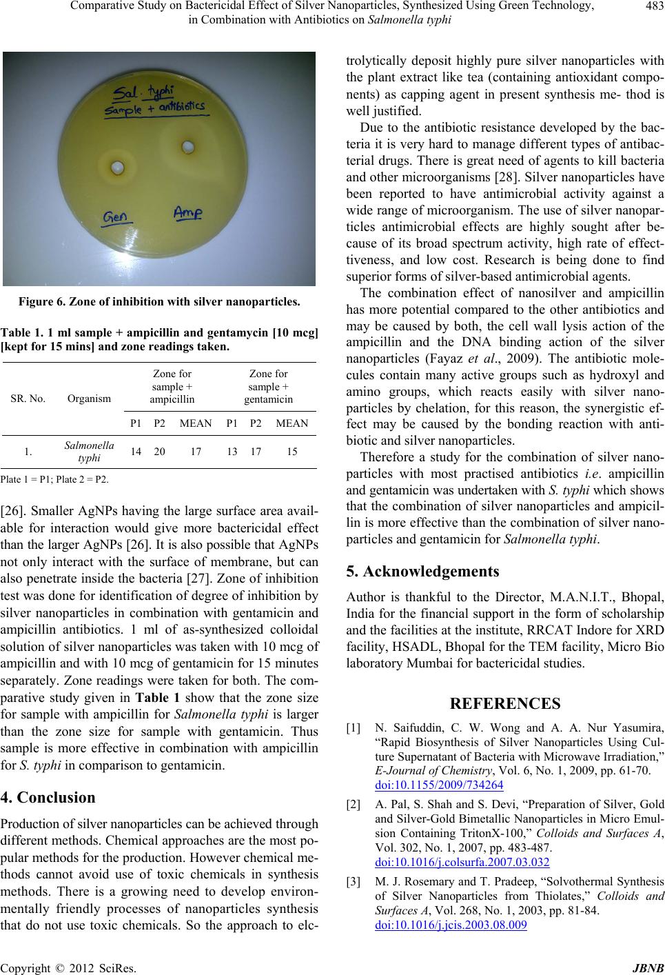 Comparative Study on Bactericidal Effect of Silver Nanoparticles, Synthesized Using Green Technology, in Combination with Antibiotics on Salmonella typhi 483 Figure 6. Zone of inhibition with silver nanoparticles. Table 1. 1 ml sample + ampicillin and gentamycin [10 mcg] [kept for 15 mins] and zone readings taken. Zone for sample + ampicillin Zone for sample + gentamicin SR. No. Organism P1 P2MEAN P1 P2MEAN 1. Salmonella typhi 14 2017 13 1715 Plate 1 = P1; Plate 2 = P2. [26]. Smaller AgNPs having the large surface area avail- able for interaction would give more bactericidal effect than the larger AgNPs [26]. It is also possible that A gNP s not only interact with the surface of membrane, but can also penetrate inside the bacteria [27]. Zone of inh ibition test was done for identification of degree of inh ibition by silver nanoparticles in combination with gentamicin and ampicillin antibiotics. 1 ml of as-synthesized colloidal solution of silver nanoparticles was taken with 10 mcg of ampicillin and with 10 mcg of gentamicin for 15 minutes separately. Zone readings were taken for both. The com- parative study given in Table 1 show that the zone size for sample with ampicillin for Salmonella typhi is larger than the zone size for sample with gentamicin. Thus sample is more effective in combination with ampicillin for S. typhi in comparison to gentamicin. 4. Conclusion Production of silver nanoparticles can be achieved through different methods. Chemical approaches are the most po- pular methods for the production. However chemical me- thods cannot avoid use of toxic chemicals in synthesis methods. There is a growing need to develop environ- mentally friendly processes of nanoparticles synthesis that do not use toxic chemicals. So the approach to elc- trolytically deposit highly pure silver nanoparticles with the plant extract like tea (containing antioxidant compo- nents) as capping agent in present synthesis me- thod is well justified. Due to the antibiotic resistance developed by the bac- teria it is very hard to manage different types of antibac- terial drugs. There is great need of agents to kill bacteria and other microorganisms [28]. Silver nanoparticles have been reported to have antimicrobial activity against a wide range of microorganism. The use of silver nanopar- ticles antimicrobial effects are highly sought after be- cause of its broad spectrum activity, high rate of effect- tiveness, and low cost. Research is being done to find superior forms of silver-based antimicrobial agents. The combination effect of nanosilver and ampicillin has more potential compared to the other antibiotics and may be caused by both, the cell wall lysis action of the ampicillin and the DNA binding action of the silver nanoparticles (Fayaz et al., 2009). The antibiotic mole- cules contain many active groups such as hydroxyl and amino groups, which reacts easily with silver nano- particles by chelation, for this reason, the synergistic ef- fect may be caused by the bonding reaction with anti- biotic and silver nanoparticles. Therefore a study for the combination of silver nano- particles with most practised antibiotics i.e. ampicillin and gentamicin was undertaken with S. typhi which shows that the combination of silver nanoparticles and ampicil- lin is more effective than th e combination of silver nano- particles and gentamicin for Salmonella typhi. 5. Acknowledgements Author is thankful to the Director, M.A.N.I.T., Bhopal, India for the financial support in the form of scholarship and the facilities at the institute, RRCAT Indore for XRD facility, HSADL, Bhopal for the TEM facility, Micro Bio laboratory Mumbai for bactericidal studies. REFERENCES [1] N. Saifuddin, C. W. Wong and A. A. Nur Yasumira, “Rapid Biosynthesis of Silver Nanoparticles Using Cul- ture Supernatant of Bacteria with Microwave Irradiation,” E-Journal of Chemistry, Vol. 6, No. 1, 2009, pp. 61-70. doi:10.1155/2009/734264 [2] A. Pal, S. Shah and S. Devi, “Preparation of Silver, Gold and Silver-Gold Bimetallic Nanoparticles in Micro Emul- sion Containing TritonX-100,” Colloids and Surfaces A, Vol. 302, No. 1, 2007, pp. 483-487. doi:10.1016/j.colsurfa.2007.03.032 [3] M. J. Rosemary and T. Pradeep, “Solvothermal Synthesis of Silver Nanoparticles from Thiolates,” Colloids and Surfaces A, Vol. 268, No. 1, 2003, pp. 81-84. doi:10.1016/j.jcis.2003.08.009 Copyright © 2012 SciRes. JBNB  Comparative Study on Bactericidal Effect of Silver Nanoparticles, Synthesized Using Green Technology, in Combination with Antibiotics on Salmonella typhi 484 [4] Y. Xie, R. Ye and H. Liu, “Synthesis of Silver Nanoparti- cles in Reverse Micelles Stabilized by Natural Biosurfac- tant,” Colloids and Surfaces A, Vol. 279, No. 1, 2006, pp. 175-178. doi:10.1016/j.colsurfa.2005.12.056 [5] M. Maillard, S. Giorgo and M. P. Pileni, “Silver Nano Disks,” Advanced Materials, Vol. 14, No. 15, 2002, pp. 1084-1086. doi:10.1002/1521-4095(20020805)14:15<1084::AID-AD MA1084>3.0.CO;2-L [6] Z. S. Pillai and P. V. Kamat, “What Factors Control the Size and Shape of Silver Nanoparticles in the Citrate Ion Reduction Method?” The Journal of Physical Chemistry B, Vol. 108, No. 3, 2004, pp. 945-951. doi:10.1021/jp037018r [7] K. Patel, S. Kapoor, D. P. Dave and T. Murherjee, “Syn- thesis of Nanosized Silver Colloids by Microwave Di- electric Heating,” Journal of Chemical Sciences, Vol. 117, No. 1, 2005, pp. 53-60. doi:10.1007/BF02704361 [8] R. A. Salkar, P. Jeevanandam, S. T. Aruna, Y. Koltypin and A. Gedanken, “The Sonochemical Preparation of Amorphous Silver Nanoparticles,” Journal of Materials Chemistry, Vol. 9, No. 6, 1999, pp. 1333-1335. doi:10.1039/a900568d [9] B. Soroushian, I. Lampre, J. Belloni and M. Mostafavi, “Radiolysis of Silver Ion Solutions in Ethylene Glycol: Solvated Electron and Radical Scavenging Yields,” Ra- diation Physics and Chemistry, Vol. 72, No. 2-3, 2005, pp. 111-118. doi:10.1016/j.radphyschem.2004.02.009 [10] M. Starowicz, B. Stypula and J. Banaoe, “Electrochemi- cal Method for the Synthesis of Silver Nanoparticles,” Journal of Electrochemistry Communications, Vol. 8, No. 2, 2006, pp. 227- 230. doi:10.1016/j.elecom.2005.11.018 [11] J. J. Zhu, X. H. Liao, X. N. Zhao and H. Y. Hen, “Prepa- ration of Silver Nanorods by Electrochemicall Methods,” Materials Letters, Vol. 49, No. 2, 2001, pp. 91-95. doi:10.1016/S0167-577X(00)00349-9 [12] S. Liu, S. Chen, S. Avivi and A. Gendanken, “Synthesis of X-Ray Amorphous Silver Nanoparticles by the Pulse Sonoelectrochemical Method,” Journal of Non-Crystal- line Solids, Vol. 283, No. 1, 2001, pp. 231-236. doi:10.1016/S0022-3093(01)00362-3 [13] D.-C. Tien, C.-Y. Liao, J.-C. Huang, K.-H. Tseng, J.-K. Lung, T.-T. Tsung1, W.-S. Kao. T.-H. Tsai, T.-W. Cheng, B.-S. Yu, H.-M. Lin and L. Stobinski, “Novel Technique for Preparing a Nano Silver Water Suspension by Arc- Discharge Method,” Reviews on Advanced Materials Science, Vol. 18, No. 8, 2008, pp. 750-756. [14] D. Manish, B. Seema and B. S. kushwaha, “Green Syn- thesis of Nanosilver Particles from Extract of Eucalyptus Hybrida (Safeda) Leaf Digest,” Journal of Nanomaterials and Biostructures, Vol. 4, No. 3, 2009, pp. 537-543. [15] S. P. Chandran, M. Chaudhary and R. Pasricha, “Synthe- sis of Gold Nanotriangles and Silver Nanoparticles Using Aloe Vera Plant Extract,” Biotechnology Progress, Vol. 22, No. 2, 2006, pp. 577-583. doi:10.1021/bp0501423 [16] I. Pastoriza-Santos and L. M. Liz-Marzán, “Formation and Characterization of Silver Nanoparticles in DMF with Shape Control,” 2005. http://nanospainconf.org/2005/Files/Orales/Nanochemistr y/Pastoriza_Isabel.pdf [17] N. Nino-Martinez, G. A. Martinez-Castanon, A. Aragon- Pina, F. Martinez-Gutierrez, J. R. Martinez-Mendoza and F. Ruiz, “Characterization of Silver Nanoparticles Syn- thesized on Titanium Dioxide Fine Particles,” Nanotech- nology, Vol. 19, No. 6, 2008, pp. 065711/1-065711/8. doi:10.1088/0957-4484/19/6/065711 [18] V. Alt, T. Bechert, P. Stei nrücke, M. Wagener, P. Seidel, E. Dingeldein, et al., “An In Vitro Assessment of the An- tibacterial Properties and Cytotoxicity of Nanoparticulate Silver Bone Cement,” Biomaterials, Vol. 25, No. 18, 2004, pp. 4383-4391. doi:10.1016/j.biomaterials.2003.10.078 [19] A. D. Russell and W. B. Hugo, “7 Antimicrobial Activity and Action of Silver,” Progress in Medicinal Chemistry, Vol. 31, 1994, pp. 351-370. doi:10.1016/S0079-6468(08)70024-9 [20] H. Y. Lee, H. K. Park, Y. M. Lee, K. Kim and S. B. Park, “A Practical Procedure for Producing Silver Nanocoated Fabric and Its Antibacterial Evaluation for Biomedical Applications,” Chemical Communications, Vol. 28, 2007, pp. 2959-2961. doi:10.1039/b703034g [21] S. Jeong, S. Yeo and S. Yi, “The Effect of Filler Particle Size on the Antibacterial Properties of Compounded Polymer/Silver Fibers,” Journal of Materials Science, Vol. 40, No. 20, 2005, pp. 5407-5411. doi:10.1007/s10853-005-4339-8 [22] W.-L. Chou, D.-G. Yu and M.-C. Yang, “The Preparation and Characterization of Silver-Loading Cellulose Acetate Hollow Fiber Me mbrane for Water Treatme nt,” Polymers for Advanced Technologies, Vol. 16, No. 8, 2005, pp. 600-607. doi:10.1002/pat.630 [23] M. Jin, X. Zhang, S. Nishimoto, Z. Liu, D. A. Tryk, A. V. Emeline, et al., “Light-Stimulated Composition Conver- sion in TiO2-Based Nanofibers,” Journal of Physical Chemistry C, Vol. 111, No. 2, 2007, pp. 658-665. doi:10.1021/jp065590n [24] Q. Chen, L. Yue, F. Xie, M. Zhou, Y. Fu, Y. Zhang, et al., “Preferential Facet of Nanocrystalline Silver Embedded in Polyethylene Oxide Nanocomposite and Its Antibiotic Behaviors,” Journal of Physical Chemistry C, Vol. 112, No. 27, 2008, pp. 10004-10007. doi:10.1021/jp800306c [25] E. Parameswari, C. Udayasoorian, S. P. Sebastian and R. M. Jayabalakrishnan, “The Bactericidal Potential of Sil- ver Nanoparticles,” International Research Journal of Biotechnology, Vol. 1, No. 3, 2010, pp. 044-049. [26] L. Kvitek, A. Panacek, J. Soukupova, M. Kolar, R. Ve- cerova, R. Prucek, et al., “Effect of Surfactants and Polymers on Stability and Antibacterial Activity of Silver Nanoparticles (NPs),” Journal of Physical Chemistry C, Vol. 112, No. 15, 2008, pp. 5825-5834. doi:10.1021/jp711616v [27] J. R. Morones, J. L. Elechiguerra, A. Camacho, K. Holt, J. Kouri, J. T. Ramirez, et al., “The Bactericidal Effect of Silver Nanoparticles,” Nanotechnology, Vol. 16, No. 10, 2005, p. 2346. doi:10.1088/0957-4484/16/10/059 Copyright © 2012 SciRes. JBNB  Comparative Study on Bactericidal Effect of Silver Nanoparticles, Synthesized Using Green Technology, in Combination with Antibiotics on Salmonella typhi Copyright © 2012 SciRes. JBNB 485 [28] D. Suchitra, A. B. N. Nageswara Rao, A. Ravindranath, S. Sakunthala Madhavendra and V. Jayathirtha Rao, “Silver Nanoparticle Synthesis From Lecanicillium Lecanii and Evalutionary Treatment on Cotton Fabrics by Measuring Their Improved Antibacterial Activity with Antibiotics against Staphylococcus Aureus (ATCC 29213) and E. coli (ATCC 25922) Strains,” International Journal of Pharmacy and Pharmaceutical Sciences, Vol. 3, No. 4, 2011, pp. 190-195. |