

Paper Menu >>
Journal Menu >>
 Advances in Bioscience and Biotechnology, 2012, 3, 679-685 ABB http://dx.doi.org/10.4236/abb.2012.326088 Published Online October 2012 (http://www.SciRP.org/journal/abb/) Implications of apoptosis in cancer immunotherapy* Gregory Lee, Bixia Ge UBC Centre for Reproductive Health, University of British Columbia, Vancouver, Canada Email: [email protected] Received 14 August 2012; revised 20 September 2012; accepted 28 September 2012 ABSTRACT Inhibitions or blockages of ligand-receptor interac- tions on cancer cell surfaces by exogenous compete- tors or antibodies often result in apoptosis or “pro- grammed cell death”. The underlying mechanisms of action for cellular apoptosis depend greatly on the molecular nature of specific ligand-receptor interac- tions and the signal transduction pathways involved. Two such unrelated systems which are potentially involved in apoptosis of cancer cells are described in this review. They are, respectively, gonadotropin- releasing hormone (GnRH) receptor and cancerous immunoglobulins, or CA215, both of which are widely expressed on the surface of cancer cells from diversi- fied tissue origins. Bindings of GnRH or its decapep- tide analogs as ligands to GnRH receptor were known to induce apoptosis of several extrapituitary cell types in gonadal tissues, as well as different cancer cells. Monoclonal antibodies against the GnRH receptor of cancer cells were shown to induce apoptosis, similar to the action of GnRH analogs. In contrast, RP215 monoclonal antibody reacts specifically with the car- bohydrate-associated epitope of cancerous immu- noglobulins and is known to induce apoptosis of can- cer cells in vitro. It also causes growth inhibition of tumor cells in nude mouse experimental models. Elu- cidations of the specific mechanisms of apoptosis in cancer cells of these two molecular interaction sys- tems will not only lead to a better understanding of cancer biology but also benefit patients in cancer monitoring and therapy. Keywords: Apoptosis; GHR106; RP215; CA215; Pan-Cancer Biomarker; GnRH Receptor 1. INTRODUCTION 1.1. Gonadotropin-Releasing Hormone versus Gonadotropin-Releasing Hormone Receptor in Cancer Cells It has been known for decades that gonadotropin-releas- ing hormone (GnRH), from the hypothalamus, acts as the ligand to the GnRH receptor, located in the anterior pi- tuitary gland, to trigger the release of luteinizing hor- mone (LH) and follicle stimulating hormone (FSH) [1]. Both LH and FSH are essential hormones for sexual maturation and differentiation of gonadal tissues in both sexes, which result in the production and release of either sperm (male) or oocytes (female) for reproduction [1]. It was later observed that GnRH receptor was also found in a number of extrapituitary tissues/organs, espe- cially in reproductive tissues. This extrapituitary GnRH/ GnRH receptor system serves completely different phy- siological functions [1,2]. Basically, GnRH and GnRH receptor in these tissues or cells are expressed in auto- crine/paracrine regulations of cellular growth, apoptosis or differentiation [3]. Under certain normal physiological conditions, apoptosis can be induced through combined expressions of GnRH and GnRH receptor in a given cell type in response to the stimuli of surrounding cellular conditions [3,4]. In the case of cancer cells, GnRH and GnRH receptor are universally expressed and regulated irrespective of their tissue origins [3-5]. Apoptosis can be readily induced with exogeneous GnRH or its analogs to cancer cells [1,2]. In view of the fact that the native neuroendocrine hormones, GnRH I and GnRH II, are relatively short in circulation half-life (~minutes), GnRH analogs were synthesized and utilized as a substitute of native GnRH and demonstrated a somewhat longer half-life (~hours) in circulation. These GnRH analogs were further classified as agonists and antagonists, depending on their hormone actions in the pituitary gland [5,6]. Although both the agonists and antagonists have com- parable or better affinities to GnRH receptor or longer half-lives than that of GnRH, their respective biological actions are completely opposite to each other. While GnRH agonists show similar functions as those of GnRH, GnRH antagonists can function to inhibit or down-regu- late LH and FSH release from the pituitary cells [7]. However, in the case of cancer cells, apoptosis can be induced whether by native GnRH, GnRH agonists, or GnRH antagonists, in vitro [8,9]. *Conflict of interest: Gregory Lee is a co-founder of Vancouver Bio- tech Ltd. Recently, monoclonal antibodies (mAbs) against the OPEN ACCESS  G. Lee, B. Ge / Advances in Bioscience and Biotechnology 3 (2012) 679-685 680 GnRH receptor, especially those specific to the extra- cellular domain of N1-29 synthetic peptide, have been successfully generated and characterized [10,11]. These mAbs can be considered as long acting and high mo- lecular weight GnRH analogs with an average half-life of 5 - 21 days in circulation. Through extensive comparative studies with GnRH analogs, it was concluded that anti-GnRH receptor mAb, GHR106, was essentially identical to GnRH or its ana- logs in terms of their respective biological actions [8,12]. Apoptosis can be induced to cancer cells in vitro upon incubation of GHR106 mAb in cultured cancer cells [13]. The apoptotic mechanisms of action of GHR106 mAb were studied through gene expression and regulation studies [12]. The results were then compared with those observed previously for the GnRH analogs. The study results are summarized in this review. Our aim is to evaluate if GHR106 mAb can be a suitable substitute for GnRH agonists and can be used as a long-acting anti- cancer drug, following humanization and clinical studies [13]. 1.2. Cancerous Immunoglobulins versus RP215 in Cancer Cells It has been known for two decades [14,15] that immu- noglobulins are expressed by cancer cells and certain normal cells other than B cells in humans [16]. It was later demonstrated in several laboratories that cancer cell-expressed immunoglobulins are required for the growth and proliferation of cancer cells in vitro and in vivo [14,15]. However, the etiology of the expression of cancerous immunoglobulins is still not fully understood. In 2008, a mAb designated as RP215 was shown to react mainly with the carbohydrate-associated epitope of can- cer cell-expressed immunoglobulins [17,18]. Through extensive matrix-assisted laser desorption/ionization time- of-flight mass spectrometry (MALDI-TOF MS) and pro- tein BLAST studies, it is now established that RP215 mAb’s specific “sugar” epitope can be detected specifi- cally in a number of glycoproteins, generally known as CA215. CA215 represent a group of glycoproteins con- sisting of immunoglobulins (42%), T-cell receptors (6%) [19], cell adhesion cells (8%), MHC molecules (5%) and several others [12,18]. It was further demonstrated that the RP215-specific epitope is not found in immunoglobu- lins derived from normal B cells [18,20]. CA215 glycoproteins consist of both membrane-bound and soluble (secreted) forms in cancer cells [18]. The RP215-specific epitope found in CA215 was shown to be universally expressed on almost all cancer cells [20]. Similar to the relationship between GHR106 mAb and GnRH receptor in cancer cells, RP215 mAb was shown to induce apoptosis and complement-dependent cytotox- icity (CDC) reactions to many types of cancer cells [19]. Attempts have been made to elucidate the mechanisms of action regarding the induction of apoptosis through gene regulation and expression studies [12]. These results are briefly summarized in this review. 2. APOPTOSIS AND GENE REGULATION OF CANCER CELLS BY GHR106 MONOCLONAL ANTIBODY Comparable Biological Activities between GHR106 and Gonadotropin-Releasing Hormone Analogs As explained in the previous section, GHR106 mAb was generated against the extracellular domain (N1-29 amino acid residue) of the human GnRH receptor [10]. It is in fact a high molecular weight equivalent of GnRH ana- logs (80 kDa vs 1.2 kDa) in terms of its relative bio- logical actions [11], except with a longer half-life than the latter (5 - 21 days vs hours). Similar to GnRH or its analogs, apoptosis of cancer cells can be induced upon treatment with GHR106 mAb for 24 to 48 hours. Dose-dependent growth inhibitions of implanted tumors was also demonstrated with GHR106 mAb, as well as GnRH analogs in nude mouse animal models [13]. The mechanisms of actions of GnRH or its analogs (agonists and antagonists) to induce apoptosis of cancer cells have been studied extensively since two decades ago [1-4]. Generally speaking, upon bindings of GHR106 or GnRH analog to GnRH receptor in cancer cells, major changes in the gene expressions of OC-3-VGH ovarian cancer cells were observed [11,21]. The results of such parallel studies with a number of genes involved in the growth regulations of cancer cells are summarized in Table 1 for comparison. Based on the results of this study, both GHR106 mAb and GnRH antagonist (Antide, in this case) were found to exhibit identical biological actions on the expressions of a number of genes which are involved in the growth and proliferation of cancer cells [22]. Interestingly, both GHR106 mAb and Antide (GnRH antagonist) cause con- sistent down-regulation of the epidermal growth factor (EGF) [8], the cell cycle regulator, cyclin D, and some of the ribosomal genes involved in cellular protein synthesis, such as L37 and P0, whereas up-regulation of another cell cycle regulator, P21, was observed. Terminal deoxynucleotidyl transferase dUTP nick end labeling (TUNEL) assay [9] has been well established to assess induced apoptosis of cultured cancer cells upon incubation with either GHR106 mAb or GnRH analogs (Antide) [12,13]. A number of cancer cell lines were used for comprehensive analysis, including those of the pros- tate (PC-3 and DU145), lung (A549), and breast (MDA- Copyright © 2012 SciRes. OPEN ACCESS 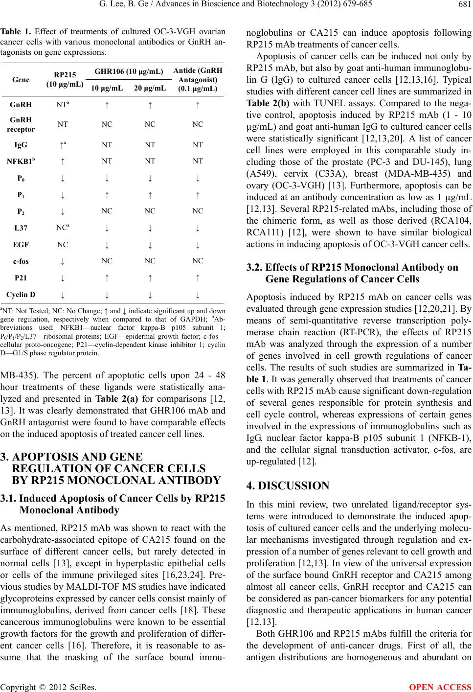 G. Lee, B. Ge / Advances in Bioscience and Biotechnology 3 (2012) 679-685 681 Table 1. Effect of treatments of cultured OC-3-VGH ovarian cancer cells with various monoclonal antibodies or GnRH an- tagonists on gene expressions. GHR106 (10 μg/mL) Gene RP215 (10 μg/mL) 10 μg/mL 20 μg/mL Antide (GnRH Antagonist) (0.1 μg/mL) GnRH NTa ↑ ↑ ↑ GnRH receptor NT NC NC NC IgG ↑a NT NT NT NFKB1b ↑ NT NT NT P0 ↓ ↓ ↓ ↓ P1 ↓ ↑ ↑ ↑ P2 ↓ NC NC NC L37 NCa ↓ ↓ ↓ EGF NC ↓ ↓ ↓ c-fos ↓ NC NC NC P21 ↓ ↑ ↑ ↑ Cyclin D ↓ ↓ ↓ ↓ aNT: Not Tested; NC: No Change; ↑ and ↓ indicate significant up and down gene regulation, respectively when compared to that of GAPDH; bAb- breviations used: NFKB1—nuclear factor kappa-B p105 subunit 1; P0/P1/P2/L37—ribosomal proteins; EGF—epidermal growth factor; c-fos— cellular proto-oncogene; P21—cyclin-dependent kinase inhibitor 1; cyclin D—G1/S phase regulator protein. MB-435). The percent of apoptotic cells upon 24 - 48 hour treatments of these ligands were statistically ana- lyzed and presented in Table 2(a) for comparisons [12, 13]. It was clearly demonstrated that GHR106 mAb and GnRH antagonist were found to have comparable effects on the induced apoptosis of treated cancer cell lines. 3. APOPTOSIS AND GENE REGULATION OF CANCER CELLS BY RP215 MONOCLONAL ANTIBODY 3.1. Induced Apoptosis of Cancer Cells by RP215 Monoclonal Antibody As mentioned, RP215 mAb was shown to react with the carbohydrate-associated epitope of CA215 found on the surface of different cancer cells, but rarely detected in normal cells [13], except in hyperplastic epithelial cells or cells of the immune privileged sites [16,23,24]. Pre- vious studies by MALDI-TOF MS studies have indicated glycoproteins expressed by cancer cells consist mainly of immunoglobulins, derived from cancer cells [18]. These cancerous immunoglobulins were known to be essential growth factors for the growth and proliferation of differ- ent cancer cells [16]. Therefore, it is reasonable to as- sume that the masking of the surface bound immu- noglobulins or CA215 can induce apoptosis following RP215 mAb treatments of cancer cells. Apoptosis of cancer cells can be induced not only by RP215 mAb, but also by goat anti-human immunoglobu- lin G (IgG) to cultured cancer cells [12,13,16]. Typical studies with different cancer cell lines are summarized in Table 2(b) with TUNEL assays. Compared to the nega- tive control, apoptosis induced by RP215 mAb (1 - 10 µg/mL) and goat anti-human IgG to cultured cancer cells were statistically significant [12,13,20]. A list of cancer cell lines were employed in this comparable study in- cluding those of the prostate (PC-3 and DU-145), lung (A549), cervix (C33A), breast (MDA-MB-435) and ovary (OC-3-VGH) [13]. Furthermore, apoptosis can be induced at an antibody concentration as low as 1 µg/mL [12,13]. Several RP215-related mAbs, including those of the chimeric form, as well as those derived (RCA104, RCA111) [12], were shown to have similar biological actions in inducing apoptosis of OC-3-VGH cancer cells. 3.2. Effects of RP215 Monoclonal Antibody on Gene Regulations of Cancer Cells Apoptosis induced by RP215 mAb on cancer cells was evaluated through gene expression studies [12,20,21]. By means of semi-quantitative reverse transcription poly- merase chain reaction (RT-PCR), the effects of RP215 mAb was analyzed through the expression of a number of genes involved in cell growth regulations of cancer cells. The results of such studies are summarized in Ta- ble 1. It was generally observed that treatments of cancer cells with RP215 mAb cause significant down-regulation of several genes responsible for protein synthesis and cell cycle control, whereas expressions of certain genes involved in the expressions of immunoglobulins such as IgG, nuclear factor kappa-B p105 subunit 1 (NFKB-1), and the cellular signal transduction activator, c-fos, are up-regulated [12]. 4. DISCUSSION In this mini review, two unrelated ligand/receptor sys- tems were introduced to demonstrate the induced apop- tosis of cultured cancer cells and the underlying molecu- lar mechanisms investigated through regulation and ex- pression of a number of genes relevant to cell growth and proliferation [12,13]. In view of the universal expression of the surface bound GnRH receptor and CA215 among almost all cancer cells, GnRH receptor and CA215 can be considered as pan-cancer biomarkers for any potential diagnostic and therapeutic applications in human cancer [12,13]. Both GHR106 and RP215 mAbs fulfill the criteria for the development of anti-cancer drugs. First of all, the antigen distributions are homgeneous and abundant on o Copyright © 2012 SciRes. OPEN ACCESS 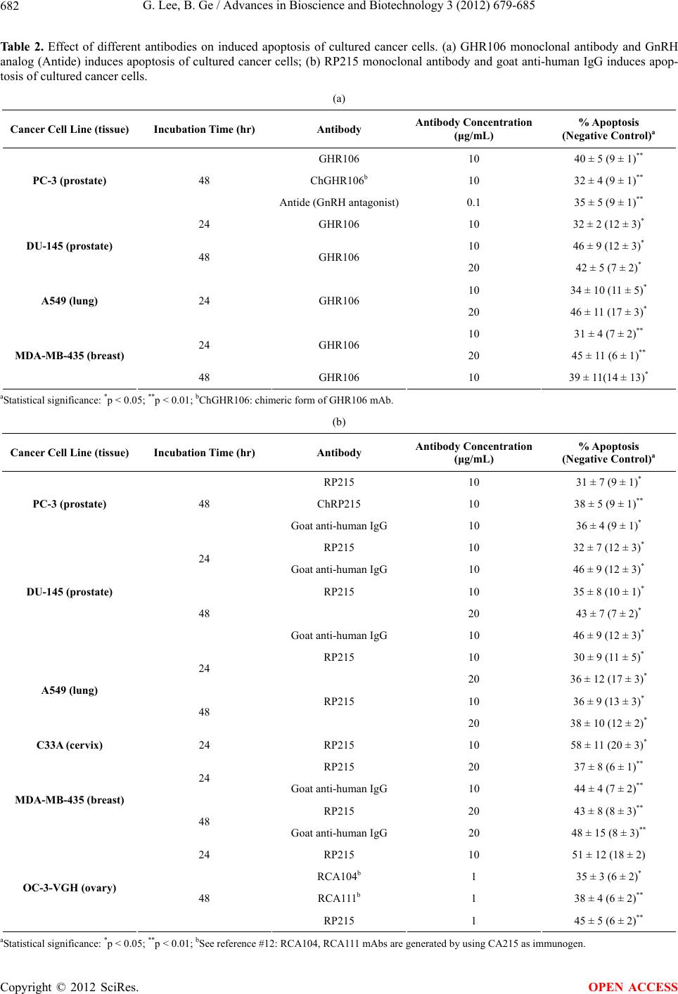 G. Lee, B. Ge / Advances in Bioscience and Biotechnology 3 (2012) 679-685 Copyright © 2012 SciRes. 682 OPEN ACCESS Table 2. Effect of different antibodies on induced apoptosis of cultured cancer cells. (a) GHR106 monoclonal antibody and GnRH analog (Antide) induces apoptosis of cultured cancer cells; (b) RP215 monoclonal antibody and goat anti-human IgG induces apop- tosis of cultured cancer cells. (a) Cancer Cell Line (tissue) Incubation Time (hr) Antibody Antibody Concentration (μg/mL) % Apoptosis (Negative Control)a GHR106 10 40 ± 5 (9 ± 1)** ChGHR106b 10 32 ± 4 (9 ± 1)** PC-3 (prostate) 48 Antide (GnRH antagonist)0.1 35 ± 5 (9 ± 1)** 24 GHR106 10 32 ± 2 (12 ± 3)* 10 46 ± 9 (12 ± 3)* DU-145 (prostate) 48 GHR106 20 42 ± 5 (7 ± 2)* 10 34 ± 10 (11 ± 5)* A549 (lung) 24 GHR106 20 46 ± 11 (17 ± 3)* 10 31 ± 4 (7 ± 2)** 24 GHR106 20 45 ± 11 (6 ± 1)** MDA-MB-435 (breast) 48 GHR106 10 39 ± 11(14 ± 13)* aStatistical significance: *p < 0.05; **p < 0.01; bChGHR106: chimeric form of GHR106 mAb. (b) Cancer Cell Line (tissue) Incubation Time (hr) Antibody Antibody Concentration (μg/mL) % Apoptosis (Negative Control)a RP215 10 31 ± 7 (9 ± 1)* ChRP215 10 38 ± 5 (9 ± 1)** PC-3 (prostate) 48 Goat anti-human IgG 10 36 ± 4 (9 ± 1)* RP215 10 32 ± 7 (12 ± 3)* 24 Goat anti-human IgG 10 46 ± 9 (12 ± 3)* RP215 10 35 ± 8 (10 ± 1)* 20 43 ± 7 (7 ± 2)* DU-145 (prostate) 48 Goat anti-human IgG 10 46 ± 9 (12 ± 3)* RP215 10 30 ± 9 (11 ± 5)* 24 20 36 ± 12 (17 ± 3)* RP215 10 36 ± 9 (13 ± 3)* A549 (lung) 48 20 38 ± 10 (12 ± 2)* C33A (cervix) 24 RP215 10 58 ± 11 (20 ± 3)* RP215 20 37 ± 8 (6 ± 1)** 24 Goat anti-human IgG 10 44 ± 4 (7 ± 2)** RP215 20 43 ± 8 (8 ± 3)** MDA-MB-435 (breast) 48 Goat anti-human IgG 20 48 ± 15 (8 ± 3)** 24 RP215 10 51 ± 12 (18 ± 2) RCA104b 1 35 ± 3 (6 ± 2)* RCA111b 1 38 ± 4 (6 ± 2)** OC-3-VGH (ovary) 48 RP215 1 45 ± 5 (6 ± 2)** aS tatistical significance: *p < 0.05; **p < 0.01; bSee reference #12: RCA104, RCA111 mAbs are generated by using CA215 as immunogen. 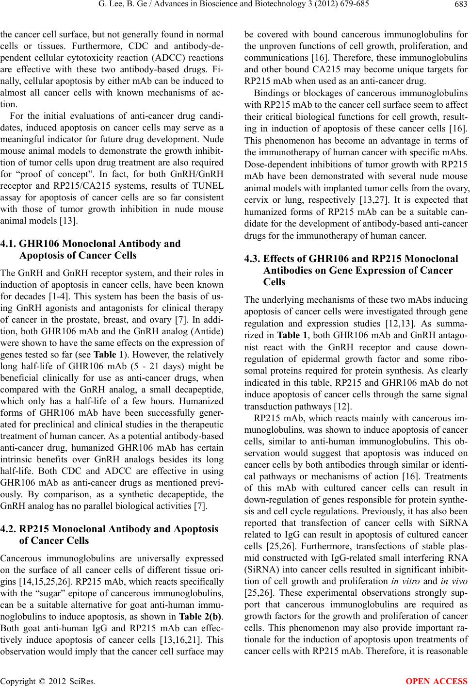 G. Lee, B. Ge / Advances in Bioscience and Biotechnology 3 (2012) 679-685 683 the cancer cell surface, but not generally found in normal cells or tissues. Furthermore, CDC and antibody-de- pendent cellular cytotoxicity reaction (ADCC) reactions are effective with these two antibody-based drugs. Fi- nally, cellular apoptosis by either mAb can be induced to almost all cancer cells with known mechanisms of ac- tion. For the initial evaluations of anti-cancer drug candi- dates, induced apoptosis on cancer cells may serve as a meaningful indicator for future drug development. Nude mouse animal models to demonstrate the growth inhibit- tion of tumor cells upon drug treatment are also required for “proof of concept”. In fact, for both GnRH/GnRH receptor and RP215/CA215 systems, results of TUNEL assay for apoptosis of cancer cells are so far consistent with those of tumor growth inhibition in nude mouse animal models [13]. 4.1. GHR106 Monoclonal Antibody and Apoptosis of Cancer Cells The GnRH and GnRH receptor system, and their roles in induction of apoptosis in cancer cells, have been known for decades [1-4]. This system has been the basis of us- ing GnRH agonists and antagonists for clinical therapy of cancer in the prostate, breast, and ovary [7]. In addi- tion, both GHR106 mAb and the GnRH analog (Antide) were shown to have the same effects on the expression of genes tested so far (see Table 1). However, the relatively long half-life of GHR106 mAb (5 - 21 days) might be beneficial clinically for use as anti-cancer drugs, when compared with the GnRH analog, a small decapeptide, which only has a half-life of a few hours. Humanized forms of GHR106 mAb have been successfully gener- ated for preclinical and clinical studies in the therapeutic treatment of human cancer. As a potential antibody-based anti-cancer drug, humanized GHR106 mAb has certain intrinsic benefits over GnRH analogs besides its long half-life. Both CDC and ADCC are effective in using GHR106 mAb as anti-cancer drugs as mentioned previ- ously. By comparison, as a synthetic decapeptide, the GnRH analog has no parallel biological activities [7]. 4.2. RP215 Monoclonal Antibody and Apoptosis of Cancer Cells Cancerous immunoglobulins are universally expressed on the surface of all cancer cells of different tissue ori- gins [14,15,25,26]. RP215 mAb, which reacts specifically with the “sugar” epitope of cancerous immunoglobulins, can be a suitable alternative for goat anti-human immu- noglobulins to induce apoptosis, as shown in Table 2(b). Both goat anti-human IgG and RP215 mAb can effec- tively induce apoptosis of cancer cells [13,16,21]. This observation would imply that the cancer cell surface may be covered with bound cancerous immunoglobulins for the unproven functions of cell growth, proliferation, and communications [16]. Therefore, these immunoglobulins and other bound CA215 may become unique targets for RP215 mAb when used as an anti-cancer drug. Bindings or blockages of cancerous immunoglobulins with RP215 mAb to the cancer cell surface seem to affect their critical biological functions for cell growth, result- ing in induction of apoptosis of these cancer cells [16]. This phenomenon has become an advantage in terms of the immunotherapy of human cancer with specific mAbs. Dose-dependent inhibitions of tumor growth with RP215 mAb have been demonstrated with several nude mouse animal models with implanted tumor cells from the ovary, cervix or lung, respectively [13,27]. It is expected that humanized forms of RP215 mAb can be a suitable can- didate for the development of antibody-based anti-cancer drugs for the immunotherapy of human cancer. 4.3. Effects of GHR106 and RP215 Monoclonal Antibodies on Gene Expression of Cancer Cells The underlying mechanisms of these two mAbs inducing apoptosis of cancer cells were investigated through gene regulation and expression studies [12,13]. As summa- rized in Table 1, both GHR106 mAb and GnRH antago- nist react with the GnRH receptor and cause down- regulation of epidermal growth factor and some ribo- somal proteins required for protein synthesis. As clearly indicated in this table, RP215 and GHR106 mAb do not induce apoptosis of cancer cells through the same signal transduction pathways [12]. RP215 mAb, which reacts mainly with cancerous im- munoglobulins, was shown to induce apoptosis of cancer cells, similar to anti-human immunoglobulins. This ob- servation would suggest that apoptosis was induced on cancer cells by both antibodies through similar or identi- cal pathways or mechanisms of action [16]. Treatments of this mAb with cultured cancer cells can result in down-regulation of genes responsible for protein synthe- sis and cell cycle regulations. Previously, it has also been reported that transfection of cancer cells with SiRNA related to IgG can result in apoptosis of cultured cancer cells [25,26]. Furthermore, transfections of stable plas- mid constructed with IgG-related small interfering RNA (SiRNA) into cancer cells resulted in significant inhibit- tion of cell growth and proliferation in vitro and in vivo [25,26]. These experimental observations strongly sup- port that cancerous immunoglobulins are required as growth factors for the growth and proliferation of cancer cells. This phenomenon may also provide important ra- tionale for the induction of apoptosis upon treatments of cancer cells with RP215 mAb. Therefore, it is reasonable Copyright © 2012 SciRes. OPEN ACCESS  G. Lee, B. Ge / Advances in Bioscience and Biotechnology 3 (2012) 679-685 684 to assume that humanized forms of RP215 mAb may have the potential to be developed as an effective anti- body-based anti-cancer drug with multi-indication for different types of cancer in humans. 5. CONCLUSION GHR106 and RP215 mAbs have been evaluated, with respect to their actions, to induce apoptosis on cultured cancer cells, of which the GnRH/GnRH receptor and RP215/CA215 systems are universally and abundantly present. It remains to be shown if the efficacy of these two mAbs in humanized forms can be demonstrated through extensive clinical studies for cancer therapy in humans in the near future. 6. ACKNOWLEDGEMENTS This work was supported in parts by NSERC-IRAP of Canada. The excellent technical support of Suefay Liu from McGill University and Vivian Wang from the University of British Columbia is acknowledged. REFERENCES [1] Millar, R.P. (2005) GnRHs and GnRH receptors. Animal Reproduction Science, 88, 5-28. doi:10.1016/j.anireprosci.2005.05.032 [2] Harrison, G.S., Wierman, M.E., Nett, T.M. and Glode, L.M. (2004) Gonadotropin-releasing hormone and its receptor in normal and malignant cells. Endocrine- Related Cancer, 11, 725-748. doi:10.1677/erc.1.00777 [3] Cheng, C.K. and Leung P.C.K. (2005) Molecular biology of gonadotropin-releasing hormone (GnRH)-I, GnRH-II, and their receptors in humans. Endocrine Reviews, 26, 283-306. doi:10.1210/er.2003-0039 [4] Flanagan, C., Millar, R. and Illing, N. (1997) Advances in understanding gonadotrophin-releasing hormone receptor structure and ligand interactions. Reviews of Reproduction, 2, 113-120. doi:10.1530/ror.0.0020113 [5] Grundker, C. and Emons G. (2003) Role of gonadotropin- releasing hormone (GnRH) in ovarian cancer. Reproduc- tive Biology and Endocrinology, 1, 65. http://www.RBEj.com/content/1/1/65 [6] Limonta, P., Moretti, R.M., Marelli, M.M. and Motta, M. (2003) The biology of gonadotropin hormone-releasing hormone: Role in the control of tumor growth and progression in humans. Frontiers in Neuroendocrinology, 24, 279-295. doi:10.1016/j.yfrne.2003.doi:10.003 [7] Felberbaum, R.E., Ludwig, M. and Diedrich, K. (2000) Clinical application of GnRH-antagonists. Molecular and Cellular Endocrinology, 166, 9-14. doi:10.1016/s0303-7207(00)00291-4 [8] Gründker, C., Völker, P., Schulz, K.-D. and Emons, G. (2000) Luteinizing hormone-releasing hormone agonist triptorelin and antagonist cetrorelix inhibit EGF-induced c-fos expression in human gynecological cancers. Gyne- cologic Oncology, 78, 194-202. doi:10.1006/gyno.2000.5863 [9] Hong, I.-S., Cheung, A.P. and Leung, P.C.K. (2008) Gon- adotropin-releasing hormones I and II induce apoptosis in human granulosa cells. Journal of Clinical Endocrinology and Metabolism, 93, 3179-3185. doi:10.1210/jc.2008-0127 [10] Lee, C.-Y.G., Ho, J., Chow, S.-N., Yasojima, K., Schwab, C. and McGeer, P.L. (2000) Immunoidentification of gonadotropin releasing hormone receptor in human sperm, pituitary and cancer cells. American Journal of Repro- ductive Immunology, 44, 170-177. doi:10.1111/j.8755-8920.2000.440307.x [11] Lee, G. and Ge, B. (2010) Growth inhibition of tumor cells in vitro by using monoclonal antibodies against gonadotropin-releasing hormone receptor. Cancer Immu- nology, Immunotherapy, 59, 1011-1019. doi:10.1007/s00262-010-0823-3 [12] Lee, G., Zhu, M. and Ge, B. (2012). Potential monoclonal antibody therapy for the treatment of ovarian cancer. In: Farghaly, S.A., Ed., Ovarian Cancer—Basic Science Per- spective, InTech, Vancouver, 385-406. [13] Lee, G., Cheung, A., Ge, B., Zhu, M., Giolma, B., Li, B., et al. (2012) CA215 and GnRH receptor as targets for cancer therapy. Cancer Immunology, Immunotherapy, 61, 1805-1817. doi:10.1007/s00262-012-1230-8 [14] Chen, Z., Qiu, X. and Gu, J. (2009) Immunoglobulin expression in non-lymphoid lineage and neoplastic cells. American Journal of Pathology, 174, 1139-1148. doi:10.2353/ajpath.2009.080879 [15] Hu, D., Zheng, H., Liu, H., Li, M., Ren, W., Liao, W., et al. (2008) Immunoglobulin expression and its biological significance in cancer cells. Cellular and Molecular Immunology, 5, 319-324. doi:10.1038/cmi.2008.39 [16] Qiu, X., Zhu, X., Zhang, L., Mao, Y., Zhang, J., Hao, P., et al. (2003) Human epithelial cancers secrete immu- noglobulin G with unidentified specificity to promote growth and survival of tumor cells. Cancer Research, 63, 6488-6495. http://cancerres.aacrjournals.org/content/63/19/6488.abstr act [17] Lee, C.Y., Chen, K.W., Sheu, F.S., Tsang, A., Chao, K.C. and Ng, H.T. (1992) Studies of a tumor-associated antigen, COX-1, recognized by a monoclonal antibody. Cancer Immunology, Immunotherapy, 35, 19-26. doi:10.1007/BF01741050 [18] Lee, G., Laflamme, E., Chien, C.-H. and Ting, H.H. (2008) Molecular identity of a pan cancer marker, CA215. Cancer Biology and Therapy, 7, 2007-2014. doi:10.4161/cbt.7.12.6984 [19] Lee, G. and Ge, B. (2009) Cancer cell expressions of immunoglobulin heavy chains with unique carbohydrate- associated biomarker. Cancer Biomarkers, 5, 177-188. doi:10.3233/cbm-2009-0102 [20] Lee, G., Zhu, M., Ge, B. and Potzold, S. (2012) Wide- spread expressions of immunoglobulin superfamily proteins in cancer cells. Cancer Immunology, Immuno- therapy, 61, 89-99. doi:10.1007/s00262-011-1088-1 [21] Lee, G. and Ge B. (2010) Inhibition of in vitro tumor cell Copyright © 2012 SciRes. OPEN ACCESS  G. Lee, B. Ge / Advances in Bioscience and Biotechnology 3 (2012) 679-685 Copyright © 2012 SciRes. 685 OPEN ACCESS growth by RP215 monoclonal antibody and antibodies raised against its anti-idiotype antibodies. Cancer Immu- nology, Immunotherapy, 59, 1347-1356. doi:10.1007/s00262-010-0864-7 [22] Kang, S.K., Tai, C.-J., Nathwani, P.S. and Leung, P.C.K. (2001) Differential regulation of two forms of gonado- tropin-releasing hormone messenger ribonucleic acid in human granulosa-luteal cells. Endocrinology, 142, 182- 192. doi:10.1210/en.142.1.182 [23] Niu, N., Zhang, J., Guo, Y., Zhao, Y., Korteweg, C. and Gu, J. (2011) Expression and distribution of immuno- globulin G and its receptors in the human nervous system. International Journal of Biochemistry and Cell Biology, 43, 556-563. doi:10.1016/j.biocel.20doi:10.12.012 [24] Niu, N., Zhang, J., Wang, S., Sun, Y., Korteweg, C., Gao, W., et al. (2011) Expression and distribution of immuno- globulin G and its receptors in an immune privileged site: The eye. Cellular and Molecular Life Sciences, 68, 2481- 2492. doi:10.1007/s00018-010-0572-7 [25] Hu, D., Duan, Z., Li, M., Jiang, Y., Liu, H., Zheng, H., et al. (2011) Heterogeneity of aberrant immunoglobulin expression in cancer cells. Cellular and Molecular Immu- nology, 8, 479-485. doi:10.1038/cmi.2011.25 [26] Li, M., Zheng, H., Duan, Z., Liu, H., Hu, D., Bode, A., et al. (2012) Promotion of cell proliferation and inhibition of ADCC by cancerous immunoglobulin expressed in cancer cell lines. Cellular and Molecular Immunology, 9, 54-61. doi:10.1038/cmi.2011.40 [27] Lee, G., Chu, R.-A. and Ting, H.H. (2009) Preclinical assessment of anti-cancer drugs by using RP215 mono- clonal antibody. Cancer Biology & Therapy, 8, 161-166. http://www.landesbioscience.com/journals/cbt/article/711 7/ |