

Paper Menu >>
Journal Menu >>
 Open Journal of Apoptosis, 2012, 1, 19-24 http://dx.doi.org/10.4236/ojapo.2012.13003 Published Online October 2012 (http://www.SciRP.org/journal/ojapo) Redox Factor-1 Mediates Inflammatory Response during Tumor Promotion in Skin Epidermal JB6 Cells Delira Robbins1, Wenjuan Li1, Katie Humphrey2, Yunfeng Zhao1* 1Department of Pharmacology, Toxicology & Neuroscience, Louisiana State University Health Sciences Center, Shreveport, USA 2Southwood High School, Shreveport, USA Email: [email protected] Received August 20, 2012; revised September 30, 2012; accepted October 19, 2012 ABSTRACT Recently, redox factor-1 (Ref-1) has received considerable attention as an enzyme for stimulating tumor cell growth. We hypothesized that Ref-1 is upregulated during the early stage of tumorigenesis. Utilizing both tumor promotion sen- sitive P+ and promotion resistant P− mouse skin epidermal JB6 cells, we found that Ref-1 expression was only induced in tumor promotion sensitive P+ cells following TPA treatment. Consistent with that, Ref-1 knockdown suppressed skin cell transformation. Interestingly, Ref-1 knockdown suppressed nuclear translocation of NF-kappaB subunit p65, and inhibited production of proinflammatory cytokines. These results suggest Ref-1 may promote early tumorigenesis and serve as a target for chemoprevention. Keywords: Ref-1; Tumorigenesis; JB6 Cells; Tumor Promotion; Chemoprevention 1. Introduction Apurinic/apyrimidinic (AP) sites can potentially block DNA replication and are cytotoxic and mutagenic to cells [1,2]. Human apurinic (apyrimidinic) endonuclease 1 (APE1) plays a key role in the base excision repair path- way (BER), and is a major repair protein for abasic sites [3]. APE1 was later coined redox factor-1 (Ref-1) due to its ability to sense changes in the intracellular redox status and activate transcription factors of the stress re- sponse pathway. APE1/Ref-1 is a ubiquitously expressed multifunctional protein also known to regulate the tranp- scription of stress inducible genes such as NF- B, p53, HIF-1 and AP-1. APE1/Ref1 expression levels have been found to be elevated in a number of cancers such as ovarian, cervical, prostate, rhabdomyosarcoma and germ cell tumors and correlated with the radiosensitivity of cervical cancers [4]. It has been suggested that APE1/Ref-1 provides an elo- quent link between cancer, DNA repair, transcription factor regulation, oxidative signaling and cell cycle con- trol which suggest APE1/Ref-1 as a potential chemopre- ventive target in tumorigenesis. In order to determine the role of Ref-1 in tumor promotion, we used tumor promo- tion-sensitive JB6 P+ and promotion-resistant JB6 P− mouse skin epidermal cells to observe the differential expression of APE1/Ref-1 following TPA treatment, and to determine how Ref-1 plays its role in early tumori- genesis. 2. Materials and Methods 2.1. Cell line, Reagents, Treatment The murine skin epidermal tumor promotion sensitive JB6 P+ and promotion resistant JB6 P− cells were pur- chased from American Type Culture Collection (ATCC, Rockville, MD). Cells were grown in EMEM medium containing 4% fetal bovine serum (Hyclone), 2 mM L-glutamine (Invitrogen), and 50 µg/ml penicillin/strep- tomycin (Invitrogen) in a 37°C incubator under 5% CO2. The tumor promoter phorbol ester, 12-O-tetradecanoyl- phorbol-13-aceteate (TPA; Sigma) was prepared as a 100 mM stock solution and directly diluted in cell culture medium, with the final concentration being 100 nM. 2.2. Total Cell Lysate Collected JB6 P+ and P− cells were suspended in 250 µl of phosphate buffered saline (PBS, pH 7.4) containing a proteinase inhibitor cocktail (Calbiochem). The JB6 cells were collected by centrifugation and resuspended in RIPA buffer (50 mM Tris, 150 mM NaCl, 0.1% SDS, 0.5% Na. deoxycholate and 1% Triton X-100) supple- mented with the proteinase cocktail (5 µg/ml each of pepstatin, leupeptin and aprotinin). Cells were sonicated on ice for two strokes (10 sec per stroke) using a Fisher *Corresponding author. C opyright © 2012 SciRes. OJApo D. ROBBINS ET AL. 20 Sonic Dismembrator (Model 100, Scale 4). After incu- bating on ice for 30 min, cell lysate was centrifuged at 18,000 × g for 20 min, and the supernatant was collected and designated as Total Cell Lysate. 2.3. Western Blot Analysis Protein concentrations of the samples were determined using a colorimetric assay (BioRad Laboratories). Thirty micrograms of total cell lysate were separated on 10% SDS-PAGE gels. Proteins were then transferred to nitro- cellulose membranes. The membranes were incubated against p65, Ref-1, β-actin, and SDHB proteins (Santa Cruz Biotechnology) to obtain the results. 2.4. Ref-1 siRNA Transfection Cells were seeded at 2 × 10⁵ cells per well in six-well tissue culture plates. The cells were incubated at 37˚C in a 5% CO2 incubator until they became 70% - 80% con- fluent. For each transfection, 3 µl of the Ref-1 siRNA du- plex (Santa Cruz Biotechnology,) were diluted into 100 µl of siRNA transfection medium (sc-36868, Santa Cruz Biotechnology) and labeled as Solution A. Solution B consisted of 6 µl of transfection reagent (Santa Cruz Biotechnology) diluted into 100 µl of siRNA transfection medium. Solution A and B were mixed gently and incu- bated for 30 minutes at room temperature. The cells were washed once with 2 ml of siRNA transfection medium. For each transfection, 0.8 ml of siRNA transfection me- dium were added to each tube containing the solution A/B mixture, mixed and directly added to the washed cells. Cells were incubated for 24 hours at 37˚C in a 5% CO2 incubator. Immediately following, the transfection mixture was removed and replaced with 2 ml of 1 × nor- mal growth medium. The cells were incubated for an additional 24 h and assayed via Western blot analysis. Fluorescein conjugated control siRNA (Santa Cruz Bio- technology) was used to monitor transfection efficiency. 2.5. Soft Agar Assay The soft agar cell transformation assay was carried out in six-well plates. The bottom agar was made using 1.25% agar, 2 × EMEM medium, 10% FBS, PBS, glutamine, and penicillin and was incubated in a hot water bath for 15 min. The mix was then divided and treated with vari- ous treatments. In each well, 3.5 mL of the agar mix was added and allowed to harden for 30 min. The top agar mix contained 0.5% bottom agar mix and 2 × 10⁵ of cells. The cell treatments were added at 2 × concentration to the top agar mix and 1 ml of each was added to each well. The agar was allowed to solidify and incubated in a 37˚C incubator under 5% CO2 for 14 d. Cells were stained with neutral red dye (0.25 mg/ml) 2-(p-Iodophenyl)-3- (p-nitrophenyl)-5-phenyl tetrazolium chloride hydrate (Aldrich #I-1,040-6) containing 1 mL per well. The dye was sonicated before added to cells. Cells were allowed to stain for twenty-four hours. 2.6. Quantification of Cytokine and Chemokine Proteins JB6 P+ cells were transfected with siRNA to Ref-1 as previously mentioned. Cells were collected and lysed using RIPA buffer to isolate the total cell lysate as pre- viously mentioned. One hundred micrograms of each cell lysate was used for cytokine and chemokine detection and quantification by the Quantibody Mouse Cytokine Array 1 kit (RayBiotech, Inc.) according to the manufac- turer’s protocol. The signal (Cy3) was captured using an Axon Genepix laser scanner (The Research Core Fa- cility at Louisiana State University Health Sciences Cen- ter, Shreveport, LA). Quantitative data and statistical analysis were performed using Prism 3.0 software. 2.7. Statistical Analysis One-way ANOVA followed by the Newman-Keuls post- test were used for multi-group comparisons. Experiments were repeated at least three times. Data were reported as mean ± standard error (S.E.M.) p < 0.05 was considered statistically significant. 3. Results 3.1. Ref-1 Expression Was Only Induced in Promotable JB6 P+ cells Following TPA Treatment We first tested whether the tumor promoting phorbol ester, TPA, could induce Ref-1 expression in mouse skin epidermal tumor promotion sensitive P+ and tumor pro- motion resistant P− cells. JB6 P+ and P− cells were treated with TPA (100 nM) for 24 hours. Ref-1 expres- sion was assessed via Western blot analysis. Our results (Figure 1) demonstrated that Ref-1 expression was only induced in JB6 P+ cells compared to JB6 P− cells as early as 24 h post TPA treatment. 3.2. Ref-1 Knockdown Suppressed TPA-Induced Skin Cell Transformation in Tumor Promotion-Sensitive Jb6 P+ Cells via Suppression of TPA-Induced P65 Nuclear Translocation To determine whether APE1/Ref-1 plays an important role in skin cell transformation, we knocked down APE1/Ref-1 utilizing siRNA and observed the effects of APE1/Ref-1 knockdown on anchorage-independent gro- wth in soft agar. We found that knockdown of APE1/ Copyright © 2012 SciRes. OJApo 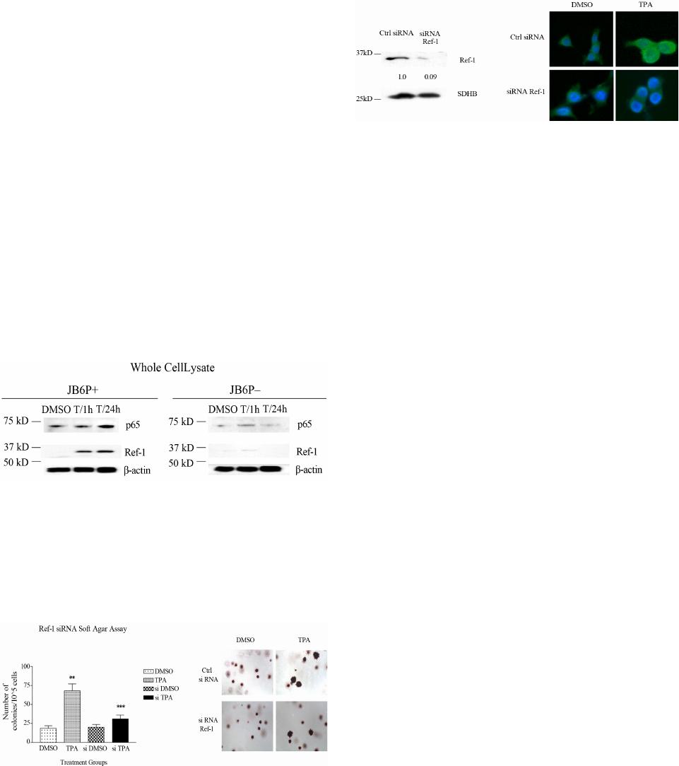 D. ROBBINS ET AL. 21 Ref-1 expression significantly decreased TPA-induced skin cell transformation (Figure 2). Ref-1 is known as a redox-sensitive upstream regulator of NF- B. We investigated the effect of TPA-induced Ref-1 expression on p65 expression, the transactivational subunit of NF- B. Similar to Ref-1 expression, we observed higher levels of p65 protein expression in JB6 P+ cells compared to P− cells. In Figure 3, we labeled phosphorylated p65 (Ser 536) with FITC staining and stained the nucleus with DAPI to detect p65 nuclear translocation induced by TPA treatment. To further reveal the potential mecha- nism of Ref-1 involvement in early stage TPA-induced tumor promotion, we knocked down Ref-1 expression via siRNA and investigated its effect on phosphorylated p65 TPA-induced nuclear translocation. We found fol- lowing TPA 24 h treatment that phosphorylated p65 (Ser536) translocated to the nucleus. Interestingly, Ref-1 knockdown suppressed phosphorylated p65 nuclear tran- slocation. Figure 1. TPA-induced Ref-1 activation was higher in pro- motable JB6 P+ cells compared to non-promotable JB6 P− cells. JB6 P+ and P− cells were treated with TPA (100 nM) for 1 or 24 h (T/1h; T/24 h). Whole cell lysate was prepared for the experiments. DMSO: 0.1% DMSO for 24 h. Western blot analysis of Ref-1, and p65 protein expression. β-actin, was used as a loading control. Experiments were repeated three times and a representative result is shown. Figure 2. Knockdown of Ref-1 suppressed TPA-induced skin cell transformation. (A) Soft agar colony formation assay was performed using siRNA-transfected cells. (B) Quantification of soft agar assay. siDMSO: siRNA Ref-1 transfected cells treated with 0.1% DMSO; siTPA: siRNA Ref-1 transfected cells treated with TPA (5 nM). * p< 0.05 significantly different from DMSO treatment group; ** p< 0.05. significantly different from TPA treatment group. Experiments were repeated three times and a representa- tive result is shown. Figure 3. Knockdown of Ref-1 suppressed TPA-induced phosphorylated p65 nuclear translocation. JB6 P+ cells were treated with TPA (100 nM) for 24 h. (A) Western blot analysis of Ref-1 siRNA transfection. (B) Immunofluores- cence of JB6 P+ cells. FITC green (phosphorylated p65); DAPI (nuclear staining). Experiments were repeated three times and a representative result is shown. 20X magnifica- tion. 3.3. Ref-1 Knockdown Suppressed Cytokine Expression in Tumor Promotion-Sensitive JB6 P+ Cells Previous reports that investigated the influence of in- flammation on carcinogenesis were concerned with the early stages of tumor development (i.e. tumor initiation and promotion). In addition, the pathogenesis of tumor development has been generally thought to occur either intrinsically through genetic alterations or extrinsically through external factors to stimulate tumor cells to re- lease inflammatory mediators [5]. To gain further insight into how APE1/Ref-1 may affect early stage tumor pro- motion, we progressed forward by assessing the effects of APE1/Ref-1 knockdown on NF- B-mediated cyto- kine/chemokine expression following TPA treatment in tumor promotion-sensitive JB6 P+ cells. Interestingly, we found that TPA treatment induced an inflammatory response of pro-inflammatory cytokines/chemokines, ma- inly those involved in leukocyte infiltration. In Figure 4, TPA 24 h treatment significantly increased KC, mono- cyte chemotactic protein-1 (MCP-1) and Regulated upon Activation, Normal T-cell Expressed and Secreted (RAN TES), IL-3 and IL-4. KC, also known as CXC1, is se- creted by human melanoma cells and has mitogenic properties. This cytokine is a main attractor of neutron- phils and expression is mediated by NF- B in mice [6]. MCP-1 expression has been observed in various inflame- matory diseases [7-9] and tumors [10]. This chemokine is directly induced by TPA treatment and plays a role in the chemotaxis of macrophages [11]. Lastly RANTES, is a chemokine for T cells, eosinophils and basophils and plays an active role in reactive oxygen species produc- ntion and recruitment of leukocytes to inflammatory sites [12]. Interestingly, knockdown of Ref-1 in both DMSO and TPA treated groups significantly suppressed the ex- pression of these cytokines/chemokines. Nevertheless, knockdown of Ref-1 significantly suppressed expression of both proinflammatory cytokines/chemokines and de- reased anchorage-independent growth of JB6 P+ cells. c Copyright © 2012 SciRes. OJApo  D. ROBBINS ET AL. Copyright © 2012 SciRes. OJApo 22 Figure 4. Knockdown of Ref-1 suppressed TPA-induced cytokine release in tumor promotable JB6 P+ cells. Quantification of pro-inflammatory cytokines/chemokines. siDMSO: siRNA Ref-1 transfected cells treated with 0.1% DMSO; siTPA: siRNA Ref-1 transfected cells treated with TPA (5 nM). *p< 0.05 significantly different from DMSO treatment group; **p< 0.05 sig- nificantly different from TPA treatment group. Experiments were repeated at least three times and a representative result is shown. 4. Discussion cal cysteine residues of NF- B [17], we further investi- gated the correlation of APE1/Ref-1 and NF- B expres- sion in early stage skin tumor promotion by comparing the expression of the NF- B transactivation subunit p65 in both promotion-sensitive (P+) and promotion-resistant (P−) cells. Elevated levels of p65 following TPA treat- ment were consistent with elevated levels of APE1/Ref-1 that occurred only in promotion-sensitive P+ cells, sug- gesting that APE1/Ref-1 and NF- B are involved in early stage tumor promotion in skin carcinogenesis. We demonstrated that targeting APE1/Ref-1 in early stage tumor promotion can have direct effects on proinflam- matory signaling and skin cell transformation. In addi- tion, knockdown of APE1/Ref-1 significantly reduced the expression of cytokines/chemokines and the colony forming ability of promotion-sensitive (P+) cells follow- ing TPA treatment. There are two common possibilities to explain the differential expression of APE1/Ref-1 in promotion-sensitive (P+) and promotion-resistant (P−) cells and the ability of APE1/Ref-1 knockdown to de- crease TPA-induced colony formation: 1) the current antioxidant status of the cell type; 2) a decrease in the stress response. Our previous studies have shown that chemoprevention via an antioxidant approach is a novel yet practical strategy to suppress early stage skin car- cinogenesis [18]. In addition, we have shown that pro- motion-resistant P− cells have higher MnSOD expression APE1/Ref-1 is ubiquitously ex pressed in cells with cell type specific cellular localization [13]. In addition, both expression and subcellular localization of APE1/Ref-1 are altered in hyperproliferative disorders such as aging and tumors. It has also been suggested that APE1/Ref-1 expression is inversely correlated with apoptosis sug- gesting that APE1/Ref-1 may contribute to early stage tumor promotion. We chose to assess the redox activity of APE1/Ref-1 in modulating NF- B nuclear transloca- tion and its effect on cytokine release and skin cell transformation. We utilized the murine JB6 skin epider- mal cell system to determine the role of APE1/Ref-1 in early stage tumorigenesis and whether APE1/Ref-1 is a potential novel target for chemoprevention. The mouse Balb/C JB6 cells [14] are the only well-characterized model to study tumor promotion. JB6 cells have two clone variants: tumor promotion sensitive P+ cells and promotion resistant P− cells. Herein, we found that tumor promotion sensitive JB6 P+ cells expressed higher APE1/ Ref-1 protein levels following TPA treatment which cor- relates with the current literature that suggests that APE1/Ref-1 expression is higher in cancer cells com- pared to their normal counterparts [15,16]. NF- B is a known transcription factor that is redox regulated by APE1/Ref-1 through reduction of the criti-  D. ROBBINS ET AL. 23 and activity when compared with promotion-sensitive P+ cells. In addition, several studies have shown that MnSOD may act as a novel tumor suppressor gene and that over- expression of MnSOD can significantly decrease tumor promotion and key oncogenic survival and pro-inflam- matory signaling pathways such as activator protein 1 (AP-1) and NF- B [19-21]. Therefore, regulating the int- racellular redox status has the potential to augment the effects of current chemotherapeutic treatments and also block survival and pro-inflammatory pathways in tumor promotion sensitive cancer cells. On the contrary, several studies have suggested that knockdown of APE1/Ref-1 can lead to apoptosis. APE1 /Ref-1 is known to be induced by oxidative stress [22]. However, APE1/Ref-1 expression has been shown to be inversely correlated with apoptosis, suggesting an anti- apoptotic function of the multifunctional protein [23]. Unnikrishnan and colleagues presented data that oxida- tive stress can alter the function of APE1/Ref-1 and the apoptotic response was increased in APE/Ref-1 haploin- sufficient mice [24]. Within this study heterozygous de- letion of APE1/Ref-1 resulted in decreased NF- B DNA binding activity, accompanied by increases in GADD45 gene expression, p53 stability and caspase activity [24]. Previous studies have also shown that loss of APE1 /Ref-1 resulted in increased TNF-induced apoptosis [25]. In addition, homozygous deletion of the APE1/Ref-1 gene is embryonic lethal, suggesting the importance of APE1/Ref-1 expression in cell survival. Therefore, mo- dulating APE1/Ref-1 expression can play either a cell survival or apoptotic role in tumorigenesis, but yet iden- tifies APE/Ref-1 as a key controller of intracellular redox changes in chemoprevention. Clinically, APE1/Ref-1 va- riants have been identified in the human population [26]; and these variants have been associated with increased cancer risk. Together, these data point toward an ap- proach to improve drug target specificity and personal- ized medicine in the prevention and treatment of tumors with upregulated APE1/Ref-1 expression. Nevertheless, further studies are ongoing to determine whether the sig- nificant decrease seen in the colony-forming ability of JB6 P+ cells was due to apoptosis or changes in mito- chondrial function. So far, we have not found any associ- ated toxicity following siRNA transfection and no indi- cations of an apoptotic phenotype in cells that underwent transfection. Moreover, we will continue to assess the role of APE1/Ref-1 redox activity in early tumor promo- ntion by utilizing known antioxidant-inducing natural products to determine whether antioxidant expression can modulate the redox functions of APE1/Ref-1 in early stage skin carcinogenesis. In conclusion, our results sug- gest that Ref-1 promotes early tumorigenesisby via con- tributing to NF-κB activation and the pro-inflammatory response. Therefore, Ref-1 might be able to serve as a novel target for chemoprevention. REFERENCES [1] L. A. Loeb and B. D. Preston, “Mutagenesis by Apu- rinic/Apyrimidinic Sites,” Annual Review of Genetics, Vol. 20, 1986, pp. 201-230. doi:10.1146/annurev.ge.20.120186.001221 [2] R. M. Schaaper, T. A. Kunkel and L. A. Loeb, “Infidelity of DNA Synthesis Associated with Bypass of Apurinic Sites,” Proceedings of the National Academy of Sciences of the United States, Vol. 80, No. 2, 1983, pp. 487-491. doi:10.1073/pnas.80.2.487 [3] D. M. Wilson and D. Barsky, “The Major Human Abasic Endonuclease: Formation, Consequences and Repair of Abasic Lesions in DNA,” Mutation Research, Vol. 485, No. 4, 2001, pp. 283-307. doi:10.1016/S0921-8777(01)00063-5 [4] A. R. Evans, M. Limp-Foster and M. R. Kelley, “Going APE over Ref-1,” Mutation Research, Vol. 461, No. 2, 2000, pp. 83-108. doi:10.1016/S0921-8777(00)00046-X [5] B. Muller-Hubenthal, M. Azemar, D. Lorenzen, M. Huber, M. A. Freudenberg, et al., “Tumor Biology: Tumour- Associated Inflammation versus Antitumor Immunity,” Anticancer Research, Vol. 29, No. 11, 2009, pp. 4795- 4806. [6] M. H. Han, W. K. Yon, H. Lee, et al., “Topical Applica- tion of Silymarin Reduces Chemical-Induced Irritant Contact Dermatitis in BALB/c Mice,” International Im- munopharmacology, Vol. 7, No. 13, 2007, pp. 1651-1658. doi:10.1016/j.intimp.2007.08.019 [7] P. C. Taylor, A. M. Peters, E. Paleolog, et al., “Reduction of Chemokine Levels and Leukocyte Traffic to Joints by Tumor Necrosis Factor Alpha Blockade in Patients with Rheumatoid Arthritis,” Arthritis & Rheumatism, Vol. 43, No. 1, 2000, pp. 38-47. doi:10.1002/1529-0131(200001)43:1<38::AID-ANR6>3. 0.CO;2-L [8] E. Van Coillie, J. Van Damme and G. Opdenakker, “The MCP/Eotaxin Subfamily of CC Chemokines,” Cytokine Growth Factor Reviews, Vol. 10, No. 1, 1999, pp. 61-86. doi:10.1016/S1359-6101(99)00005-2 [9] M. Tucci, C. Quatraro, M. A. Frassanito and F. Silvestris, “Deregulated Expression of Monocyte Chemoattractant Protein-1 (MCP-1) in Arterial Hypertension: Role of En- dothelial Inflammation and Atheromasia,” Journal of Hypertension, Vol. 24, 2006, pp. 1307-1318. doi:10.1097/01.hjh.0000234111.31239.c3 [10] M. O’Hayre, C. L. Salanga, T. M. Handel and S. J. Allen, “Chemokines and Cancer: Migration, Intracellular Sig- naling and Intercellular Communication in the Microen- vironment,” Biochemical Journal, Vol. 409, No. 3, 2008, pp. 635-649. doi:10.1042/BJ20071493 [11] J. Zhang, L. Chen, M. Xiao, et al., “FSP1+ Fibroblasts Promote Skin Carcinogenesis by Maintaining MCP-1 Mediated Macrophage Infiltration and Chronic Inflamma- tion,” American Journal of Pathology, Vol. 178, No. 1, 2011, pp. 382-390. doi:10.1016/j.ajpath.2010.11.017 [12] A. Kapp, G. Zeck-Kapp, W. Czech and E. Schopf, “The Chemokine RANTES is More than a Chemoattractant: Characterization of Its Effect on Human Eosinophil Oxi- Copyright © 2012 SciRes. OJApo  D. ROBBINS ET AL. Copyright © 2012 SciRes. OJApo 24 dative Metabolism and Morphology in Comparison with IL-5 and GM-CSF,” Journal of Investigative Dermatol- ogy, Vol. 102, No. 6, 1994, pp. 906-914. doi:10.1111/1523-1747.ep12383399 [13] S. Kakolyris, L. Kaklamanis, A. Giatromanolaki, M. Koukourakis, S. I. D. Hickson, et al., “Expression and Subcellular Localization of Human AP Endonuclease 1 (HAP1/Ref-1) Protein: A Basis for Its Role in Human Disease,” Histopath, Vol. 33, No. 6, 1998, pp. 561-569. doi:10.1046/j.1365-2559.1998.00541.x [14] N. H. Colburn, B. F. Former, K. A. Nelson and S. H. Yuspa, “Tumour Promoter Induces Anchorage Inde- pendence Irreversibly,” Nature, Vol. 281, 1979, pp. 589- 591. doi:10.1038/281589a0 [15] Y. Xu, D. H. Moore, J. Broshears, L. Liu, T. M. Wilson, et al., “The Apurinic/Apyrimidinic Endonuclease (APE/ Ref-1) DNA Repair Enzyme Is Elevated in Pre-Malignant and Malignant Cervical Cancer,” Anticancer Research, Vol. 17, No. 5B, 1997, pp. 3713-3719. [16] D. H. Moore, H. Michael, R. Tritt, S. H. Parsons and M. R. Kelley, “Alterations in the Expression of the DNA Repair/Redox Enzyme APE/Ref-1 in Epithelial Ovarian Cancers,” Clinical Cancer Research, Vol. 6, No. 2, 2000, pp. 602-609. [17] K. Ando, S. Hirao, Y. Kabe, Y. Ogura, I. Sato, Y. Yama- guchi, T. Wada and H. Handa, “A New APE1/Ref-1- Dependent Pathway Leading to Reduction of NF-Kappab and AP-1, and Activation of Their DNA-Binding Activ- ity,” Nucleic Acids Research, Vol. 36, No. 13, 2008, pp. 4327-4336. doi:10.1093/nar/gkn416 [18] J. Liu, X. Gu, D. Robbins, G. Li, R. Shi, et al., “Pro- tandim, a Fundamentally New Antioxidant Approach in Chemoprevention Using mouse Two-Stage Skin Car- cinogenesis as a Model,” PLoS One, Vol. 4, No. 4, 2009, p. e5284. doi:10.1371/journal.pone.0005284 [19] Y. Zhao, L. Chaiswing, T. D. Oberley, I. Batinic-Harberle, W. St. Clair, et al., “A Mechanism-Based Antioxidant Approach for the Reduction of Skin Carcinogenesis,” Cancer Research, Vol. 65, No. 4, 2005, pp. 1401-1405. doi:10.1158/0008-5472.CAN-04-3334 [20] Y. Zhao, Y. Xue, T. D. Oberley, K. K. Kiningham, S. M. Lin, et al., “Overexpression of Manganese Superoxide Dismutase Suppresses Tumor Formation by Modulation of Activator Protein-1 Signaling in a Multistage Skin Car- cinogenesis Model,” Cancer Research, Vol. 61, No. 16, 2001, pp. 6082-6088. [21] Y. Zhao, K. K. Kiningham, S. M. Lin and D. K. St. Clair, “Overexpression of MnSOD Protects Murine Fibrosar- coma Cells (FSa-II) from Apoptosis and Promotes a Dif- ferentiation Program upon Treatment with 5-Azacytidine: Involvement of MAPK and NFkappaB Pathways,” Anti- oxidants & Redox Signaling, Vol. 3, No. 3, 2001, pp. 375-386. doi:10.1089/15230860152409022 [22] S. Grosch, G. Fritz and B. Kaina, “Apurinic Endonucle- ase (Ref-1) Is Induced in Mammalian Cells by Oxidative Stress and Is Involved in Clastogenic Adaptation,” Can- cer Research, Vol. 58, No. 19, 1998, pp. 4410-4416. [23] C. V. Ramana, I. Boldogh, T. Izumi and S. Mitra, “Acti- vation of Apurinic/Apyrimidinic Endonuclease in Hu- man Cells by Reactive Oxygen Species and Its Correla- tion with Their Adaptive Response to Genotoxicity of Free Radicals,” Proceedings of the National Academy of Sciences of United States, Vol. 95, No. 9, 1998, pp. 5061- 5066. doi:10.1073/pnas.95.9.5061 [24] A. Unnikrishnan, J. J. Raffoul, H. V. Patel, T. M. Prychitko, N. Anyangwe, et al., “Oxidative Stress Alters Base Excision Repair Pathway and Increases Apoptotic Response in Apurinic/Apyrimidinic Endonuclease 1/Re- dox Factor-1 Haploinsufficient Mice,” Free Radical Bi- ology & Medicine, Vol. 46, No. 11, 2009, pp. 1488-1499. [25] J. L. Hall, X. Wang, V. Adamson, Y. Zhao and G. H. Gibbons, “Overexpression of Ref-1 Inhibits Hypoxia and Tumor Necrosis Factor-Induced Endothelial Cell Apop- tosis through Nuclear Factor-κB-Independent and -De- pendent Pathways,” Circulation Research, Vol. 88, 2001, pp. 1247-1253. doi:10.1161/hh1201.091796 [26] M. Z. Hadi, M. A. Coleman, K. Fidelis, H. W. Mohren- weiser and D. M. Wilson III, “Functional characterization of Ape1 variants identified in the human population,” Nucleic Acids Research, Vol. 28, No. 20, 2000, pp. 3871- 3879. doi:10.1093/nar/28.20.3871 Abbreviations AP, Apurinic/apyrimidinic; AP-1, activator protein 1; APE1, apurinic (apyrimidinic) endonuclease 1; DMSO, dimethyl sulfoxide; FBS, fetal bovine serum; MCP-1, monocyte chemotactic protein-1; MnSOD, manganese superoxide dismutase; NF-κB, nuclear factor kappa B; PBS, phosphate buffered saline; Ref-1, redox factor-1; ROS, reactive oxygen species; SDHB, succinate dehy- drogenase subunit B; siRNA, small interfering RNA; TNF, tumor necrosis factor; TPA, 12-O-tetradecanoy- lphorbol-13-aceteate. |