

Paper Menu >>
Journal Menu >>
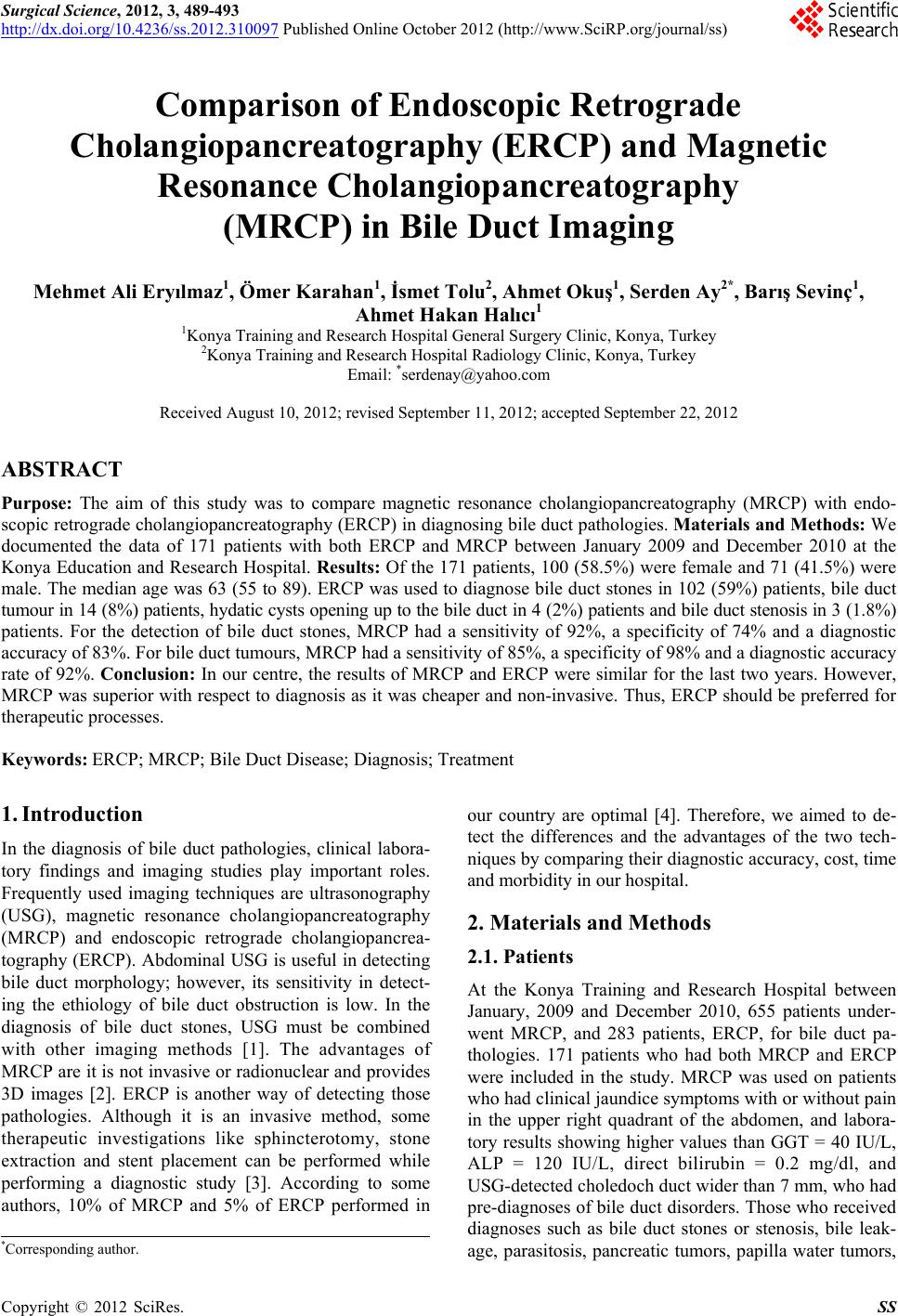 Surgical Science, 2012, 3, 489-493 http://dx.doi.org/10.4236/ss.2012.310097 Published Online October 2012 (http://www.SciRP.org/journal/ss) Comparison of Endoscopic Retrograde Cholangiopancreatography (ERCP) and Magnetic Resonance Cholangiopancreatography (MRCP) in Bile Duct Imaging Mehmet Ali Eryılmaz1, Ömer Karahan1, İsmet Tolu2, Ahmet Okuş1, Serden Ay2*, Barış Sevinç1, Ahmet Hakan Halıcı1 1Konya Training and Research Hospital General Surgery Clinic, Konya, Turkey 2Konya Training and Research Hospital Radiology Clinic, Konya, Turkey Email: *[email protected] Received August 10, 2012; revised September 11, 2012; accepted September 22, 2012 ABSTRACT Purpose: The aim of this study was to compare magnetic resonance cholangiopancreatography (MRCP) with endo- scopic retrograde cholangiopancreatography (ERCP) in diagnosing bile duct pathologies. Materials and Methods: We documented the data of 171 patients with both ERCP and MRCP between January 2009 and December 2010 at the Konya Education and Research Hospital. Results: Of the 171 patients, 100 (58.5%) were female and 71 (41.5%) were male. The median age was 63 (55 to 89). ERCP was used to diagnose bile duct stones in 102 (59%) patients, bile duct tumour in 14 (8%) patients, hydatic cysts opening up to the bile duct in 4 (2%) patients and bile duct stenosis in 3 (1.8%) patients. For the detection of bile duct stones, MRCP had a sensitivity of 92%, a specificity of 74% and a diagnostic accuracy of 83%. For bile duct tumours, MRCP had a sensitivity of 85%, a specificity of 98% and a diagnostic accuracy rate of 92%. Conclusion: In our centre, the results of MRCP and ERCP were similar for the last two years. However, MRCP was superior with respect to diagnosis as it was cheaper and non-invasive. Thus, ERCP should be preferred for therapeutic processes. Keywords: ERCP; MRCP; Bile Duct Disease; Diagnosis; Treatment 1. Introduction In the diagnosis of bile duct pathologies, clinical labora- tory findings and imaging studies play important roles. Frequently used imaging techniques are ultrasonography (USG), magnetic resonance cholangiopancreatography (MRCP) and endoscopic retrograde cholangiopancrea- tography (ERCP). Abdominal USG is useful in detecting bile duct morphology; however, its sensitivity in detect- ing the ethiology of bile duct obstruction is low. In the diagnosis of bile duct stones, USG must be combined with other imaging methods [1]. The advantages of MRCP are it is not invasive or radionuclear and provides 3D images [2]. ERCP is another way of detecting those pathologies. Although it is an invasive method, some therapeutic investigations like sphincterotomy, stone extraction and stent placement can be performed while performing a diagnostic study [3]. According to some authors, 10% of MRCP and 5% of ERCP performed in our country are optimal [4]. Therefore, we aimed to de- tect the differences and the advantages of the two tech- niques by comparing their diagnostic accuracy, cost, time and morbidity in our hospital. 2. Materials and Methods 2.1. Patients At the Konya Training and Research Hospital between January, 2009 and December 2010, 655 patients under- went MRCP, and 283 patients, ERCP, for bile duct pa- thologies. 171 patients who had both MRCP and ERCP were included in the study. MRCP was used on patients who had clinical jaundice symptoms with or without pain in the upper right quadrant of the abdomen, and labora- tory results showing higher values than GGT = 40 IU/L, ALP = 120 IU/L, direct bilirubin = 0.2 mg/dl, and USG-detected choledoch duct wider than 7 mm, who had pre-diagnoses of bile duct disorders. Those who received diagnoses such as bile duct stones or stenosis, bile leak- age, parasitosis, pancreatic tumors, papilla water tumors, *Corresponding author. C opyright © 2012 SciRes. SS 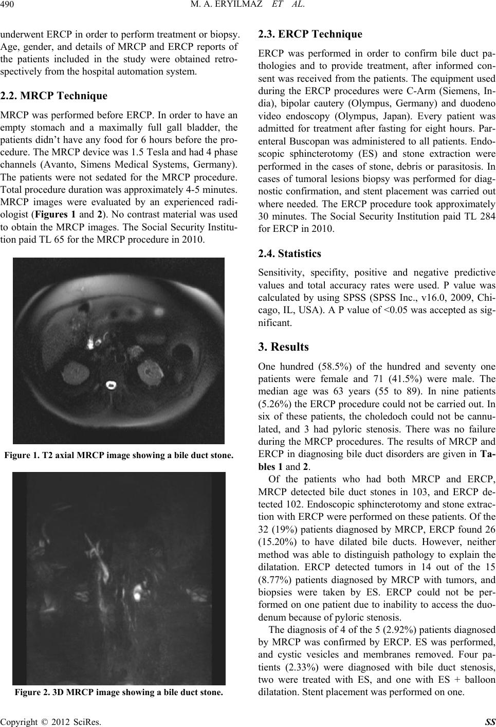 M. A. ERYILMAZ ET AL. 490 underwent ERCP in order to perform treatment or biopsy. Age, gender, and details of MRCP and ERCP reports of the patients included in the study were obtained retro- spectively from the hospital automation system. 2.2. MRCP Technique MRCP was performed before ERCP. In order to have an empty stomach and a maximally full gall bladder, the patients didn’t have any food for 6 hours before the pro- cedure. The MRCP device was 1.5 Tesla and had 4 phase channels (Avanto, Simens Medical Systems, Germany). The patients were not sedated for the MRCP procedure. Total procedure duration was approximately 4-5 minutes. MRCP images were evaluated by an experienced radi- ologist (Figures 1 and 2). No contrast material was used to obtain the MRCP images. The Social Security Institu- tion paid TL 65 for the MRCP procedure in 2010. Figure 1. T2 axial MRCP image showing a bile duct stone. Figure 2. 3D MRCP image showing a bile duct stone. 2.3. ERCP Technique ERCP was performed in order to confirm bile duct pa- thologies and to provide treatment, after informed con- sent was received from the patients. The equipment used during the ERCP procedures were C-Arm (Siemens, In- dia), bipolar cautery (Olympus, Germany) and duodeno video endoscopy (Olympus, Japan). Every patient was admitted for treatment after fasting for eight hours. Par- enteral Buscopan was administered to all patients. Endo- scopic sphincterotomy (ES) and stone extraction were performed in the cases of stone, debris or parasitosis. In cases of tumoral lesions biopsy was performed for diag- nostic confirmation, and stent placement was carried out where needed. The ERCP procedure took approximately 30 minutes. The Social Security Institution paid TL 284 for ERCP in 2010. 2.4. Statistics Sensitivity, specifity, positive and negative predictive values and total accuracy rates were used. P value was calculated by using SPSS (SPSS Inc., v16.0, 2009, Chi- cago, IL, USA). A P value of <0.05 was accepted as sig- nificant. 3. Results One hundred (58.5%) of the hundred and seventy one patients were female and 71 (41.5%) were male. The median age was 63 years (55 to 89). In nine patients (5.26%) the ERCP procedure could not be carried out. In six of these patients, the choledoch could not be cannu- lated, and 3 had pyloric stenosis. There was no failure during the MRCP procedures. The results of MRCP and ERCP in diagnosing bile duct disorders are given in Ta- bles 1 and 2. Of the patients who had both MRCP and ERCP, MRCP detected bile duct stones in 103, and ERCP de- tected 102. Endoscopic sphincterotomy and stone extrac- tion with ERCP were performed on these patients. Of the 32 (19%) patients diagnosed by MRCP, ERCP found 26 (15.20%) to have dilated bile ducts. However, neither method was able to distinguish pathology to explain the dilatation. ERCP detected tumors in 14 out of the 15 (8.77%) patients diagnosed by MRCP with tumors, and biopsies were taken by ES. ERCP could not be per- formed on one patient due to inability to access the duo- denum because of pyloric stenosis. The diagnosis of 4 of the 5 (2.92%) patients diagnosed by MRCP was confirmed by ERCP. ES was performed, and cystic vesicles and membranes removed. Four pa- tients (2.33%) were diagnosed with bile duct stenosis, two were treated with ES, and one with ES + balloon dilatation. Stent placement was performed on one. Copyright © 2012 SciRes. SS 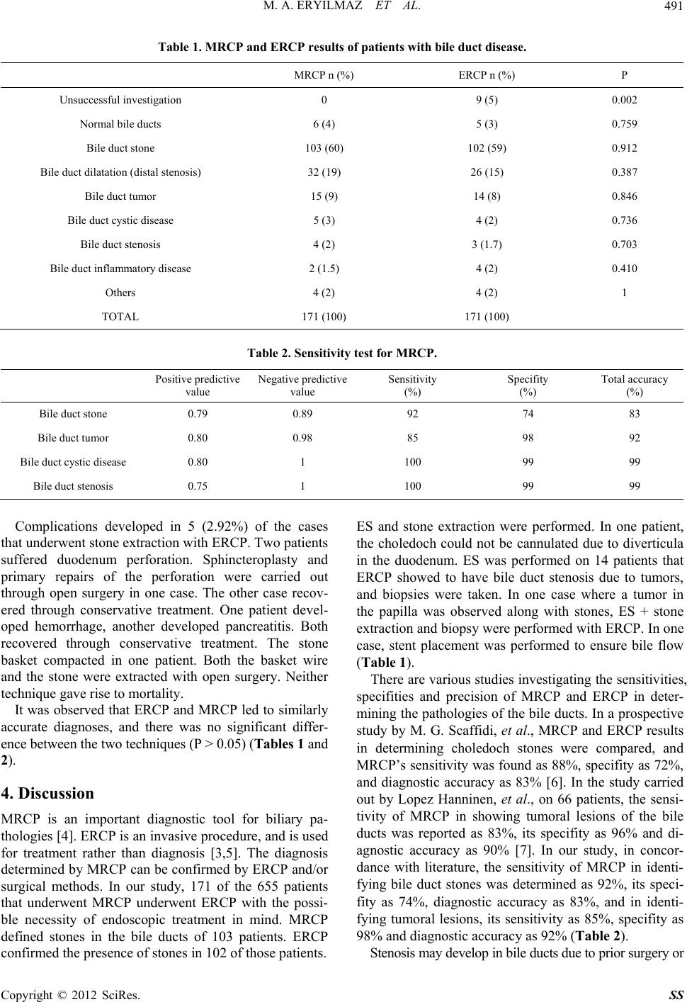 M. A. ERYILMAZ ET AL. Copyright © 2012 SciRes. SS 491 Table 1. MRCP and ERCP results of patients with bile duct disease. MRCP n (%) ERCP n (%) P Unsuccessful investigation 0 9 (5) 0.002 Normal bile ducts 6 (4) 5 (3) 0.759 Bile duct stone 103 (60) 102 (59) 0.912 Bile duct dilatation (distal stenosis) 32 (19) 26 (15) 0.387 Bile duct tumor 15 (9) 14 (8) 0.846 Bile duct cystic disease 5 (3) 4 (2) 0.736 Bile duct stenosis 4 (2) 3 (1.7) 0.703 Bile duct inflammatory disease 2 (1.5) 4 (2) 0.410 Others 4 (2) 4 (2) 1 TOTAL 171 (100) 171 (100) Table 2. Sensitivity test for MRCP. Positive predictive value Negative predictive value Sensitivity (%) Specifity (%) Total accuracy (%) Bile duct stone 0.79 0.89 92 74 83 Bile duct tumor 0.80 0.98 85 98 92 Bile duct cystic disease 0.80 1 100 99 99 Bile duct stenosis 0.75 1 100 99 99 Complications developed in 5 (2.92%) of the cases that underwent stone extraction with ERCP. Two patients suffered duodenum perforation. Sphincteroplasty and primary repairs of the perforation were carried out through open surgery in one case. The other case recov- ered through conservative treatment. One patient devel- oped hemorrhage, another developed pancreatitis. Both recovered through conservative treatment. The stone basket compacted in one patient. Both the basket wire and the stone were extracted with open surgery. Neither technique gave rise to mortality. It was observed that ERCP and MRCP led to similarly accurate diagnoses, and there was no significant differ- ence between the two techniques (P > 0.05) (Tables 1 and 2). 4. Discussion MRCP is an important diagnostic tool for biliary pa- thologies [4]. ERCP is an invasive procedure, and is used for treatment rather than diagnosis [3,5]. The diagnosis determined by MRCP can be confirmed by ERCP and/or surgical methods. In our study, 171 of the 655 patients that underwent MRCP underwent ERCP with the possi- ble necessity of endoscopic treatment in mind. MRCP defined stones in the bile ducts of 103 patients. ERCP confirmed the presence of stones in 102 of those patients. ES and stone extraction were performed. In one patient, the choledoch could not be cannulated due to diverticula in the duodenum. ES was performed on 14 patients that ERCP showed to have bile duct stenosis due to tumors, and biopsies were taken. In one case where a tumor in the papilla was observed along with stones, ES + stone extraction and biopsy were performed with ERCP. In one case, stent placement was performed to ensure bile flow (Table 1). There are various studies investigating the sensitivities, specifities and precision of MRCP and ERCP in deter- mining the pathologies of the bile ducts. In a prospective study by M. G. Scaffidi, et al., MRCP and ERCP results in determining choledoch stones were compared, and MRCP’s sensitivity was found as 88%, specifity as 72%, and diagnostic accuracy as 83% [6]. In the study carried out by Lopez Hanninen, et al., on 66 patients, the sensi- tivity of MRCP in showing tumoral lesions of the bile ducts was reported as 83%, its specifity as 96% and di- agnostic accuracy as 90% [7]. In our study, in concor- dance with literature, the sensitivity of MRCP in identi- fying bile duct stones was determined as 92%, its speci- fity as 74%, diagnostic accuracy as 83%, and in identi- fying tumoral lesions, its sensitivity as 85%, specifity as 98% and diagnostic accuracy as 92% (Table 2). Stenosis may develop in bile ducts due to prior surgery or  M. A. ERYILMAZ ET AL. 492 to primary sclerosing cholangitis. According to literature, MRCP’s sensitivity in showing the localization and length of the stenosis is 100% its specifity is 91%, and diagnos- tic accuracy is 95% [8]. Our study results are similar. In this field, we found MRCP’s sensitivity to be 100%, specifity, 99% and diagnostic accuracy, 99%. Bile duct obstruction due to hydatic cysts opening into the bile duct can be seen in 5% - 25% of patients. ERCP and ES are one of the treatment methods [9]. In our study, hydatic cyst lesions opening into the bile duct were iden- tified in 4 (2.33%) patients. ES was performed by ERCP, and the vesicles and membranes of the cyst were re- moved. In our study, the sensitivity of MRCP was deter- mined as 100%, specifity as 99%, and diagnostic accu- racy as 99%. There was no significant difference be- tween MRCP and ERCP (P > 0.05). Stones that are smaller than the section thickness of the MR device may give false negative results, and in patients where there is a gap of more than 72 hours be- tween the MRCP and the ERCP, although MRCP has indicated stones, ERCP may not find stones due to their spontaneous passing [10]. In such cases, ES is a thera- peutical procedure [11]. In our study, 32 patients by MRCP and 26 patients by ERCP were detected to have dilated bile ducts that could not be explained by any ob- structive pathology such as stones or tumors, and ES was performed. MRCP and diagnostic ERCP were compared for cost effectiveness through a literature scan over 28 articles by Kaltenthaler et al., and it was decided that MRCP was more economical [12]. In our study, it was determined that the cost of one MRCP event cost TL 65, while one ERCP event cost approximately TL 284. Even in experienced hands, ERCP has a cannulation failure rate of up to 3% - 9% [13]. In our study, it has been observed that ERCP didn’t give satisfactory results in 5.26% of the cases. ERCP, considered a reference method presently, is accepted to have a morbidity rate of 0.8% - 10% [14]. Morbidity was 2.92% in our study. In one case where a bile duct stone was detected, the mor- bidity was due to basket compaction. Two cases had perforations, one had a hemorrhage and one had pa- creatitis. T. Obana et al., have observed no mortalities connected to the ERCP procedure [15]. However, low that it may be, ERCP has a mortality rate of 0.7% - 0.9% [16]. There were no mortalities in our study. Our study has shown the ERCP procedures which are routinely carried out by gastroenterologists can be carried out successfully by experienced General Surgeons. 10% of the MRCPs and 5% of the ERCPs carried out in our country in the beginning of the 2000s were optimally successful [11]. In the 2010 plus years, MRCP can be carried out on all cases where clinicians deem it neces- sary without the need for sedation, and ERCP is carried out with success rates of up to 95% (Table 1). Our study has some shortcomings. While MRCP was carried out on all patients with bile duct pathologies, se- lected patients underwent ERCP. ERCP and MRCP were not carried out on the same day. The diagnosis reached with MRCP was confirmed with ERCP. Surgery was performed in cases where ERCP was unsuccessful. In our country, the cost of MRCP and ERCP are based upon the unit price paid by the social security institution, and may differ from other countries. 5. Conclusion In the identification of bile duct pathologies, MRCP has accuracy values near to that of ERCP. It is approximately four times more economical, and takes far less time. Therefore, MRCP should be used for diagnosis, and ERCP should be performed for treatment, if necessary. REFERENCES [1] E. Elüdokuz, E. Tankurt, H. Akbaylar, E. İğci, O. Dicle, İ. Şimşek, et al., “The Sensitivity of Ultrasonography ın Cases Choledocholithiasis Diagnosed with ERCP,” Tur- kiye Klinikleri Journal of Gastroenterohepatology, Vol. 7, 1996, pp. 108-110. [2] P. D. Prabhakar, A. M. Prabhakar, H. B. Prabahakar and D. Sahani, “Magnetic Resonance Cholangiopancreato- graphy of Benign Disorders of the Biliary System,” Mag- netic Resonance Imaging Clinics of North America, Vol. 18, No. 3, 2010, pp. 497-514. doi:10.1016/j.mric.2010.08.007 [3] G. Ersöz, “Endoscopic Retrograde Cholangiopancreato- graphy in Bile Duct Disorders,” Türkiye Klinikleri Jour- nal of Medical Sciences, Vol. 3, 2007, pp. 12-16. [4] H. Şentürk, B. Canbakan and E. Özden, “ERCP: Indica- tions and Risks,” Series of Clinical Approach in Gastro- enterology, Vol. 30, 2004, pp. 101-109. [5] F. Maccioni, M. Martinelli, N. Al Ansari, A. Kagarmanova, et al., “Magnetic Resonance Cholangiography: Past, Pre- sent and Future: A Review,” European Review for Medi- cal and Pharmacological Sciences, Vol. 14, No. 8, 2010, pp. 721-725. [6] E. C. Kaltenthaler, S. J. Walters, J. Chilcott, A. Blake- borough, Y. B. Vergel and S. Thomas, “MRCP Compared to Diagnostic ERCP for Diagnosis When Biliary Obstruc- tion Is Suspected: A Systematic Review,” BMC Medical Imaging, Vol. 6, 2006, pp. 9-24. doi:10.1186/1471-2342-6-9 [7] M. G. Scaffidi, C. Luigiano, P. Consolo, R. Pellicano, G. Giacobbe, M. Gaeta, et al., Magnetic Resonance Cholan- giopancreatography versus Endoscopic Retrograde Cho- langiopancreatography in the Diagnosis of Common Bile Duct Stones: A Prospective Comparative Study,” Miner- va Medica, Vol. 100, No. 5, 2009, pp. 341-348. [8] E. L. Hänninen, H. Amthauer, N. Hosten, J. Ricke, M. Böhmig, J. Langrehr, et al., “Prospective Evaluation of Pancreatic Tumours: Accuracy of MR Imaging with MR Copyright © 2012 SciRes. SS  M. A. ERYILMAZ ET AL. Copyright © 2012 SciRes. SS 493 Cholangiopancreatography and MR Angiography,” Radi- ology, Vol. 224, No. 1, 2002, pp. 34-41. doi:10.1148/radiol.2241010798 [9] P. Angulo, D. H. Pearce, C. D. Johnson, J. J. Henry, N. F. LaRusso, B. T. Petersen, et al., “Magnetic Resonance Cholangiopancreatography in Patients with Biliary Dis- ease: Its Role in Primary Sclerosing Cholangitis,” Journal of Hepatology, Vol. 33, No. 4, 2000, pp. 520-527. doi:10.1016/S0168-8278(00)80002-1 [10] G. Galati, A. V. Sterpetti, M. Caputo, M. Adduci, G. Lu- candri, S. Brozzetti, et al., “Endoscopic Retrograde Cho- langiography for Intrabiliary Rupture of Hydatic Cyst,” The American Journal of Surgery, Vol. 191, No. 2, 2006, pp. 206-210. doi:10.1016/j.amjsurg.2005.09.014 [11] M. M. Calvo, L. Bujanda, A. Calderon, J. L. Cabrıada, A. Bernal, V. Orive, et al., “Role of Magnetic Resonance Cholangiopancreatography in Patients with Suspected Choledocholithiasis,” Mayo Clinic Proceedings, Vol. 77, No. 5, 2002, pp. 422-428. doi:10.1016/S0025-6196(11)62210-6 [12] E. Kaltenthaler, Y. B. Vergel, J. Chilcott, S. Thomas, T. Blakeborough, S. J. Waters, et al., “A Systematic Review and Economic Evaluation of Magnetic Resonance Cholan- giopancreatography Compared with Diagnostic Endoscopic Retrograde Cholangiopancreatography,” Health Technol- ogy Assessment, Vol. 8, No. 10, 2004, pp. 1-89. [13] R. Schöfl, “Diagnostic Endoscopic Retrograde Cholan- giopancreatography,” Endoscopy, Vol. 33, No. 2, 2001, pp. 147-157. doi:10.1055/s-2001-11667 [14] M. L. Freeman, D. B. Nelson, S. Sherman, G. B. Haber, M. E. Herman, P. J. Dorsher, et al., “Complications of Endoscopic Biliary Sphincterotomy,” The New England Journal of Medicine, Vol. 335, No. 13, 1996, pp. 909-919. doi:10.1056/NEJM199609263351301 [15] T. Obana, N. Fujita, Y. Noda, G. Kobayashi, K. Ito, J. Horaguchi, et al., “Efficacy and Safety of Therapeutic ERCP for the Elderly with Choledocholithiasis: Compa- rison with Younger Patients,” Internal Medicine, Vol. 49, No. 18, 2010, pp. 1935-1941. doi:10.2169/internalmedicine.49.3660 [16] K. Hekimoğlu, Y. Üstündağ, A. Duşak, Z. Erdem, B. Karademir, S. Aydemir, et al., “MRCP vs ERCP in the Evaluation of Biliary Pathologies: Review of Current Lit- erature,” Journal of Digestive Diseases, Vol. 9, No. 3, 2008, pp. 162-169. doi:10.1111/j.1751-2980.2008.00339.x |