

Paper Menu >>
Journal Menu >>
 Open Journal of Veterinary Medicine, 2012, 2, 158-162 http://dx.doi.org/10.4236/ojvm.2012.23025 Published Online September 2012 (http://www.SciRP.org/journal/ojvm) Tacrolimus for the Treatment of Inflammatory B owel Disease in a Dog Masashi Yuki*, Yosiaki Oota, Noriaki Nagata Yuki Animal Hospital, Nagoya, Japan Email: *[email protected] Received July 12, 2012; revised August 18, 2012; accepted August 24, 2012 ABSTRACT A 2-year-old intact female Toy poodle was referred with a 2-week history of diarrhea. Blood examination findings in- dicated thrombocytosis, severe hypoproteinemia, and hypoalbuminemia; endoscopy revealed duodenal mucosal irregu- larity and increased graininess. Based on these results and additional histopathological findings, we made a diagnosis of protein-losing enteropathy caused by lymphocytic-plasmacytic enteritis with lymphangiectasia. The dog was initially treated with prednisolone. Improvement was only observed with high-dose prednisolone; its dose could not be reduced without relapse. When cyclosporin, methotrexate, and chlorambucil were combined with prednisolone, no further bene- ficial effect was observed. When tacrolimus was combined with prednisolone, improvement was seen and the dose of prednisolone could be reduced. Tacrolimus is both a calcineurin inhibitor and a multi-drug-resistant inhibitor, so it may be an effective treatment choice for a dog refractory to standard inflammatory bowel disease treatment. This is the first report of tacrolimus for the treatment of inflammatory bowel disease in dogs. Keywords: Dog; Inflammatory Bowel Disease; Tacrolimus 1. Introduction Canine inflammatory bowel disease (IBD) is a chronic, immunologically mediated intestinal disorder resulting from the complex interaction of environmental, genetic, and immune factors [1]. Variations in the histologic ap- pearance of the inflammation suggest that IBD is not a single disease entity and the nomenclature merely re- flects the predominant cell type present. Lymphocytic- plasmacytic enteritis (LPE) is the most common form reported, eosinophilic gastroenteritis is less common, and granulomatous enteritis and neutrophilic infiltration are rare in IBD [2]. Protein-losing enteropathy (PLE) may occur secondary to conditions such as chronic inflamma- tion in small intestinal diseases and is often caused by intestinal lymphangiectasia following LPE [3]. The development of LPE is thought to originate as a consequence of a deregulation of mucosal immunity in predisposed animals. The immune-mediated basis of the disease can be inferred by the response to the administra- tion of immunosuppressive drugs [4]. Immunosuppres- sive drugs such as prednisolone, azathioprine, chloram- bucil, cyclosporin, and methotrexate are often used in the treatment of this disorder [5], but achievement and main- tenance of remission may be difficult using these agents. Tacrolimus is an immunosuppressive macrolide iso- lated from the fermentation broth of Streptomyces tsu- kubaensis. It potently inhibits helper T lymphocyte acti- vation [6]. Tacrolimus is the primary immunosuppressive agent developed for organ transplantation. In recent re- views of human refractory IBD, tacrolimus had therapeu- tic efficacy, whereas prednisolone did not [7]. Recently, in veterinary medicine, tacrolimus in ointment form has begun to be used in the treatment of immune-mediated skin diseases and keratoconjunctive disease [8-12]. In the present case, despite using a combination of prednisolone and cyclosporin, methotrexate, and chlo- rambucil, which is a potent, immunosuppressive agent, the expected effect was not obtained. However, improve- ment was confirmed when tacrolimus was substituted for the trio of cyclosporin, methotrexate, and chlorambucil, and rapid remission was achieved and maintained. 2. Case Description A 3.5 kg, 2-year-old intact female Toy poodle was re- ferred to our hospital with a 2-week history of diarrhea. Upon physical examination, the dog had a temperature of 38.1˚C, a heart rate of 144 beats per minute, and a respi- ratory rate of 30 breaths per minute; all were within normal limits. The dog had a body condition score of 2 out of 5 and revealed a pendulous abdomen. Examina- tions of the mucus membranes and heart sounds were *Corresponding author. C opyright © 2012 SciRes. OJVM 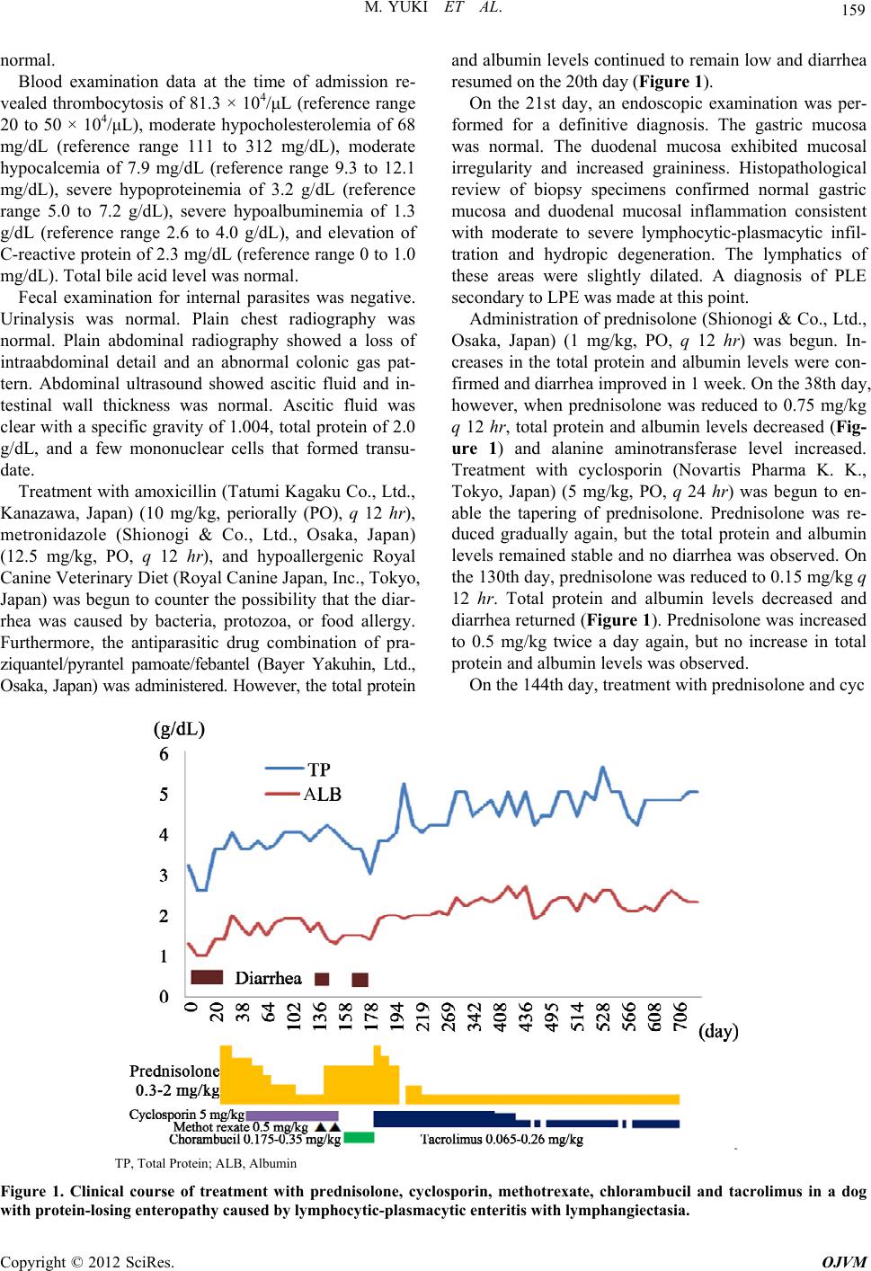 M. YUKI ET AL. 159 normal. Blood examination data at the time of admission re- vealed thrombocytosis of 81.3 × 10⁴/μL (reference range 20 to 50 × 104/μL), moderate hypocholesterolemia of 68 mg/dL (reference range 111 to 312 mg/dL), moderate hypocalcemia of 7.9 mg/dL (reference range 9.3 to 12.1 mg/dL), severe hypoproteinemia of 3.2 g/dL (reference range 5.0 to 7.2 g/dL), severe hypoalbuminemia of 1.3 g/dL (reference range 2.6 to 4.0 g/dL), and elevation of C-reactive protein of 2.3 mg/dL (reference range 0 to 1.0 mg/dL). Total bile acid level was normal. Fecal examination for internal parasites was negative. Urinalysis was normal. Plain chest radiography was normal. Plain abdominal radiography showed a loss of intraabdominal detail and an abnormal colonic gas pat- tern. Abdominal ultrasound showed ascitic fluid and in- testinal wall thickness was normal. Ascitic fluid was clear with a specific gravity of 1.004, total protein of 2.0 g/dL, and a few mononuclear cells that formed transu- date. Treatment with amoxicillin (Tatumi Kagaku Co., Ltd., Kanazawa, Japan) (10 mg/kg, periorally (PO), q 12 hr), metronidazole (Shionogi & Co., Ltd., Osaka, Japan) (12.5 mg/kg, PO, q 12 hr), and hypoallergenic Royal Canine Veterinary Diet (Royal Canine Japan, Inc., Tokyo, Japan) was begun to counter the possibility that the diar- rhea was caused by bacteria, protozoa, or food allergy. Furthermore, the antiparasitic drug combination of pra- ziquantel/pyrantel pamoate/febantel (Bayer Yakuhin, Ltd., Osaka, Japan) was administered. However, the total protein and albumin levels continued to remain low and diarrhea resumed on the 20th day (Figure 1). On the 21st day, an endoscopic examination was per- formed for a definitive diagnosis. The gastric mucosa was normal. The duodenal mucosa exhibited mucosal irregularity and increased graininess. Histopathological review of biopsy specimens confirmed normal gastric mucosa and duodenal mucosal inflammation consistent with moderate to severe lymphocytic-plasmacytic infil- tration and hydropic degeneration. The lymphatics of these areas were slightly dilated. A diagnosis of PLE secondary to LPE was made at this point. Administration of prednisolone (Shionogi & Co., Ltd., Osaka, Japan) (1 mg/kg, PO, q 12 hr) was begun. In- creases in the total protein and albumin levels were con- firmed and diarrhea improved in 1 week. On the 38th day, however, when prednisolone was reduced to 0.75 mg/kg q 12 hr, total protein and albumin levels decreased (Fig- ure 1) and alanine aminotransferase level increased. Treatment with cyclosporin (Novartis Pharma K. K., Tokyo, Japan) (5 mg/kg, PO, q 24 hr) was begun to en- able the tapering of prednisolone. Prednisolone was re- duced gradually again, but the total protein and albumin levels remained stable and no diarrhea was observed. On the 130th day, prednisolone was reduced to 0.15 mg/kg q 12 hr. Total protein and albumin levels decreased and diarrhea returned (Figure 1). Prednisolone was increased to 0.5 mg/kg twice a day again, but no increase in total protein and albumin levels was observed. On the 144th day, treatment with prednisolone and cyc A TP, Total Protein; ALB, Albumin Figure 1. Clinical course of treatment with prednisolone, cyclosporin, methotrexate, chlorambucil and tacrolimus in a dog with protein-losing enteropathy caused by lymphocytic-plasmacytic enteritis with lymphangiectasia. Copyright © 2012 SciRes. OJVM  M. YUKI ET AL. 160 losporine with methotrexate (Wyeth, Tokyo, Japan) (0.5 mg/kg, intramuscularly, once a week) was started. Diar- rhea improved, although no increases in total protein and albumin levels were observed. Vomiting, which is a side effect of methotrexate, was observed. On the 151st day, cyclosporin was discontinued because it did not produce sufficient effect and its cost was a problem. On the 158th day, chlorambucil (Glaxo Smith Kline, Bern, Switzerland) (0.35 mg/kg, PO, q 24 hr) was substituted for meth- otrexate. Vomiting, which is a side effect of chloram- bucil, and diarrhea were observed. On the 178th day, a severe decrease in total protein and albumin levels was seen, tacrolimus (Astellas Pharma Inc., Tokyo, Japan) (0.13 mg/kg, PO, q 12 hr) was sub- stituted for chlorambucil, and prednisolone was increased to 1 mg/kg q 12 hr again. The increase in the total protein and albumin levels was confirmed and diarrhea improved after 1 week (Figur e 1). On the 194th day, moreover, pred- nisolone was discontinued 4 days into the course due to a bite wound, no decrease in total protein and albumin lev- els was seen, and a temporary increase of globulin was observed (Figure 1). On the 219th day, prednisolone dosing was gradually reduced to a low-dose level (0.15 mg/kg, PO, q 12 hr) while maintaining an increase of total protein and albumin levels. In addition, the trough level of the tacrolimus at this point was below 2 ng/mL (Mitsubishi Chemical Medience Corporation, Tokyo, Japan). On the 290th day, the total protein and albumin levels reached the reference ranges and it was possible to get a complete remission (Figure 1). Tacrolimus dosing was gradually reduced to 0.065 mg/kg every other day from the 401st day to the 467th day. However, decreases in total protein and albumin levels were observed clearly (Figure 1). On the 467th day, tacrolimus dosing was increased again to 0.065 mg/kg q 24 hr. On the 509th day, total protein and albu- min levels improved into the reference ranges. On the 538th day, tacrolimus dosing was reduced to every other day again to relieve the burden of its cost. However, a decrease of total protein and albumin levels was ob- served and tacrolimus was again increased to q 24 hr (Figure 1). More than 800 days have passed since tac- rolimus treatment was begun; no adverse effect has been observed and treatment progress is good. 3. Discussion The mechanism of action of tacrolimus is similar to that of cyclosporin, even though their chemical structures differ greatly [6]. Tacrolimus binds to immunophilins, which are cytoplasmic binding proteins. While tacrolimus binds to immunophilins called FK-binding proteins (FKBPs), cyc- losporin binds to immunophilins called cyclophilins. The immunophilin-drug complex binds competitively to and inhibits calcineurin, a phosphatase whose activity is de- pendent on its being bound to calcium and calmodulin. Inhibition of calcineurin is believed to mediate the im- munosuppressive activity of both tacrolimus and cyc- losporine [6]. Tacrolimus has been shown to inhibit the transcription of the early activation genes for cytokines such as interleukin 2, tumor necrosis factor α, and interferon γ in T cells [7]. Although its mode of action is similar to that of cyclosporin, the immunosuppressive effect of tacrolimus is 30 - 100 times greater in vitro and 10 - 20 times greater in vivo than that of cyclosporin [7]. In recent reviews of human refractory IBD, the cal- cineurin inhibitor tacrolimus had therapeutic efficacy [7]. In recent reviews of veterinary medicine, the calcineurin inhibitor cyclosporin was also effective in dogs with re- fractory IBD. The anti-inflammatory effect of cyclosporin in human and dogs IBD are belived to be due to suppres- sion of activated T cells infiltrating the mucosa [13]. In this case, it was insufficient, but the use of tac- rolimus with the same mechanism was tried because some effect of cyclosporin was seen. In human IBD, tac- rolimus is used at a rate of 0.025 mg/kg given orally every 12 hours, with a trough level of 5 - 15 ng/mL [7]. In dogs, a high dose of 0.4 - 2.0 mg/kg seems to be needed to get this trough level [14,15]. However, severe adverse effects including body weight loss and pneumo- nia may result from the high dose of 0.4 - 2.0 mg/kg [15]. Clinically, the therapeutic dose of tacrolimus for dogs has not been determined, so we used as a reference the dosage in human organ transplantation [6]. The trough level in this case was below 2 ng/mL, low- er than the recommended therapeutic dose for human IBD. However, effectiveness was observed. All cytokine production was inhibited completely at a blood level of 1 ng/mL in a basic study of tacrolimus in humans; the 50% inhibitory concentration was 0.02 - 0.11 ng/mL and an inhibitory effect was indicated by a low concentration [16]. The trough level was not recorded in a report on tacrolimus for the treatment of sterile panniculitis in a dog, but the effect was observed at a low dose of 0.06 mg/kg/day [17]. Therefore, there is a possibility that even a low dose and a low trough level can be effective in dogs. However, because in this case, tacrolimus was given every other day, the resulting lower total protein and albumin levels indicated the need for daily admini- stration. Recently, the high expression of p-glycoprotein was confirmed in lymphocytes from duodenum mucous mem- brane lamina propria after treatment with prednisolone in dogs with IBD and possible drug resistance by p-glyco- protein was suggested [18]. Presently, p-glycoprotein and cytochrome P-450 3A4, 3A5 manifested in the small intestine and liver appear to be cooperatively functioning as the cause of the absorption barrier of tacrolimus [19]. On the other hand, cyclosporin and tacrolimus can block Copyright © 2012 SciRes. OJVM  M. YUKI ET AL. 161 the p-glycoprotein of resistant cells and are viewed as multi-drug-resistant inhibitors that obstruct the multi- drug resistance function [20]. It is thought that calcineurin inhibitors have different actions and that these actions are manifested early at low concentrations [20,21]. An insuf- ficient effect was observed from the use of cyclosporin in this case. However, with tacrolimus, an immediate effect was observed with its low dose, so its multi-drug resis- tant inhibitor actions may be involved in this case. In humans, the most common adverse effects seen with tacrolimus include headache, tremors, insomnia, hyperesthesia, and musculoskeletal complaints [6]. In this case, low-dose tacrolimus has been given for more than 2 years, but no adverse effect has been observed. However, attention will be necessary for the sign because adverse effect include body weight loss and pneumonia in the dogs [15]. In conclusion, because tacrolimus is both a calcineurin inhibitor and a multi-drug-resistant inhibitor, it may be an effective treatment choice for dogs refractory to IBD treatment. More studies about the effect on IBD and the clinical dose of tacrolimus in dogs are needed. To the best of our knowledge, this is the first report of tac- rolimus for the treatment of IBD in dogs. REFERENCES [1] A. E. Jergens, “Inflammatory Bowel Disease: Current Perspectives,” Veterinary Clinics of North America: Small Animal Practice, Vol. 29, No. 2, 1999, pp. 501-521. [2] S. J. Ettinger and E. C. Feldman, “Disease of the Small Intestine,” In: E. D. Hall and A. J. German, Eds., Text- book of Veterinary Internal Medicine, Saunders Co., London, 2010, pp. 1526-1572. [3] P. B. Peterson and M. D. Willard, “Protein-Losing En- teropathies,” Veterinary Clinics of North America: Small Animal Practice, Vol. 33, No. 5, 2003, pp. 1061-1082. doi:10.1016/S0195-5616(03)00055-X [4] M. Cerquetella, A. Spaterna, F. Laus, B. Tesei, G. Rossi, E. Antonelli, V. Villanacci and G. Bassotti, “Inflamma- tory Bowel Disease in the Dog: Differences and Similari- ties with Humans,” World Journal of Gastroenterology, Vol. 16, No. 9, 2010, pp. 1050-1056. doi:10.3748/wjg.v16.i9.1050 [5] S. J. Ettinger and E. C. Feldman, “Disease of the Large Intestine,” In: K. Allenspach, Ed., Textbook of Veterinary Internal Medicine, Saunders Co., Philadelphia, 2010, pp. 1573-1594. [6] P. A. Kelly, G. J. Burckart and R. Venkataramanan, “Ta- crolimus: A New Immunosuppressive Agent,” American Journal of Health-System Pharmacy, Vol. 52, No. 14, 1995, pp. 1521-1535. [7] H. Ogata, T. Matsui, M. Nakamura, M. Iida, M. Takazoe, Y. Suzuki and T. Hibi, “A Randomised Dose Finding Study of Oral Tacrolimus (FK506) Therapy in Refractory Ulcerative Colitis,” An International Journal of Gastro- enterology & Hepatology, Vol. 55, No. 11, 2006, pp. 1255-1262. doi:10.1136/gut.2005.081794 [8] B. S. Misseghers, A. G. Binnington and K. A. Mathews, “Clinical Observations of the Treatment of Canine Peri- anal Fistulas with Topical Tacrolimus in 10 Dogs,” Ca- nadian Veterinary Journal, Vol. 41, No. 8, 2000, pp. 623- 627. [9] J. D. Griffies, C. L. Mendelsohn, W. S. Rosenkrantz, R. Muse, M. J. Boord and C. E. Griffin, “Topical 0.1% Tac- rolimus for the Treatment of Discoid Lupus Erythemato- sus and Pemphigus Erythematosus in Dogs,” Journal of the American Animal Hospital Association, Vol. 40, No. 1, 2004, pp. 29-41. [10] R. Marsella, C. F. Nicklin, S. Saglio and J. Lopez, “In- vestigation on the Clinical Efficacy and Safety of 0.1% Tacrolimus Ointment (Protopic) in Canine Atopic Der- matitis: A Randomized, Double-Blinded, Placebo-Con- trolled, Cross-Over Study,” Veterinary Dermatology, Vol. 15, No. 5, 2004, pp. 294-303. doi:10.1111/j.1365-3164.2004.00397.x [11] A. Berdoulay, R. V. English and B. Nadelstein, “Effect of Topical 0.02% Tacrolimus Aqueous Suspension on Tear Production in Dogs with Keratoconjunctivitis Sicca,” Ve- terinary Ophthalmology, Vol. 8, No. 4, 2005, pp. 225- 232. doi:10.1111/j.1463-5224.2005.00390.x [12] B. J. Stanley and J. G. Hauptman, “Long-Term Prospec- tive Evaluation of Topically Applied 0.1% Tacrolimus Ointment for Treatment of Perianal Sinuses in Dogs,” Journal of the American Veterinary Medical Association, Vol. 235, No. 4, 2009, pp. 397-404. doi:10.2460/javma.235.4.397 [13] K. Allenspach, S. Rufenacht, S. Sauter, A. Gröne, J. Steffan, G. Strehlau and F. Gaschen, “Pharmacokinetics and Clin- ical Efficacy of Cyclosporin Treatment of Dogs with Steroid-Refractory Inflammatory Bowel Disease,” Jour- nal of Veterinary Internal Medicine, Vol. 20, No. 2, 2006, pp. 239-244. doi:10.1111/j.1939-1676.2006.tb02852.x [14] S. Todo, Y. Ueda, J. A. Demetris, O. Imventarza, M. Nalesnik, R. Venkataramanan, L. Makowka and T. E. Starzl, “Immunosuppression of Canine, Monkey, and Baboon Allografts by FK 506: With Special Reference to Synergism with Other Drugs and to Tolerance Induction,” Surgery, Vol. 104, No. 2, 1988, pp. 239-249. [15] T. Fujisawa, Y. Saitoh, N. Urabe, T. Takeda, Y. Sekine, M. Baba and Y. Yamaguchi, “Dose Study of the Immu- nosuppression of FK 506 in Canine Lung Allo-Trans- plantation,” Surgery Today, Vol. 23, No. 4, 1993, pp. 338-343. doi:10.1007/BF00309052 [16] S. Sakuma, Y. Higashi, N. Sato, T. Sasakawa, T. Sengoku, Y. Ohkubo, T. Amaya and T. Goto, “Tacrolimus Sup- pressed the Production of Cytokines Involved in Atopic Dermatitis by Direct Stimulation of Human PBMC Sys- tem (Comparison with Steroids),” International Im- munopharmacology, Vol. 1, No. 6, 2001, pp. 1219-1226. doi:10.1016/S1567-5769(01)00059-5 [17] R. Kano, K. Okabayashi, Y. Nakamura, K. Fujiwara, A. Hasegawa and Y. Sasaki, “Systemic Treatment of Sterile Panniculitis with Tacrolimus and Prednisolone in Dogs,” Journal of Veterinary Medical Science, Vol. 68, No. 1, Copyright © 2012 SciRes. OJVM  M. YUKI ET AL. Copyright © 2012 SciRes. OJVM 162 2006, pp. 95-96. doi:10.1292/jvms.68.95 [18] K. Allenspach, P. J. Bergman, S. Sauter, A. Gröne, M. G. Doherr and F. Gaschen, “P-Glycoprotein Expression in Lamina Propria Lymphocytes of Duodenal Biopsy Sam- ples in Dogs with Chronic Idiopathic Enteropathies,” Journal of Comparative Pathology, Vol. 134, No. 1, 2006, pp. 1-7. doi:10.1016/j.jcpa.2005.06.003 [19] S. Masuda and K. Inui, “An Up-Date Review on Indi- vidualized Dosage Adjustment of Calcineurin Inhibitors in Organ Transplant Patients,” Pharmacology & Thera- peutics, Vol. 112, No. 1, 2006, pp. 184-198. doi:10.1016/j.pharmthera.2006.04.006 [20] K. Suzuki, K. Saito, S. Tsujimura, S. Nakayamada, K. Yamaoka, N. Sawamukai, S. Iwata, M. Nawata, K. Na- kano and Y. Tanaka, “Tacrolimus, a Calcineurin Inhibitor, Overcomes Treatment Unresponsiveness Mediated by P-Glycoprotein on Lymphocytes in Refractory Rheuma- toid Arthritis,” The Journal of Rheumatology, Vol. 37, No. 3, 2010, pp. 512-520. doi:10.3899/jrheum.090048 [21] S. Tsujimura, K. Saito, S. Nakayamada, K. Nakano, J. Tsukada, K. Kohno and Y. Tanaka, “Transcriptional Re- gulation of Multidrug Resistance-1 Gene by Interleukin-2 in Lymphocytes,” Genes to Cells, Vol. 9, No. 12, 2004, pp. 1265-1273. doi:10.1111/j.1365-2443.2004.00803.x |