

Paper Menu >>
Journal Menu >>
 Open Journal of Veterinary Medicine, 2012, 2, 89-97 http://dx.doi.org/10.4236/ojvm.2012.23016 Published Online September 2012 (http://www.SciRP.org/journal/ojvm) Safety and Efficacy of Cefovecin (Convenia®) as an Adjunctive Treatment of Periodontal Disease in Dogs Henry Giboin, Csilla Becskei, Jacky Civil, Michael R. Stegemann* Veterinary Medicine Research & Development, Pfizer Animal Health, Zaventem, Belgium Email: *[email protected] Received April 30, 2012; revised June 8, 2012; accepted June 15, 2012 ABSTRACT This study was designed to determine the safety and efficacy of cefovecin (Convenia®; Pfizer Animal Health) when compared to clindamycin (Antirobe®; Pfizer Animal Health) as an adjunctive therapy to periodontal scaling or surgery for severe periodontal disease in dogs. A multi-centre, double-masked, randomised study was conducted in 299 dogs with severe periodontal disease. Clindamycin, administered once daily at 11 mg/kg bodyweight orally for 10 days fol- lowing dental surgery was compared with a single, subcutaneous injection of cefovecin (8 mg/kg bodyweight) adminis- tered at the time of dental surgery. The primary efficacy parameter assessed was percentage of tooth-root sites bleeding when probed (an indicator of gingival inflammation) 42 days after surgery. Two-hundred and ninety-one (291) dogs were included in the efficacy assessments. Cefovecin was shown to be non-inferior to clindamycin. The percentage of sites bleeding on probing was reduced from 54.3% to 20.3% for the cefovecin group (53.1% reduced to 17.4% for the clindamycin group). There were no suspected adverse drug experiences attributed to treatment with cefovecin or clin- damycin. Cefovecin was shown to be as effective and safe adjunctive treatment for severe periodontal disease in dogs undergoing periodontal scaling and surgery as clindamycin. Keywords: Canine; Antibiotics; Dentistry; Porphyromonas gulae; Prevotella intermedia 1. Introduction Periodontal disease is the most common dental infection in dogs [1]. It is caused by the accumulation of plaque and an associated change in periodontal bacterial flora (from commensal aerobic to pathogenic anaerobic bacte- ria such as Porphyromonas spp. and Prevotella spp) [2-5]. Periodontal disease is a collective term for a num- ber of inflammatory conditions affecting the periodon- tium around the tooth (attached gingiva, periodontal liga- ment, cementum of the root and alveolar bone). It pro- gresses from reversible gingivitis, characterised by in- flamed and often bleeding gingiva, to periodontitis with the associated inflammatory tissue damage, the formation of deep periodontal pockets or gingival recession, loss of epithelial attachment and bone resorption. The end result of periodontitis is loss of the tooth due to progressive destruction of its periodontium [6]. As periodontal disease disturbs the integrity of mucous membranes, periodontal pathogens can be exported via the blood stream. In severe periodontal disease, bacte- raemia may even occur during minimal mechanical dis- turbance, such as normal mastication, without any pro- fessional tooth cleaning or surgery [7,8]. Periodontal dis- ease is associated with myocardial infarction and stroke in humans [9] and with abscesses in organs, endocarditis and glomerulonephritis in dogs [10,11]. In dogs, prevention of periodontal disease by home applied hygiene as in humans is rarely possible and the effect of poor plaque control is often only managed by periodontal treatment (e.g. debridement, scaling, polish- ing). However, mechanical periodontal therapy alone will not adequately reduce the periodontopathogen load [12,13]. In severe and progressive cases adjunctive anti- biotic treatment is justified to augment a reduction in periodontopathogens and physiological bacterial flora, thereby facilitating detoxification of the periodontium from detrimental bacterial toxins and aiding gingival healing [13-16]. Results from clinical studies in dogs support this notion, as systemic treatment with clinda- mycin in periodontal disease significantly decreased plaque scores, gingivitis and pocket depths compared to placebo when used as adjunctive therapy to ultrasonic scaling, root planing and polishing [17-19]. Neither in human nor in veterinary dentistry is there a consensus in the choice of antimicrobial agent or in the minimum duration required to successfully treat perio- *Corresponding author. C opyright © 2012 SciRes. OJVM  H. GIBOIN ET AL. 90 dontal infections [13]. Here we show that in a large clinical study, a single subcutaneous administration of cefovecin at 8 mg/kg effectively aided healing of the periodontal tissues after professional periodontal treat- ment and surgery in dogs. Cefovecin is an extended-spectrum cephalosporin, ap- proved for veterinary use in dogs and cats. It is formu- lated as an injectable solution containing 80 mg/ml cefo- vecin sodium. Following subcutaneous administration in dogs, cefovecin has a long elimination half-life (5.5 days), low clearance (0.76 ml/hour/kg) and therapeutic tissue concentrations are maintained for approximately 14 days [20]. As a consequence, prolonged therapeutic efficacy can be maintained through injections adminis- tered at 14-day intervals. Cefovecin, administered as a subcutaneous injection at 14-day intervals, was highly effective in the treatment of both superficial and deep canine pyoderma [21,22] and also of wounds and ab- scesses in both cats [23,24] and dogs [21,22]. Further- more, cefovecin was demonstrated to be an effective and safe treatment for urinary tract infections in dogs [25] and cats [26]. 2. Materials and Methods 2.1. General Study Design This multi-centre study was conducted in compliance with VICH guidelines for Good Clinical Practice [27] (International Co-operation on Harmonisation of Tech- nical Requirements for Registration of Veterinary Medi- cal Products) in veterinary practices in Belgium (n = 5) and France (n = 15). At each site one veterinarian, ex- perienced in veterinary dentistry made all the observa- tions, who received training in the procedures before study start. Approval was obtained from the appropriate regulatory authorities and the study conformed to local animal welfare standards. Informed consent was obtained from the owners of all dogs participating in the study. Dogs were randomised in a 1:1 ratio to treatment with either cefovecin or clindamycin in a double-masked study. 2.2. Selection of Animals Only dogs, which were assessed by the veterinarian to have advanced severe periodontal disease requiring sys- temic antimicrobial therapy for at least 10 days as an adjunct to professional periodontal treatment, were con- sidered for the study. Inclusion also required that dogs had at least one tooth site of gingival bleeding in addition to either a gingival pocket of at least 4.0 mm deep and/or a gingival recession of at least 1.0 mm. Pocket depth was defined as the distance (mm) between the margin of the gingiva and the bottom of the deepest pocket at that site. Gingival recession was defined as the distance (mm) from the gingival margin to the associated tooth’s apical cemento-enamel junction. Dogs that had been treated with local or systemic an- timicrobial agents or long acting corticosteroids within the previous 4 weeks, with short acting corticosteroids within the previous week, and dogs being treated with an oral antiseptic or an anti-plaque agent were excluded. Concomitant administration of local or systemic anti- microbials or corticosteroids was not permitted. 2.3. Clinical Examination and Parameter Measurements Prior to treatment (day 0) and at study completion (day 42) all dogs were subjected to a detailed mouth examina- tion under general anaesthesia. First halitosis and general oral health were assessed on a visual analogue scale (VAS, healthy to extremely unhealthy). Then before any invasive procedures, any two most severely affected teeth (i.e. not necessarily the same teeth on day 0 and 42) were selected for subgingival bacteriological sampling using sterile paper points (Nº. 40, Roeko, Dentsply Bel- gium). The 2 samples from the same dog were pooled and later analyzed together. Following sampling, the Gingival Bleeding Index (GBI) was assessed whilst probing the tooth-root sites with a pressure sensitive probe (Florida Probe, Florida Probe Corporation, Gainesville, Florida) to measure the gingival pocket depth and to identify any evidence of gingival recession. For each site, normal gingiva was gi- ven a GBI score of 0, mild inflammation without bleeding was scored 1, moderate inflammation with bleeding with- in 30 seconds was scored 2, whilst severely inflamed gingiva which spontaneously bled on probing scored 3. All measurements were undertaken at 50 pre-determined tooth-root sites, which are reported to be most frequently and most severely affected in dogs [6]. This included all labial roots (both mesial and distal of multiple-rooted teeth) of all investigated teeth and both labial and palatal side of the upper canine teeth. No root-sites were consid- ered for any of the first premolar, the second molar and the third molar teeth. In addition, the height of the upper canine tooth (mm) was recorded to allow adjustments of the total mouth periodontal scores for periodontitis (TMPS-P) for the size of the animal [28]. After all measurements taken on day 0, dental proce- dures were completed as necessary, including ultrasonic supragingival scale, subgingival debridement, dental po- lish and extraction. Further VAS assessments of halitosis and general oral health were recorded on day 14 on un- anaesthetized animals. Veterinarians and owners were requested to report all suspected adverse events for all treated dogs. Copyright © 2012 SciRes. OJVM  H. GIBOIN ET AL. 91 2.4. Laboratory Examination A single laboratory in Belgium (Katholieke Universiteit Leuven, Leuven) was used to evaluate bacteriological samples. These were transported by courier in a cool box to the laboratory. Upon arrival, the samples were plated on agar plates and incubated for growth. After incubation, total aerobic colony forming units (cfu) and total anaero- bic cfu were counted. In addition, within the anaerobic strains, the black pigmented strains were identified mor- phologically, and the Porphyromonas gulae and Prevotell a intermedia were identified via enzymatic tests. All strain identification was performed by the same laboratory technician. If present and identified, one strain per pre-treatment sample of P. gulae and P. intermedia was tested for an- timicrobial sensitivity using agar dilution minimum in- hibitory concentration (MIC) methodology (supplement- ed Brucella Blood agar) in accordance with CLSI guide- lines M31-A3 and M11-A8. Antimicrobials tested were cefovecin, clindamycin, metronidazole and amoxicil- lin/clavulanate 2 to 1 ratio. 2.5. Investigational Treatment Administration As 10 days of antimicrobial treatment was required, dogs randomised to receive cefovecin received a single sub- cutaneous injection of cefovecin (8 mg/kg bodyweight; Convenia®, Pfizer Animal Health) followed by 10 days of oral placebo capsules. Dogs randomised to receive clindamycin capsules received a placebo subcutaneous injection followed by 10 days of oral clindamycin cap- sules (11 mg/kg bodyweight; Antirobe®, Pfizer Animal Health). The veterinarian administered the subcutaneous injections to dogs after periodontal treatment on day 0 and the owners administered the capsules once daily. Owners kept a diary in which capsule administration was recorded and compliance with treatment could then be determined. 2.6. Assessment Criteria The primary efficacy criterion was the percentage of tooth sites which bled when probed (GBI > 1). This was measured using a clear and reproducible, GBI scoring system, validated for veterinary use [28]. Clinically, bleeding on probing is a well accepted and objective measure of active gingivitis and current periodontal pocket inflammation. Statistically, because each dog is assessed at numerous sites to generate an overall per- centage, the power for comparison is superior to a vari- able such as clinical success or bacteriological cure. Fur- ther, the good reproducibility of the score between dogs and clinics ensures that high quality data is generated, permitting robust statistical comparison between treat- ment groups. Therefore, for both clinical and statistical reasons, percentage of bleeding on probing was consid- ered the most suitable parameter for a primary efficacy endpoint. Gingival pocket depth, total mouth periodontal scores (TMPS), the presence of pathogens before and after treatment, halitosis and general oral health at each ex- amination were considered as secondary efficacy end- points. TMPS for gingivitis (TMPS-G) was calculated ac- cording Harvey et al. [28] and was a composite of all the GBI scores for a particular dog, weighted according to the circumference of the cemento-enamel junction at each site, giving a final individual score for each dog of between 0 and 3. TMPS-P was a composite of all the pocket depth measurements for a particular dog, weight- ed according to the root surface area at each site and normalised according to the length of the upper canine. This allowed comparison of periodontitis in dogs of dif- ferent sizes. 2.7. Statistical Analysis For each assessment criterion, two analyses were con- ducted. One analysis included all treated dogs which completed the study on day 42 (Intent to Treat analysis: ITT). A second analysis (Per Protocol analysis: PP) ex- cluded all animals for which procedures (including treatment administration and efficacy measurements) were not conducted to a sufficient standard to enable a fair comparison. Results are presented for the PP analysis only unless otherwise stated. As recommended by EMEA guidelines [29] a non-inferiority approach to compare cefovecin with clindamycin was selected for the primary efficacy crite- rion. For each animal, the percentage of tooth sites with a GBI > 1 was calculated for both day 0 and 42. Data were analysed using a mixed linear model using the day 0 re- sults as a covariate. For the non-inferiority test, the dif- ference in the mean percentage between the two treat- ments (cefovecin minus clindamycin) on day 42 was calculated together with a 95 percent two-sided confi- dence interval. Thus, if the upper confidence bound on the difference was less than 10 percentage points, then cefovecin was considered non-inferior to clindamycin. The 10% margin was justified based on pilot data which indicated that 4 weeks after adjunctive treatment with cefovecin, the percentage of bleeding on probing was reduced by a further 14% than for surgery alone (data not shown). Thus it was reasoned that to be clinically rele- vant, the non-inferiority margin should exclude the effect of surgery alone; i.e. to be less than 14%. Power calcula- tions based on preliminary data indicated that a mini- mum of 50 dogs per treatment group were needed to Copyright © 2012 SciRes. OJVM  H. GIBOIN ET AL. Copyright © 2012 SciRes. OJVM 92 3.2. Gingival Bleeding Index demonstrate non-inferiority with at least 90% power. All secondary parameters were assessed via the calcu- lation of the treatment difference and 95% confidence intervals. The mean percentages of the GBI scores per animal are summarized on Figure 1. Before treatment more than half of the sites bled when probed, and less than 20% of the sites were considered normal. More than 20% of all sites had the highest GBI score of 3 in both groups. On day 42, the number of sites with normal gingiva had more than doubled in both treatment groups, whilst the number of most severely affected sites (GBI = 3) was reduced by more than 75%. Overall, there was a reduc- tion in the number of bleeding sites in both treatment groups with no significant difference between cefovecin and clindamycin (Table 1). Therefore, cefovecin suc- cessfully achieved non-inferiority to clindamycin. 3. Results 3.1. Evaluation and Completion of Dogs In total 308 dogs were evaluated for inclusion in the study. Four dogs were excluded before dental treatment as they were unsuitable for anaesthesia (n = 2) or their owners did not want to proceed (n = 2). After periodontal treatment and bacteriological sampling, 5 dogs did not continue (2 did not meet inclusion criteria, the probe failed in 2 and 1 dog did not recover from anaesthesia). Following periodontal treatment, 150 dogs received cefovecin and 149 clindamycin. One dog did not com- plete the study due to owner non-compliance (clindamy- cin group) and one other dog was excluded because of concomitant antimicrobial therapy for a non-study re- lated adverse event (cefovecin group). Hence 297 dogs completed the study on day 42 and were included for the ITT analyses. Six further dogs were excluded from all PP analyses; four dogs were underdosed during the study (1 in cefovecin and 3 in clindamycin group), one dog re- ceived an unauthorised concomitant therapy, whilst data was missing for another (both clindamycin group). A further 25 dogs (14 in cefovecin and 11 in clindamycin group) were excluded from PP analyses of pocket depth and TMPS-P due to incorrect probe usage. 3.3. Gingival Pocket Depth Before treatment, the mean gingival pocket depth was 2.48 mm (cefovecin group) and 2.39 mm (clindamycin group). Of the total number of tooth root sites measured, 19% had a pocket depth between 3 - 5 mm in both groups and the proportion of pocket depth larger than 5 mm was 5.3% and 3.9% in the cefovecin and clindamycin group Of the 299 dogs enrolled into the study, 240 were pu- rebred (wide range of small, medium and larger breeds) and the remaining were crossbred. The ages of the dogs in both groups ranged from 2 to 17 years (mean: 9.3 and 9.2 years for the cefovecin and clindamycin group, re- spectively). The mean body-weights were 11.6 kg and 11.0 kg for the cefovecin and clindamycin group, respec- tively. There were 161 female dogs (81 intact and 80 neutered) and 138 male dogs (99 intact and 39 neutered). In Belgium, 72 dogs were enrolled from 5 practices, and in France, 227 dogs were enrolled from 15 practices. Figure 1. Mean percentage of 0, 1, 2 or 3 Gingival Bleeding Index (GBI) scores per animal in each treatment group be- fore treatment (day 0) and at the end of the study (day 42). Table 1. Summary of percentage of sites bleeding on probing before treatment (day 0) and after treatment (day 42). Treatment Cefovecin Clindamycin Treatment Comparison Day 0* Day 42* Day 0* Day 42* Difference day 42 [95% CI] Non-inferiority demonstrated? PP1 54.3% 20.3% 53.1% 17.4% 2.91% [−0.65 to 6.48] Yes (<10%) ITT2 54.5% 20.5% 52.7% 17.4% 3.16% [−0.35 to 6.67] Yes (<10%) *Day 0 is presented as a Mean and day 42 as a Least Square (LS) Mean because day 0 datum is used as a covariate in the model to compare the results from day 42. 1PP: per protocol analysis; Number of dogs in the cefovecin group: 148 and 143 in the clindamycin group. 2ITT: intent to treat; Number of dogs in the cefo- vecin group: 149 and 148 in the clindamycin group. 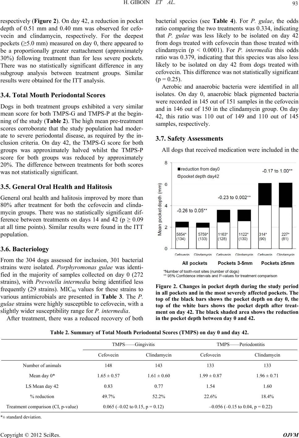 H. GIBOIN ET AL. 93 respectively (Figure 2). On day 42, a reduction in pocket depth of 0.51 mm and 0.40 mm was observed for cefo- vecin and clindamycin, respectively. For the deepest pockets (≥5.0 mm) measured on day 0, there appeared to be a proportionally greater reattachment (approximately 30%) following treatment than for less severe pockets. There was no statistically significant difference in any subgroup analysis between treatment groups. Similar results were obtained for the ITT analysis. 3.4. Total Mouth Periodontal Scores Dogs in both treatment groups exhibited a very similar mean score for both TMPS-G and TMPS-P at the begin- ning of the study (Tab le 2). The high mean pre-treatment scores corroborate that the study population had moder- ate to severe periodontal disease, as required by the in- clusion criteria. On day 42, the TMPS-G score for both groups was approximately halved whilst the TMPS-P score for both groups was reduced by approximately 20%. The difference between treatments for both scores was not statistically significant. 3.5. General Oral Health and Halitosis General oral health and halitosis improved by more than 80% after treatment for both the cefovecin and clinda- mycin groups. There was no statistically significant dif- ference between treatments on days 14 and 42 (p ≥ 0.09 at all time points). Similar results were found in the ITT population. 3.6. Bacteriology From the 304 dogs assessed for inclusion, 301 bacterial strains were isolated. Porphyromonas gulae was identi- fied in the majority of samples collected on day 0 (272 strains), with Prevotella intermedia being identified less frequently (29 strains). MIC90 values for these strains to various antimicrobials are presented in Table 3. The P. gulae strains were highly susceptible to cefovecin, with a slightly wider susceptibility range for P. intermedia. After treatment, there was a reduced recovery of both bacterial species (see Table 4). For P. gulae, the odds ratio comparing the two treatments was 0.334, indicating that P. gulae was less likely to be isolated on day 42 from dogs treated with cefovecin than those treated with clindamycin (p < 0.0001). For P. intermedia this odds ratio was 0.379, indicating that this species was also less likely to be isolated on day 42 from dogs treated with cefovecin. This difference was not statistically significant (p = 0.25). Aerobic and anaerobic bacteria were identified in all isolates. On day 0, anaerobic black pigmented bacteria were recorded in 145 out of 151 samples in the cefovecin and in 146 out of 150 in the clindamycin group. On day 42, this ratio was 110 out of 149 and 110 out of 145 samples, respectively. 3.7. Safety Assessments All dogs that received medication were included in the Figure 2. Changes in pocket depth during the study period in all pockets and in the most severely affected pockets. The top of the black bars shows the pocket depth on day 0, the top of the white bars shows the pocket depth after treat- ment on day 42. The black shaded area shows the reduction n the pocket depth between day 0 and 42. i Table 2. Summary of Total Mouth Periodontal Scores (TMPS) on day 0 and day 42. TMPS——Gingivitis TMPS——Periodontitis Cefovecin Clindamycin Cefovecin Clindamycin Number of animals 148 143 133 133 Mean day 0* 1.65 ± 0.57 1.61 ± 0.60 1.99 ± 0.87 1.96 ± 0.71 LS Mean day 42 0.83 0.77 1.54 1.60 % reduction 49.7% 52.2% 22.6% 18.4% Treatment comparison (CI, p-value) 0.065 (–0.02 to 0.15, p = 0.12) –0.056 (–0.15 to 0.04, p = 0.22) *± standard deviation. Copyright © 2012 SciRes. OJVM 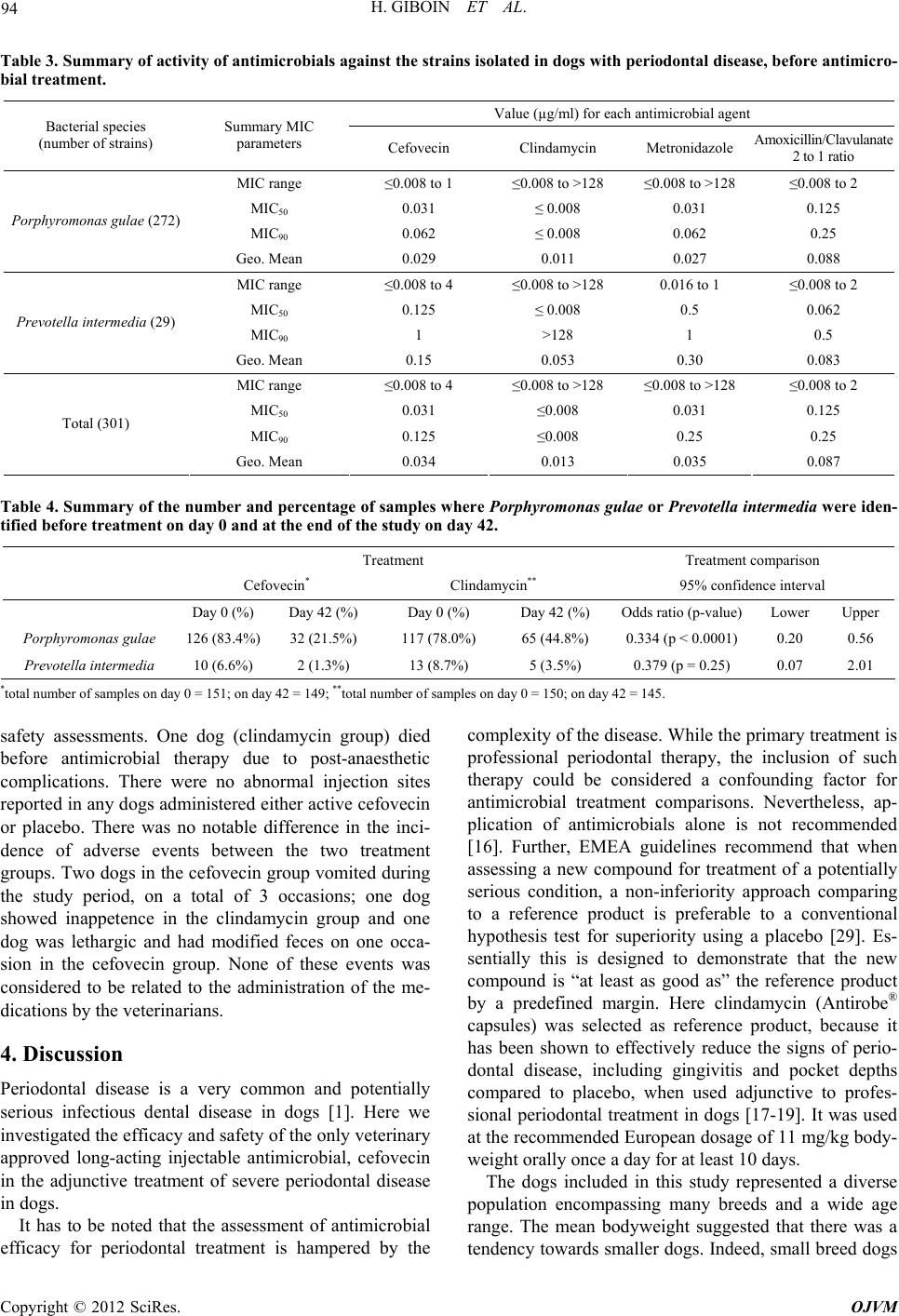 H. GIBOIN ET AL. 94 Table 3. Summary of activity of antimicrobials against the strains isolated in dogs with periodontal disease, before antimicro- bial treatment. Value (µg/ml) for each antimicrobial agent Bacterial species (number of strains) Summary MIC parameters Cefovecin Clindamycin Metronidazole Amoxicillin/Clavulanate 2 to 1 ratio MIC range ≤0.008 to 1 ≤0.008 to >128 ≤0.008 to >128 ≤0.008 to 2 MIC50 0.031 ≤ 0.008 0.031 0.125 MIC90 0.062 ≤ 0.008 0.062 0.25 Porphyromonas gulae (272) Geo. Mean 0.029 0.011 0.027 0.088 MIC range ≤0.008 to 4 ≤0.008 to >128 0.016 to 1 ≤0.008 to 2 MIC50 0.125 ≤ 0.008 0.5 0.062 MIC90 1 >128 1 0.5 Prevotella intermedia (29) Geo. Mean 0.15 0.053 0.30 0.083 MIC range ≤0.008 to 4 ≤0.008 to >128 ≤0.008 to >128 ≤0.008 to 2 MIC50 0.031 ≤0.008 0.031 0.125 MIC90 0.125 ≤0.008 0.25 0.25 Total (301) Geo. Mean 0.034 0.013 0.035 0.087 Table 4. Summary of the number and percentage of samples where Porphyromonas gulae or Prevotella intermedia were iden- tified before treatment on day 0 and at the end of the study on day 42. Treatment Treatment comparison Cefovecin* Clindamycin** 95% confidence interval Day 0 (%) Day 42 (%) Day 0 (%) Day 42 (%) Odds ratio (p-value) Lower Upper Porphyromonas gul ae 126 (83.4%) 32 (21.5%) 117 (78.0%) 65 (44.8%) 0.334 (p < 0.0001) 0.20 0.56 Prevotella intermedia 10 (6.6%) 2 (1.3%) 13 (8.7%) 5 (3.5%) 0.379 (p = 0.25) 0.07 2.01 *total number of samples on day 0 = 151; on day 42 = 149; **total number of samples on day 0 = 150; on day 42 = 145. safety assessments. One dog (clindamycin group) died before antimicrobial therapy due to post-anaesthetic complications. There were no abnormal injection sites reported in any dogs administered either active cefovecin or placebo. There was no notable difference in the inci- dence of adverse events between the two treatment groups. Two dogs in the cefovecin group vomited during the study period, on a total of 3 occasions; one dog showed inappetence in the clindamycin group and one dog was lethargic and had modified feces on one occa- sion in the cefovecin group. None of these events was considered to be related to the administration of the me- dications by the veterinarians. 4. Discussion Periodontal disease is a very common and potentially serious infectious dental disease in dogs [1]. Here we investigated the efficacy and safety of the only veterinary approved long-acting injectable antimicrobial, cefovecin in the adjunctive treatment of severe periodontal disease in dogs. It has to be noted that the assessment of antimicrobial efficacy for periodontal treatment is hampered by the complexity of the disease. While the primary treatment is professional periodontal therapy, the inclusion of such therapy could be considered a confounding factor for antimicrobial treatment comparisons. Nevertheless, ap- plication of antimicrobials alone is not recommended [16]. Further, EMEA guidelines recommend that when assessing a new compound for treatment of a potentially serious condition, a non-inferiority approach comparing to a reference product is preferable to a conventional hypothesis test for superiority using a placebo [29]. Es- sentially this is designed to demonstrate that the new compound is “at least as good as” the reference product by a predefined margin. Here clindamycin (Antirobe® capsules) was selected as reference product, because it has been shown to effectively reduce the signs of perio- dontal disease, including gingivitis and pocket depths compared to placebo, when used adjunctive to profes- sional periodontal treatment in dogs [17-19]. It was used at the recommended European dosage of 11 mg/kg body- weight orally once a day for at least 10 days. The dogs included in this study represented a diverse population encompassing many breeds and a wide age range. The mean bodyweight suggested that there was a tendency towards smaller dogs. Indeed, small breed dogs Copyright © 2012 SciRes. OJVM 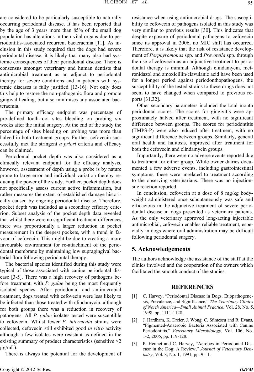 H. GIBOIN ET AL. 95 are considered to be particularly susceptible to naturally occurring periodontal disease. It has been reported that by the age of 3 years more than 85% of the small dog population has alterations in their vital organs due to pe- riodontitis-associated recurrent bacteraemia [11]. As in- clusion in this study required that the dogs had severe periodontal disease, it is likely that many also had sys- temic consequences of their periodontal disease. There is consensus amongst veterinary and human dentists that antimicrobial treatment as an adjunct to periodontal therapy for severe conditions and in patients with sys- temic diseases is fully justified [13-16]. Not only does this help to restore the non-pathogenic flora and promote gingival healing, but also minimises any associated bac- teraemia. The primary efficacy endpoint was percentage of pre-defined tooth-root sites bleeding on probing six weeks after the initial surgery. At the end of the study the percentage of sites bleeding on probing was more than halved in both treatment groups. Further, cefovecin suc- cessfully met the stringent a priori criteria and efficacy can be claimed. Periodontal pocket depth was also considered as a clinically relevant endpoint for the efficacy analysis, however, assessment of depth using a probe is by nature prone to large error and individual variation thereby re- ducing the power of the study. Further, pocket depth does not specifically assess current active inflammation, but rather measures the extent of established damage histori- cally caused by ongoing periodontal disease. Therefore, pocket depth was included as a secondary efficacy crite- rion. Subset analysis of the pocket depth data revealed that whilst there were no significant treatment differences, there was proportionally a larger reduction in pocket measurement in the deepest pockets, with a trend in fa- vour of cefovecin. This might be due to creating a more favourable environment for re-attachment of the perio- dontal membrane by maintaining low supragingival bac- terial flora following periodontal therapy. The bacterial species identified during this study were typical of those associated with canine periodontal dis- ease [3-5]. There was a high recovery of pathogens be- fore treatment, with P. gulae being the most frequently isolated species. After periodontal and antimicrobial treatment, dogs treated with cefovecin were less likely to be infected than those treated with clindamycin, although for both groups there was a reduction in recovery of pathogens. All P. gulae isolates tested were susceptible to cefovecin. Whilst fewer P. intermedia strains were collected, cefovecin still exhibited good in vitro activity although a few isolates were resistant as defined in the existing summary of product characteristics (sensitive ≤2 μg/mL). There is always the potential for the development of resistance when using antimicrobial drugs. The suscepti- bility to cefovecin of pathogens isolated in this study was very similar to previous results [30]. This indicates that despite exposure of periodontal pathogens to cefovecin since its approval in 2006, no MIC shift has occurred. Therefore, it is likely that the risk of resistance develop- ment of Porphyromon as spp. and Prevotella spp. through the use of cefovecin as an adjunctive treatment to perio- dontal therapy is minimal. Although clindamycin, met- ronidazol and amoxicillin/clavulanic acid have been used for a longer period against periodonthopathogens, the susceptibility of the tested strains to these drugs does not seem to have changed when compared to previous re- ports [31,32]. Other secondary parameters included the total mouth periodontal scores. The scores for gingivitis were ap- proximately halved after treatment, with no significant difference between groups. The scores for periodontitis (TMPS-P) were also reduced after treatment, with no significant difference between groups. Similarly, general oral health and halitosis, improved after treatment for both the cefovecin and clindamycin groups. Importantly, there were no adverse events reported due to treatment for either group. While owner diaries docu- mented a few adverse events, including gastrointestinal symptoms, these were unrelated to treatment according to the observing veterinarians. There was no injection- site reaction reported. In conclusion, cefovecin at a dose of 8 mg/kg body- weight administered once subcutaneously was safe and efficacious in the adjunctive treatment of severe perio- dontal disease in dogs presented as veterinary patients. As the only veterinary approved long-acting injectable antimicrobial, cefovecin enables reliable treatment, espe- cially in dogs where oral administration may be difficult following periodontal surgery. 5. Acknowledgements The authors acknowledge the assistance of the staff at the clinics involved and the cooperation of the owners which facilitated the smooth conduct of the studies. REFERENCES [1] C. Harvey, “Periodontal Disease in Dogs. Etiopathogene- sis, Prevalence, and Significance,” The Veterinary Clinics of North America—Small Animal Practice, Vol. 28, No. 5, 1998, pp. 1111-1128. [2] J. Hardham, K. Dreier, J. Wong, C. Sfintescu and R. Evans, “Pigmented-Anaerobic Bacteria Associated with Canine Periodontitis,” Veterinary Microbiology, Vol. 106, No. 1-2, 2005, pp. 119-128. [3] P. Hennet and C. Harvey, “Aerobes in Periodontal Dis- ease in the Dog: A Review,” Journal of Veterinary Den- tistry, Vol. 8, No. 1, 1991, pp. 9-11. Copyright © 2012 SciRes. OJVM  H. GIBOIN ET AL. 96 [4] P. Hennet and C. Harvey, “Anaerobes in Periodontal Disease in the Dog: A Review,” Journal of Veterinary Dentistry, Vol. 8, No. 2, 1991, pp. 18-21. [5] P. Hennet and C. Harvey, “Spirochetes in Periodontal Disease in the Dog: A Review,” Journal of Veterinary Dentistry, Vol. 8, No. 3, 1991, pp. 16-17. [6] P. Hennet and C. Harvey, “Natural Development of Pe- riodontal Disease in the Dog: A Review of Clinical, Ana- tomical and Histological Features,” Journal of Veterinary Dentistry, Vol. 9, No. 3, 1992, pp. 13-19. [7] L. Forner, T. Larsen, M. Kilian and P. Holmstrup, “Inci- dence of Bacteremia after Chewing, Tooth Brushing and Scaling in Individuals with Periodontal Inflammation,” Journal of Clinical Periodontology, Vol. 33, No. 6, 2006, pp. 401-407. doi:10.1111/j.1600-051X.2006.00924.x [8] M. Nieves, P. Hartwig, J. Kinyon and D. Riedesel, “Bac- terial Isolates from Plaque and from Blood during and af- ter Routine Dental Procedures in Dogs,” Veterinary Sur- gery, Vol. 26, No. 1, 1997, pp. 26-32. doi:10.1111/j.1532-950X.1997.tb01459.x [9] H. Horz and G. Conrads, “Diagnosis and Anti-Infective Therapy of Periodontitis,” Expert Review of Anti-Infective Therapy, Vol. 5, No. 4, 2007, pp. 703-715. doi:10.1586/14787210.5.4.703 [10] L. DeBowes, D. Mosier, E. Logan, C. Harvey, S. Lowry and D. Richardson, “Association of Periodontal Disease and Histologic Lesions in Multiple Organs from 45 Dogs,” Journal of Veterinary Dentistry, Vol. 13, No. 2, 1996, pp. 57-60. [11] Z. Pavlica, M. Petelin, P. Juntes, D. Erzen, D. Crossley and U. Skaleric, “Periodontal Disease Burden and Patho- logical Changes in Organs of Dogs,” Journal of Veteri- nary Dentistry, Vol. 25, No. 2, 2008, pp. 97-105. [12] M. Jorgensen, A. Aalam and J. Slots, “Periodontal An- timicrobials—Finding the Right Solutions,” International Dental Journal, Vol. 55, No. 1, 2005, pp. 3-12. [13] M. Addy and M. Martin, “Systemic Antimicrobials in the Treatment of Chronic Periodontal Diseases: A Dilemma,” Oral Diseases, Vol. 9, No. S1, 2003, pp. 38-44. doi:10.1034/j.1601-0825.9.s1.7.x [14] F. Sgolastra, R. Gatto, A. Petrucci and A. Monaco, “Ef- fectiveness of Systemic Amoxicillin/Metronidazole as Adjunctive Therapy to Scaling and Root Planing in the Treatment of Chronic Periodontitis: A Systematic Review and Meta-Analysis,” Journal of Periodontology, Vol. 83, No. 10, 2012, pp. 1257-1269. doi:10.1902/jop.2012.110625 [15] A. Haffajee, S. Dibart, R. Kent and S. Socransky, “Clini- cal and Microbiological Changes Associated with the Use of 4 Adjunctive Systemically Administered Agents in the Treatment of Periodontal Infections,” Journal of Clinical Periodontology, Vol. 22, No. 8, 1995, pp. 618-627. doi:10.1111/j.1600-051X.1995.tb00815.x [16] AVDC, “Policy Statement: The Use of Antibiotics in Ve- terinary Dentistry,” 2005. http://avdc.org/Antibiotic_Use_April_2005.pdf [17] J. Warrick, G. Inskeep, T. Yonkers, G. Stookey and T. Ewing, “Effect of Clindamycin Hydrochloride on Oral Malodor, Plaque, Calculus, and Gingivitis in Dogs with Periodontitis,” Veterinary Therapeutics, Vol. 1, No. 1, 2000, pp. 5-16. [18] K. Zetner, G. Pum, W. Rausch, X. Rausch-Fan and S. Hung, “Effect of Clindamycin Hydrochloride on Gingival Crevicular Fluid and Immune Mediators in Beagles,” Ve- terinary Therapeutics, Vol. 3, No. 2, 2002, pp. 177-188. [19] D. Nielsen, C. Walser, G. Kodan, R. D. Chaney, T. Yonkers, J. D. VerSteeg, G. Elfring and J. Slots, “Effects of Treatment with Clindamycin Hydrochloride on Pro- gression of Canine Periodontal Disease after Ultrasonic Scaling,” Veterinary Therapeutics, Vol. 1, No. 3, 2000, pp. 150-158. [20] M. R. Stegemann, J. Sherington and S. Blanchflower, “Pharmacokinetics and Pharmacodynamics of Cefovecin in Dogs,” Journal of Veterinary Pharmacology and The- rapeutics, Vol. 29, No. 6, 2006, pp. 501-511. doi:10.1111/j.1365-2885.2006.00801.x [21] R. Six, J. Cherni, R. Chesebrough, D. Cleaver, C. J. Lin- deman, G. Papp, et al., “Efficacy and Safety of Cefovecin in Treating Bacterial Folliculitis, Abscesses, or Infected Wounds in Dogs,” Journal of the American Veterinary Medical Association, Vol. 233, No. 3, 2008, pp. 433-439. doi:10.2460/javma.233.3.433 [22] M. R. Stegemann, N. Coati, C. A. Passmore and J. Sher- ington, “Clinical Efficacy and Safety of Cefovecin in the Treatment of Canine Pyoderma and Wound Infections,” The Journal of Small Animal Practice, Vol. 48, No. 7, 2007, pp. 378-386. doi:10.1111/j.1748-5827.2007.00363.x [23] M. R. Stegemann, J. Sherington and C. Passmore, “The Efficacy and Safety of Cefovecin in the Treatment of Fe- line Abscesses and Infected Wounds,” The Journal of Small Animal Practice, Vol. 48, No. 12, 2007, pp. 683-689. doi:10.1111/j.1748-5827.2007.00390.x [24] R. Six, D. M. Cleaver, C. J. Lindeman, J. Cherni, R. Chesebrough, G. Papp, et al., “Effectiveness and Safety of Cefovecin Sodium, an Extended-Spectrum Injectable Cephalosporin, in the Treatment of Cats with Abscesses and Infected Wounds,” Journal of the American Veteri- nary Medical Association, Vol. 234, No. 1, 2009, pp. 81-87. doi:10.2460/javma.234.1.81 [25] C. A. Passmore, J. Sherington and M. R. Stegemann, “Efficacy and Safety of Cefovecin (Convenia) for the Treatment of Urinary Tract Infections in Dogs,” Journal of Small Animal Practice, Vol. 48, No. 3, 2007, pp. 139-144. doi:10.1111/j.1748-5827.2006.00231.x [26] C. A. Passmore, J. Sherington and M. R. Stegemann, “Efficacy and Safety of Cefovecin for the Treatment of Urinary Tract Infections in Cats,” The Journal of Small Animal Practi c e, Vol. 49, No. 6, 2008, pp. 295-301. doi:10.1111/j.1748-5827.2008.00545.x [27] VICH, “International Cooperation on Harmonisation of Technical Requirements for Registration of Veterinary Medicinal Products,” 2000. http://www.vichsec.org/pdf/2000/Gl09_st7.pdf [28] C. Harvey, L. Laster, F. Shofer and B. Miller, “Scoring the Full Extent of Periodontal Disease in the Dog: De- velopment of a Total Mouth Periodontal Score (Tmps) System,” Journal of Veterinary Dentistry, Vol. 25, No. 3, Copyright © 2012 SciRes. OJVM  H. GIBOIN ET AL. Copyright © 2012 SciRes. OJVM 97 2008, pp. 176-180. [29] EMEA, “Guidelines on Statistical Principles for Veteri- nary Clinical Trials,” 2000. http://www.ema.europa.eu/docs/en_GB/document_library /Scientific_guideline/2010/09/WC500097081.pdf [30] M. Stegemann, C. Passmore, J. Sherington, C. Lindeman, G. Papp, D. Weigel, et al., “Antimicrobial Activity and Spectrum of Cefovecin, a New Extended—Spectrum Ce- phalosporin, against Pathogens Collected from Dogs and Cats in Europe and North America,” Antimicrobial Agents and Chemotherapy, Vol. 50, No. 7, 2006, pp. 2286-2292. doi:10.1128/AAC.00077-06 [31] C. E. Harvey, C. Thornsberry, B. R. Miller and F. S. Shofer, “Antimicrobial Susceptibility of Subgingival Bacterial Flora in Dogs with Gingivitis,” Journal of Veterinary Dentistry, Vol. 12, No. 4, 1995, pp. 151-155. [32] M. Radice, P. A. Martino and A. M. Reiter, “Evaluation of Subgingival Bacteria in the Dog and Susceptibility to Commonly Used Antibiotics,” Journal of Veterinary Dentistry, Vol. 23, No. 4, 2006, pp. 219-224. |