

Paper Menu >>
Journal Menu >>
 Advances in Infectious Diseases, 2012, 2, 82-88 http://dx.doi.org/10.4236/aid.2012.23013 Published Online September 2012 (http://www.SciRP.org/journal/aid) Heterogeneity in femA in the Indian Isolates of Staphylococcus aureus Limits Its Usefulness as a Species Specific Marker Rosy Chikkala1, Nikhil Oommen George1, Kamaraju S. Ratnakar2, Ranganathan Natarajan Iyer3, Venkataraman Sritharan1* 1Molecular Diagnostics and Biomarkers Laboratory, Department of Laboratory Medicine, Global Hospitals, Hyderabad, India; 2Global Hospitals, Hyderabad, India; 3Department of Clinical Microbiology, Global Hospitals, Hyderabad, India. Email: *[email protected] Received May 31st, 2012; revised July 1st, 2012; accepted August 3rd, 2012 ABSTRACT Increase in prevalence of MRSA worldwide and hence the need for rapid detection, have led to use of molecular meth- ods for confirmation of the species and also MRSA. Species specific markers like fem or nuc along with the methicil- lin-resistance determinant, mecA, have been used by several investigators worldwide for the identification of MRSA. In the current study, we have screened 54 microbiologically confirmed (MRSA, MSSA and CoNS) isolates for the pres- ence of mecA, 16S rRNA, femA and nuc markers. While mecAPCR and 16S rRNAPCR results were consistent with other studies, femA and nuc showed dramatic variation in detection rate (sensitivity) of S. aureus 29.6% and 53.7% re- spectively. Evidences are presented to demonstrate the absence of femA. Our attempt to amplify the complete femA gene using sequences flanking femA further confirmed these results and also indicated that variations exist even in the ge- nomic sequences around femA. Our data reveals the need for exercising care while using primers designed on se- quences of constitutive genes like femA and nuc for PCR based identification of S. aureus species. Though geograph ic variations in the genome of S. aureus have previously been reported from around the world, we present here evidence for the first time from India for absence of femA and also for prob able var iations in the seq uences around the femA gene in clinical isolates of S. aureus. Keywords: femA; mecA; nuc; PCR; S. aureus 1. Introduction Staphylococcus aureus is one of the major causes of wide spread gram positive bacterial nosocomial infections, especially the post-surgical wound infections [1]. Its d is- ease manifestations range from minor skin infections to life-threatening diseases such as pneumonia, sepsis etc. The emergence of MRSA in patients with no apparent risk factors seems to be a growing concern. In India the prevalence of MRSA is 51.8% in 20 10 [2]. Although the discovery of penicillin proved to be a major breakthrough in treating these infections, with it had also emerged a major concern; the notorious ability of Staphylococcus aureus to develop resistance to antibiotics and remain non- responsive to treatment [3]. Shortly after the introduc tion of methicillin, reports of Methicillin-resistant Staphylo- coccus aureus (MRSA) had begun to surface within hos- pitals in the early 1960s, which are now increasingly pre- valent worldwide [4,5]. Such endemic MRSA infections are difficult to eradicate and remain active reservoirs of infection thereby increasing the hospital costs, length of hospital stays, morbidity and mortality. Methicillin-resistance in staph ylococci is expressed b y the mecA gene that produces a “Penicillin Binding Pro- tein 2a” (PBP2a) [6], a modified transpeptidase. This PBP-2a has lower affinity to penicillin and its derivatives than the other PBPs [7]. The Staphylococcal Chromo- some Cassette mec (SCCmec), a mobile genetic element, is composed of the mec gene complex which includes mecA, its regulatory genes mecI, mecR and a ccr gene complex. The latter encodes site specific recombinases, namely ccrA/B, which in-turn regulate the mobility of SCCmec [8]. Significant geographic variations have been found in the structural organization of the SCCmec and these variations have been used to classify the SCCmec types [9]. However, mecA alone does not solely confer the methicillin resistance. Studies have shown that fem (factors essential for methicillin-resistance) or the auxiliary genes like fem A/B/X in addition to mecA are also impor- *Corresponding a uthor. Copyright © 2012 SciRes. AID  Heterogeneit y in femA in the Indian Isolates of Staphylococcus aureus Limits Its Usefulness as a Species Specific Marker 83 tant in the expression of methicillin resistance [10]. The femABX operon encodes factors which are responsible for the formation of pentaglycine bridges in the cell wall of Staphylococci [11]. Kirby-Bauer antibiotic testin g (disc diffusion antibiotic sensitivity test), oxacillin agar screen test, agar dilution and the Epsilometer test (E-test) are commonly used to determine the MRSA phenotype in the clinical microbe- ology laboratory. These tests have limitations and fre- quently show variations [12]. Although the detection of mecA gene remains the gold standard for detecting me- thicillin-resistance, its detection alone does not confirm the presence of Staphylococcus aureus [13] and there is no consensus on the molecular target that could be used to confirm the S. aureus species. Constitutively expressed genes such as femA, femB and nuc are being used as mo- lecular targets for the identification of Staphylococcus aureus [14,15]. Polymorphisms within these constitutive genes have been reported [16] worldwide and also fail- ures to confirm the species of S. aureus using these genes as targets for PCR detection [17]. However, from India, there are no reports about any variations in the sequences of these co ns titu tive g en es ye t and for th at matter SCCmec or the genome of S. aureus are yet to be characterised in India. The aim of our study was to optimise and estab lish a multiplex PCR protocol to screen clinical isolates for molecular markers of MRSA namely, 16S rRNA, mecA, femA or nuc. We are reporting here absence of femA gene in several clinical isolates of S. aureus (both MRSA and MSSA), poor sensitivity of nuc as a target for S. aureus identification and probable variation in the sequences flanking the femA. These findings have serious implica- tions for molecular diagnosis of S. aureus and are reflec- tive of the geographic changes occurring in the gen o me o f S. aureus. 2. Materials and Methods 54 isolates of S. aureus were collected from Jan 2012 to March 2012 in Global Hospital, Hyderabad. These strai ns were isolated on Mueller Hinton Agar (MHA) either from blood culture bottles or directly from different clinical specimens (pus, drain fluids/secretions etc). Co- agulase and catalase tests were used to confirm S. aureus in the isolates. Standard disc diffusion test usin g the anti- biotic oxacillin were used to assess th e drug sensitiv ity o f these isolates. Epsilometer test (E-test) was used to find out the MIC of the isolates. 2.1. Preparation of Bacterial DNA Lysates A single bacterial colony was scrapped off the agar and the cells washed by suspending in 100 µl of TEX buffer (10 mM Tris-HCl pH 8.5, 1 mM EDTA, 1% (w/v) Triton X-100). The suspension was vortexed to achieve a uni- form suspension and was centrifuged. The pellet was subjected to another wash in TEX buffer. Finall y t he pellet was resuspended in 100 µl of TEX buffer and was lysed by heating in a dry bath at 95˚C for 15min [18]. The lys- ate was used as the DNA template for PCR. This lysate could be stored at 2˚C - 8˚C for 2 - 3 months and at –20˚C for > 1 year. 2.2. Polymerase Chain Reaction All the primer sequences listed Table 1 were procured from Eurofins Genomics India Pvt Ltd. Multiplex-PCR mixture for mecA, femA and 16S rRNA consisted of the following; 1 × PCR mixture (Fermentas Life Sciences/ HiMedia) containing 1.5 mM MgCl2, 50 pM of each primer, 200 µM of each dNTP along with 1 U of Taq (HiMedia) and 5 µL of DNA template in 20 µL final volume. PCR reactions were initiated with a denaturation at 94˚C for 5 mins, 40 cycles of denaturation (94˚C, 30 s), annealing (55˚C, 40 s) and primer extension (72˚C, 50 s), with a final extension at 72˚C for 10mins. A femA monoplex PCR was performed under the re- ported conditions [14,19] but with 10 µL of the DNA template. Monoplex PCR was also optimized for the identification of the nuc gene in these isolates. The PCR reaction mixture contained in 20 µL final volume 50 pM of nuc pr imers and 5 µl of DNA lysate in addition to 200 µM of dNTPs, and 1 unit of Taq DNA polymerase. The PCR reactions with femA flanking primers were performed in a volume of 20 µL containing 10 µL of DNA lysate, containing 1.7 mM MgCl2, 100 pM of primers, 0.2 mM dNTPs along with 2.5 units of Taq DNA poly- merase. A 30-cycle amplification followed an initial de- naturation at 94˚C for 5 min, with d enaturation (94˚C, 30 s), annealing (45˚C, 1 min) and primer extension (72˚C, 45 s), and final extension at 72˚C for 10 mins. 3. Results 3.1. 16s rRNA, femA and mecA Multiplex PCR Of the 54 isolates which were included in the study, 21 (38%) were isolated from blood, 13 (24%) from pus, 9 (16.6%) from wound swabs and the rest from other sources such as sputum, tissue, endo-tracheal secretions and others. These isolates were identified as MRSA (26 isolates), MSSA (26 isolates) and CoNS (2 isolates). Multiplex-PCR was performed targeting 16S rRNA for the Staphylococci genus, femA for S. aureus and mecA for methicillin resistance. All the isolates tested positive for the genus specific 16S rRNA 886 bp product. The 293 bp mecA was detected in all 26 (100%) MRSA iso- lates, 16 (61.5%) MSSA isolates and 1 CoNS isolate. Copyright © 2012 SciRes. AID 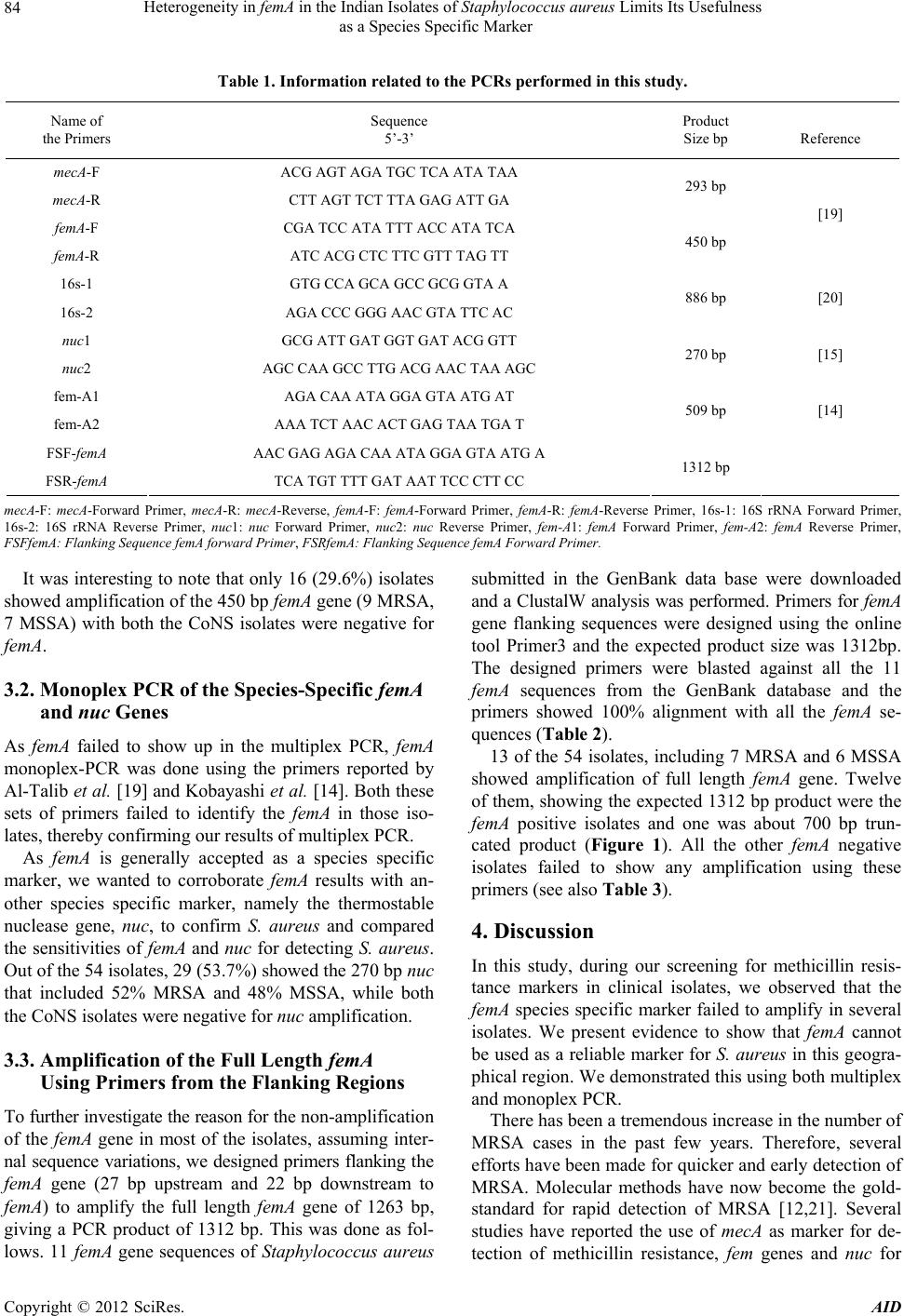 Heterogeneit y in femA in the Indian Isolates of Staphylococcus aureus Limits Its Usefulness as a Species Specific Marker Copyright © 2012 SciRes. AID 84 Table 1. Information related to the PCRs performed in this study. Name of the Primers Sequence 5’-3’ Product Size bp Reference mecA-F ACG AGT AGA TGC TCA ATA TAA mecA-R CTT AGT TCT TTA GAG ATT GA 293 bp femA-F CGA TCC ATA TTT ACC ATA TCA femA-R ATC ACG CTC TTC GTT TAG TT 450 bp [19] 16s-1 GTG CCA GCA GCC GCG GTA A 16s-2 AGA CCC GGG AAC GTA TTC AC 886 bp [20] nuc1 GCG ATT GAT GGT GAT ACG GTT nuc2 AGC CAA GCC TTG ACG AAC TAA AGC 270 bp [15] fem-A1 AGA CAA ATA GGA GTA ATG AT fem-A2 AAA TCT AAC ACT GAG TAA TGA T 509 bp [14] FSF-femA AAC GAG AGA CAA ATA GGA GTA ATG A FSR-femA TCA TGT TTT GAT AAT TCC CTT CC 1312 bp mecA-F: mecA-Forward Primer, mecA-R: mecA-Reverse, femA-F: femA-Forward Primer, femA-R: femA-Reverse Primer, 16s-1: 16S rRNA Forward Primer, 16s-2: 16S rRNA Reverse Primer, nuc1: nuc Forward Primer, nuc2: nuc Reverse Primer, fem-A1: femA Forward Primer, fem-A2: femA Reverse Primer, FSFfemA: Flanking Sequence femA forward Primer, FSRfemA: Flanking Sequence femA Forward Primer. It was interesting to note that only 16 (29.6%) isolates showed amplification of the 450 bp femA gene (9 MRSA, 7 MSSA) with both the CoNS isolates were negative for femA. 3.2. Monoplex PCR of the Species-Specific femA and nuc Genes As femA failed to show up in the multiplex PCR, femA monoplex-PCR was done using the primers reported by Al-Talib et al. [19] and Kobayashi et al. [14]. Both these sets of primers failed to identify the femA in those iso- lates, thereby conf irming our results of multiplex PCR. As femA is generally accepted as a species specific marker, we wanted to corroborate femA results with an- other species specific marker, namely the thermostable nuclease gene, nuc, to confirm S. aureus and compared the sensitivities of femA and nuc for detecting S. aureus. Out of the 54 isolates, 29 (53.7%) show ed the 270 bp nuc that included 52% MRSA and 48% MSSA, while both the CoNS isolates were negative for nuc amplification. 3.3. Amplification of the Full Length femA Using Primers from the Flanking Regions To further investigate the reason for the non -a mpl if ic at io n of the femA gene in most of the isolates, assuming inter- nal sequence variations, we designed primers flanking the femA gene (27 bp upstream and 22 bp downstream to femA) to amplify the full length femA gene of 1263 bp, giving a PCR product of 1312 bp. This was done as fol- lows. 11 femA gene sequences of Staphylococcus aureus submitted in the GenBank data base were downloaded and a ClustalW analysis was performed. Primers for femA gene flanking sequences were designed using the online tool Primer3 and the expected product size was 1312bp. The designed primers were blasted against all the 11 femA sequences from the GenBank database and the primers showed 100% alignment with all the femA se- quences (Table 2). 13 of the 54 isolates, including 7 MRSA and 6 MSSA showed amplification of full length femA gene. Twelve of them, showing the exp ected 1312 bp product were the femA positive isolates and one was about 700 bp trun- cated product (Figure 1). All the other femA negative isolates failed to show any amplification using these primers (see also Table 3). 4. Discussion In this study, during our screening for methicillin resis- tance markers in clinical isolates, we observed that the femA species specific marker failed to amplify in several isolates. We present evidence to show that femA cannot be used as a reliable marker for S. aureus in this geogra- phical region. We demonstrated this using both multiplex and monoplex PCR. There has been a tremendous increase in the number of MRSA cases in the past few years. Therefore, several efforts have been made for quicker and early detection of MRSA. Molecular methods have now become the gold- standard for rapid detection of MRSA [12,21]. Several studies have reported the use of mecA as marker for de- tection of methicillin resistance, fem genes and nuc for  Heterogeneit y in femA in the Indian Isolates of Staphylococcus aureus Limits Its Usefulness as a Species Specific Marker 85 Table 2. List of femA gene sequences reviewed from the Ge nBank Database . S.No Strain GenBank accession Gene location A: 1 MRSA252 NC_002952 1444550 - 144581 2 2 USA300_FRP3757 NC_007793 1397007 - 1398308 3 JKD6008 NC_017341 1427188 - 142845 6 4 COL NC_002951 1420810 - 142211 1 5 Newman NC_009641 1417031 - 1418293 6 ED98 NC_013450 1391969 - 1393231 7 USA300_TCH 1516 NC_010079 1410993 - 1412255 8 TCH60 NC_017342 1939657 - 1940994 9 RF122 NC_007622 1344801 - 1346102 10 MW2 NC_003923 1381193 - 1382455 11 Mu3 NC_009782 1456933 - 1458195 B: 12 JKD6159 NC_017338 1365979 - 1367241 13 N315 NC_002745 1379204 - 1380466 14 Mu50 NC_002758 1455533 - 1456795 15 T0131 NC_017347 1428806 - 1430068 16 04-02981 NC_017340 1413795 - 1415057 17 HO 5096 0412 NC_017763 1357669 - 135893 1 18 TW20 NC_017331 1470704 - 1471966 Lane 1: Low Range Ladder, Lane 2: Multiplex-PCR [16S rRNA (886bp), femA (450 bp), mecA (293bp)], Lane 3: femA-Al-Talib (450 bp), Lane 4: femA-Kobayashi (509 bp), Lane 5: nuc (270 bp), Lane 6: femA full length (1312 bp), Lane 7: femA full le ngth (mutated, approx. 700 bp), Lan e 8: Negative Control. Figure 1. PCR amplification of molecular targets for MRSA and S. aureus. Copyright © 2012 SciRes. AID  Heterogeneit y in femA in the Indian Isolates of Staphylococcus aureus Limits Its Usefulness as a Species Specific Marker 86 Table 3. Summary of results phenotype vs genotype. Phenotype MRSA (26)* MSSA (26)* CoNS (2)* Genotype + – + – + – Total (54)* + mecA 26 (100%) 0 (0%) 16 (61.5%) 10 (38.5%) 1 (50%) 1 (50%) 43 (79.6%) 16S rRNA 26 (100%) 0 (0%) 26 (100%) 0 (0%) 2 (100%) 0 (0%) 54 (100%) femA 9 (34.6%) 17 (65.4%) 7 (26.9%) 19 (73.1%) 0 (0%) 2 (100%) 16 (29.6%) nuc 15 (57.7%) 11 (42.3%) 14 (53.8%) 12 (46.2%) 0 (0%) 2 (100%) 29 (53.7%) femA full length 7 (26.9%) 19 (73.1%) 6 (23.1%) 20 (76.9%) 0 (0%) 2 (100%) 13 (24%) *Total Number of Isolates. identification of S. aureus species. Good correlation of phenotype with genotype tests were reported for MRSA isolates, which harboured mecA and femA genes [19,22]. In the study by Kobayashi et al, mecA was detected in 100% of MRSA, 16.7% of MSSA isolates. 10.6% of their S. aureus isolates did not amplify femA gene PCR product [14]. Thermostable nuclease gene nuc was re- ported to have 100% sensitivity and specificity for the identification of S. aureus isolates [15,23]. In India, only a few studies have reported the use of femA and nuc along with mecA as molecular targets for identification of S. aureus and characterisation of MRSA [24,25] and there are no reports which specifically investigated the predictive value of femA gene for the identification of S. aureus. Variations (polymorphism) in the genomic se- quences are not uncommon in S. aureus, even highly conserved and widely used species specific markers like coagulase (coa), Staphylococcal Protein A (spa) genes have shown polymorphisms. Variations in the sequence of the coa and spa genes and the hyper variable region adjacent to the mecA gene, have been the basis for the most widely used forms of PCR typing of MRSA [26-28]. Further, it is also known that sequence variations culmi- nate in changes in virulence properties of S. aureus which influence clinical disease manifestations in humans [29]. The mecA PCR in our study showed absolute correla- tion to MRSA phenotype and we observed a rather high rate (58.3%) of detection of mecA among MSSA isolates. An intriguing finding in this study was the non-amplifi- cation of femA gene in most (70.4%) of the isolates when screened with two different sets of primers for the femA gene and confirmed by non-ampli fication of the full length femA gene with the primers flanking the femA gene. Though nuc PCR was more sensitive (57.4%) compared to femA PCR (29.6%) in the detection of S. aureus we could not achieve 100% sensitivity with nuc PCR also. The femA PCR and nuc PCR showed good correlation in 15 out of 16 isolates. However, 22 S. aureus isolates were negative for both femA and nuc genetic markers. This is suggestive of likely mutations or deletions in the nuc gene also. However, further investigations are being car- ried out to understand this observation. Mutations in the aux illary genes such as the fem, cou ld explain the absence of any phenotypic expression of re- sistance in MSSA isolates though these isolates con- tained mecA gene [30,31]. We propose to examine this phenomenon in future in several MSSA isolates. All the MRSA isolates that showed positive femA PCR, had MICs greater than 256 µg/mL; yet there were 3 isolates with MICs > 256 µg/mL that showed no femA gene am- plification. It is known that methicillin resistance could manifest with or without mecA. Auxillary genes like femA influence the extent of resistance [14] implying that there could be other mechanisms that circumvent absence of femA gene to confer methicillin-resistance. Assuming that mutations in the femA gene sequence at the primer annealing sites could have resulted in non-am- plification of PCR product in some isolates, we attemp ted to amplify the full length femA gene by designing prim- ers flanking femA as described previously. Surprisingly, amplification of the whole gene was seen in only in some of those isolates which were positive with femA primers. One MSSA isolate which was negative for femA showed amplification of a truncated (smaller) (app roximately 7 00 bp instead of 1312 bp) product, sugg esting possible dele- tions in the femA gene. We are sequencing this smaller PCR product. Four femA positive isolates did not amplify the full length PCR product indicating that there are variations in primer annealing sites flanking the femA gene. Re-visit to the GenBank database for any new en- tries of femA gene, we found 7 new sequences of S. aureus that have recently been submitted in April/May 2012 (Table 2, B) which showed significant variations in their sequences compared to the 11 femA gene sequences (Ta- ble 2, A) which were used in the design of the PCR primers that we evaluated in this study. Of these se- quences, a strain JKD6159 showed two nucleotide varia- tions in the sequence flanking the femA gene (4th & 13th base downstream of femA) which could probably explain the non-amplification of the full length femA gene se- quence in our isolates also. Significant polymorphisms in the recently submitted femA gene sequences suggest the Copyright © 2012 SciRes. AID  Heterogeneit y in femA in the Indian Isolates of Staphylococcus aureus Limits Its Usefulness as a Species Specific Marker 87 femA gene variations are quite common, could be region specific, may play important role in the expression of MRSA and in the identification of S. aureus in clinical isolates. Our study shows that polymorphisms in femA gene sequences are present in both MRSA and MSSA isolates in India and requires further investigations such as sequencing to characterise the genome in general and femA in particular. We have initiated a detailed investi- gation of genetic variations in th e femA gene of S. aureus, its relevance to the drug resistance phenotype and to ex- amine any role for these genomic variatio ns in molecular epidemiology of clinical isolates of S. aureus. 5. Acknowledgements Technical assistance by Mr. T. Naresh Singh of Clinical Microbiology Laboratory is acknowledged and appreci- ated. REFERENCES [1] A. F. Shorr, “Epidemiology of Staphylococcal Resistance,” Clinical Infectious Diseases, Vol. 45, No. 3, 2007, pp. S171-S176. doi:10.1086/519473 [2] S. K. Mallick and S. Basak, “MRSA—Too Many Hurdles to Overcome: A Study from Central India,” Tropical Doc- tor, Vol. 40, No. 2, 2010, pp. 108-110. doi:10.1258/td.2010.090440 [3] H. F. Chambers and F. R. DeLeo, “Waves of Resistance: Staphylococcus aureus in the Antibiotic Era,” Nature Re- views Microbiology, Vol. 7, No. 9, 2009, pp. 629-641. doi:10.1038/nrmicro2200 [4] M. Jevons, “Celbenin-Resistant Staphylococci,” British Medical Journal, Vol. 1, No. 5219, 1961, pp. 124-125. doi:10.1136/bmj.1.5219.124-a [5] S. Stefani, D. R. Chung, J. A. Lindsay, et al., “Meticillin- Resistant Staphylococcus aureus (MRSA): Global Epi- demiology and Harmonisation of Typing Methods,” In- ternational Journal of Antimicrobial Agents, Vol. 39, No. 4, 2012, pp. 273-282. doi:10.1016/j.ijantimicag.2011.09.030 [6] W. D. Beck, B. Berger-Bachi and F. H. Kayser, “Addi- tional DNA in Methicillin-Resistant Staphylococcu s au reu s and Molecular Cloning of mec Specific DNA,” Journal of Bacteriology, Vol. 165, No. 2, 1986, pp. 373-378. [7] D. Lim and N. C. Strynadka, “Structural Basis for the Beta Lactam Resistance of PBP2a from Methicillin-Re- sistant Staphylococcus aureus,” Nature Structural & Mo- lecular Biology, Vol. 9, No. 11, 2002, pp. 870-876. [8] T. Ito, K. Okuma, X. X. Ma, H. Yuzawa and K. Hi ramatsu, “Insights on Antibiotic Resistance of Staphyloc occus aureus from Its Whole Genome: Genomic Island SCC,” Drug Resistance Updates, Vol. 6, No. 1, 2003, pp. 41-52. doi:10.1016/S1368-7646(03)00003-7 [9] A Turlej, W. Hryniewicz and J. Empel, “Staphylococcal Cassette Chromosome mec (SCCmec) Classification and Typing Methods: An Overview,” Polish Journal of Mi- crobiology, Vol. 60, No. 2, 2011, pp. 95-103. [10] S. S. Hegde and T. E. Shrader, “FemABX Family Mem- bers Are Novel Nonribosomal Peptidyltransferases and Important Pathogen-Specific Drug Targets,” The Journal of Biological Chemistry, Vol. 276, No. 10, 2001, pp. 6998- 7003. doi:10.1074/jbc.M008591200 [11] H. Ton-That, H. Labischinski, B Berger-Bachi and O. Schneewind, “Anchor Structure of Staphylococcal Surface Proteins: III Role of the femA, femB, and femX Factors in Anchoring Surface Proteins to the Bacterial Cell Wall,” The Journal of Biological Chemistry, Vol. 273, No. 44, 1998, pp. 29143-29149. doi:10.1074/jbc.273.44.29143 [12] P. U. Krishnan, K. Miles and N. Shetty, “Detection of Methicillin and Mupirocin Resistance in Staphylococcus aureus Isolates Using Conventional and Molecular Meth- ods: A Descriptive Study from a Burns Unit with High Prevalence of MRSA,” Journal of Clinical Pathology, Vol. 55, No. 10, 2002, pp. 745-748. doi:10.1136/jcp.55.10.745 [13] K. Murakami, W. Minamide, K. Wada, E. Nakamura, E. Teraoka and S. Watanabe, “Identification of Methicillin- Resistant Strains of Staphylococci by Polymerase Chain Reaction,” Journal of Clinical Microbiology, Vol. 29, No. 10, 1991, pp. 2240-2244. [14] N. Kobayashi, H. Wu, K. Kojima, et al., “Detection of mecA, femA, and femB Genes in Clinical Strains of Staphy- lococci Using Polymerase Chain Reaction,” Epidemiol- ogy & Infection, Vol. 113, No. 2, 1994, pp. 259-266. doi:10.1017/S0950268800051682 [15] O. G. Brakstad, K. Aasbakk and J. A. Maeland, “Detec- tion of Staphylococcus aureus by Polymerase Chain Re- action Amplification of the nuc Gene,” Journal of Clini- cal Microbiology, Vol. 30, No. 7, 1992, pp. 1654-1660. [16] Y. Cai, F. Kong, Q. Wang, et al., “Comparison of Single- and Multi-Locus Sequence Typing and Toxin Gene Pro- filing for Characterization of Methicillin-Resistant Staphy- lococcus aureus,” Journal of Clinical Microbiology, Vol. 45, No. 10, 2007, pp. 3302-3308. doi:10.1128/JCM.01082-07 [17] Z. Hussain and M. A. John, “Failure of a PCR Screening Method to Detect MRSA,” Infection Control and Hospi- tal Epidemiology, Vol. 21, No. 10, 2000, pp. 627-628. doi:10.1086/503240 [18] V. Sritharan and R. H. Barker Jr., “A Simple Method for Diagnosing M. Tuberculosis Infection in Clinical Samples Using PCR,” Molecular and Cellular Probes, Vol. 5, No. 5, 1991, pp. 385-395. doi:10.1016/S0890-8508(06)80011-3 [19] H. Al-Talib, Y. Y. Chan, A. Alyaa, et al., “A Pentaplex PCR Assay for the Detection of Methicillin-Resistant Sta- phylococcus aureus and Panton-Valentine Leucocidin,” BMC Microbiology, Vol. 9, No. 1, 2009, p. 113. [20] A. B. Poulsen, R. Skov and L. V. Pallesen, “Detection of Methicillin Resistance in Coagulase-Negative Staphylo- cocci and in Staphylococci Directly from Simulat ed Blood Cultures Using the EVIGENE MRSA Detection Kit,” Journal of Antimicrobial Chemotherapy, Vol. 51, No. 2, Copyright © 2012 SciRes. AID  Heterogeneit y in femA in the Indian Isolates of Staphylococcus aureus Limits Its Usefulness as a Species Specific Marker Copyright © 2012 SciRes. AID 88 2003, pp. 419-421. doi:10.1093/jac/dkg084 [21] K. C. Carroll, “Rapid Diagnostics for Methicillin-Resis- tant Staphylococcus aureus: Current Status,” Molecular Diagnosis & Therapy, Vol. 12, No. 1, 2008, pp. 15-24. doi:10.1007/BF03256265 [22] P. Vannuffel, J. Gigi, H. Ezzedine, et al., “Specific Detec- tion of Methicillin-Resistant Staphylococcus Species by Multiplex PCR,” Journal of Clinical Microbiology, Vol. 33, No. 11, 1995, pp. 2864-2867. [23] L. Louie, J. Goodfellow, P. Mathieu, A. Glatt, M. Louie and A. E. Simor, “Rapid Detection of Methicillin-Resis- tant Staphylococci from Blood Culture Bottles by Using a Multiplex PCR Assay,” Journal of Clinical Microbiology, Vol. 40, No. 8, 2002, pp. 2786-2790. doi:10.1128/JCM.40.8.2786-2790.2002 [24] A. A. Mathews, M. Thomas, B. Appalaraju and J. Jaya- lakshmi. “Evaluation and Comparison of Tests to Detect Methicillin Resistant S. aureus,” Indian Journal of Pa- thology & Microbiology, Vol. 53, No. 1, 2010, pp. 79-82. doi:10.4103/0377-4929.59189 [25] S. P. Chakraborty, S KarMahapatra, M. Bal and S. Roy, “Isolation and Identification of Vancomycin Resistant Staphylococcus aureus from Post Operative Pus Sample,” Al Ameen Journal of Medical Sciences, Vol. 4, No. 2, 2010, pp. 152-168. [26] I. Janwithayanuchit, S. Ngamululert, P Paungmoung and W. Rangsipanuratn, “Epidemiologic Study of methicillin- Resistant Staphyl ococcus aureus by Coagulase Gene Poly - morphism,” Science Asia, Vol. 32, 2006, pp. 127-132. doi:10.2306/scienceasia1513-1874.2006.32.127 [27] A. Schwarzkope and H. Karch, “Genetic Variation in Sta- phylococcus Coagulase Gene: Potential and Limits for Use as Epidemiological Marker,” Journal of Clinical Mi- crobiology, Vol. 32, No. 10, 1994, pp. 2407-2412. [28] N. Kobayashi, K. Taniguchi, K. Kojima, S. Urasawa, N. Uchara and Y. Omize, “Analysis of Methicillin-Resistant Staphylococcus aureus by a Molecular Typing Method Based on Coagulase Gene Polymorphisms,” Epidemiol- ogy & Infection, Vol. 115, No. 3, 1995, pp. 419-426. doi:10.1017/S095026880005857X [29] T. Baba, F. Takeuchi, M. Kuroda, et al., “Genome and Virulence Determinants of High Virulence Community- Acquired MRSA,” Lancet, Vol. 359, No. 9320, 2002, pp. 1819-1827. doi:10.1016/S0140-6736(02)08713-5 [30] A. Grundling, D. M. Missiakas and O. Schneewind, “ Stap hy- lococcus aureus Mutants with Increased Lypostaphin Re- sistance,” Journal of Bacteriology, Vol. 188, No. 17, 2006, pp. 6286-6297. doi:10.1128/JB.00457-06 [31] S. Giannouli, M. Labrou, A. Kyritsis, et al., “Detection of Mutations in the FemXAB Protein Family in Oxacillin- Susceptible mecA-Positive Staphylococcus aureus clinical Isolates,” Journal of Antimicrobial Chemotherapy, Vol. 65, No. 4, 2010, pp. 626-633. doi:10.1093/jac/dkq039 |