

Paper Menu >>
Journal Menu >>
 American Journal of Plant Sciences, 2012, 3, 1272-1282 http://dx.doi.org/10.4236/ajps.2012.39154 Published Online September 2012 (http://www.SciRP.org/journal/ajps) In Vitro Organogenesis of Quisqualis indica Linn. —An Ornamental Creeper Jaydip Mandal1*, Undurthy Laxminarayana2 1Department of Education in Science and Mathematics, Regional Institute of Education, National Council of Educational Research and Training, Bhopal, India; 2Department of Education, Regional Institute of Education, National Council of Educational Research and Training, Mysore, India. Email: *[email protected] Received July 7th, 2012; revised August 3rd, 2012; accepted August 13th, 2012 ABSTRACT Shoot organogenesis and plant regen eration were achiev ed on callu s derived from leaf section and stem base explants of Quisqualis indica (Combretaceae). In vitro cultures were established using nodal segments obtained from mature field-grown shrubby plants. For the development of optimized protocol, different types and concentrations of plant growth regulators were used to induce adventitious shoot regeneration via callus from leaf section and one-node stem base explants obtained from in vitro regenerated micro shoots and direct field-grown newly flush-off shoots. The TDZ was considered to be the best among the cytokinins (6-benzyladenine (BA), 6-( - , dimethylallyamino purine) (2-iP) and thidiazuron (TDZ) added to the Murashige and Skoog’s medium (MS) for adventitious shoot productions. A com- bination of 1.0 mg/L TDZ and 0.5 mg/L GA3 was most effective in stimulating callus induction and ad ventitious shoot regeneration from the leaf section derived calli with an average of 6 shoots per callus exp lant and an av erag e of 8 shoots per callus explant originated from one-node stem base explants. In vitro raised shoots were sub- cu ltu red on MS medium supplemented with 1.0 mg/L BA and 0.5 mg/L GA3 for further shoot growth. Maximum rooting of in vitro regenerated shoots was obtained on MS medium supplemented with either 0.5 mg/L indole-3-acetic acid (IAA) or indole-3-butyric acid (IBA) individually or a combination of 0.5 mg/L IAA and 0.5 mg/L IBA. Plantlets raised in vitro were acclima- tized and subsequently transferred to experimental field. Keywords: Organogenesis; Quisqualis indica; Shoot Regeneration; Tissue Culture 1. Introduction Rangoon Creeper scientifically known as Quisqualis in- dica originated from South East Asia and occurs all over Africa, Philippines, Vietnam, Malaysia, India, Bangla- desh and Thailand. It has bright colored fragrant flowers and is one of the most stunning ornamental of the family Combretaceae. Quisqualis indica is grown as an orna- mental garden plant for it’s horizontally orientation to pendulous white, pink and red flowers that give out dis- tinct perfume. The flowers con tain high quantity of poly- phenol that are believed to be strong antioxidants benefi- cial for human health [1-3]. This species is known to have free radical scavenging activity and alleviating flatulent distension of abdomen like that of the medicinal properties of Terminalia chebula, T. belerica and Em- blica officinalis [4-6]. The plant parts such as roots, flowers and seeds of the plant are used for curing diar- rhea, fever, rickets, rheumatism and nephritis [2,7]. The leaves, fruits and seeds of the plant have been used as anthelmintic for expelling round worms and thread worms [8-10]. This ornamental shrub is conventionally propagated through seeds and cuttings. However, according to Lam- bardi and Rugini [11] propagation through seeds renders undesirable variation whereas shoot cuttings of many genotypes do not respond to root inducing medium. These difficulties may be overcome using in vitro tissue culture techniques. Plant tissue culture techniques are considered as easy and reliable for rapid up-scaling of shoot regeneration of elite g enotypes independent of sea- sonal and environmental influences. In addition, produc- tion of transgenic plants relies on successful establish- ment of in vitro regeneration methods based on organo- genesis and thus can complement conventional breeding technique. Regeneration protocols have been reported for micro- propagation of many species such as Cinnamomum cam- phora ([12], Terminalia chebula [13], Saussurea obva- llata [14], Terminalia bellirica [15], Terminalia arjuna [16], Aloe polyphyla [17], Vaccinium species [18], Pha- *Corresponding author. Copyright © 2012 SciRes. AJPS  In Vitro Organogenesis of Quisqualis indica Linn.—An Ornamental Creeper 1273 seolus vulgaris [19], Punica granatum [20], Trifolium alexandrinum [21] from axillary meristems and shoot tips as well as shoot organogenesis fr om excised leaf ex- plants, cotyledon and embryo axis with different plant growth regul at ors. In vitro systems may offer the tools for rapid multipli- cation and conserv ation of genetic stocks and availability of wild Quisqualis germplasm for its medicinal and or- namental use besides its potential use in genetic engi- neering and plant breeding. However, there has been no report on the shoot organogenesis of Quisqualis indica. The aim of the present study was to establish an efficient regeneration protocol through callus mediated organo- genesis of leaf and stem base explants of Quisqualis in- dica. 2. Materials and Methods 2.1. Culture Initiation Nodal segments from newly flush off shoots of adult climber plants of Quisqualis indica were collected in December 2009, February 2010 and March 2011 and thoroughly washed under running tap water for 10 min and surface sterilized with 0.1% HgCl2 for 5 min under aseptic condition in Laminar air flow. After five wash- ings in sterile double distilled water for 10 min, the cut ends of the nodal segments were trimmed and cultured on MS [22] Murashige and Skoog, 1962) medium sup- plemented with the 1.0 mg/L benzyladenine (BA), 0.5 mg/L gibberellic acid (GA3), 3% sucrose and gelled with 0.8% agar in 20 150 mm glass tubes and 100 ml coni- cal flasks. The pH was adjusted to 5.8 with 1 M HCl or 1 M NaOH and autoclaved at 121˚C for 20 min. Cultures were incubated in a culture room maintained at 29˚C 2˚C under a 16/8 h photosynthetic photon density of 40 µmol·m–2·s–1 provided by cool white fluorescent tubes (40 W; Philips, India). Shoots of in vitro plants were cut into nodal segments and subcultured on MS medium supplemented with 1.0 mg/L 6-benzyladenine (BA) and 0.5 mg/L gibberellic acid (GA3). Every three weeks, newly formed shoots were subjected to the same proce- dure of cutting nodal segments and subculture on the fresh medium. 2.2. Callus Induction Field-grown matured plants as well as three-week old in vitro regenerated shoots were the sources of leaf section and one-node stem base explants. Explants from field grown plants were surface sterilized following the pro- cedures used above for cu lture initiation. The petiole was removed and two cuts were made on each leaf. Stem base with one-node was used as another explant. The surface sterilized leaf section explants were inoculated with ad- axial or abaxial side in contact with the medium. The stem base with one-node was inoculated with lower base inserted into the medium. The MS medium was gelled with 0.8% agar and differ ent types and concentrations of plant growth regulators such as cytokinins, 6-benzylade- nine (BA) or 6-( - , dimethylallylamino purine) (2-iP) or thidiazuron (TDZ) at the concentrations of 0.5 mg/L or 1.0 mg/L either individually or in combination with ei- ther 0.25 mg/L naphthaleneacetic acid (NAA) or 0.5 mg/L gibberellic acid (GA3). Preliminary trials suggested that there were no marked differences in the calli induc- tion potentials between the explants of field-grown and in vitro shoots, subsequent experiments based on ex- plants of field-grown plants were considered. Every three weeks the developed calli were subdivided and subcul- tured on the same medium. 2.3. Shoot Regeneration and Proliferation Isolated calli (0.5 - 1.0 cm2) from leaf section and basal portion of one-node stem base explants were placed on MS medium supplemented with 0.5 mg/L or 1.0 mg/L BA or TDZ either individually or in combination with 0.25 mg/L α-naphthaleneacetic acid (NAA) or 0.5 mg/L gibberellic acid (GA3) for regeneration of shoots for six weeks. Shoots of 5 - 8 mm in length were separated from the calli explants and subcultured on MS supplemented with 1.0 mg/L BA and 0.5 mg/L gibberellic acid (GA3) for elongation and multiple shoot regeneration for four weeks. A total of five explants per conical flasks were used and each treatment was replicated four times. The experiment was repeated three times. The explants were incubated for six weeks to the same photoperiod, light intensity and temperature used for culture initiation. Data on percentage callus induction, percentage of organo- genic calli, and number of shoots per callus explant and shoot length was recorded after four weeks of incubation and presented in Figures 1-4. 2.4. In Vitro Rooting of Shoots and Hardening Six-week old in vitro shoots (2 - 3 cm) developed from calli were cultured on MS medium supplemented with 0.5 mg/L or 1.0 mg/L IAA or IBA either individually or in combination for rooting for a period of four weeks following a procedure for rooting of micropropagated shoots of Quisqualis indica (unpublished, data not shown). After this incubation period, in vitro raised plantlets with healthy root systems and without roots were thoroughly washed under running tap water and planted in plastic cups containing a 1:1 (v/v) mixture of sterilized vermiculite and garden soil moistened with 1/2 MS strength. These cups were wrapped with polyethy- lene bags ensuring high humidity and kept under the same growth conditions as for culture initiation for fur- ther development. Thereafter, the plants were nurtured Copyright © 2012 SciRes. AJPS 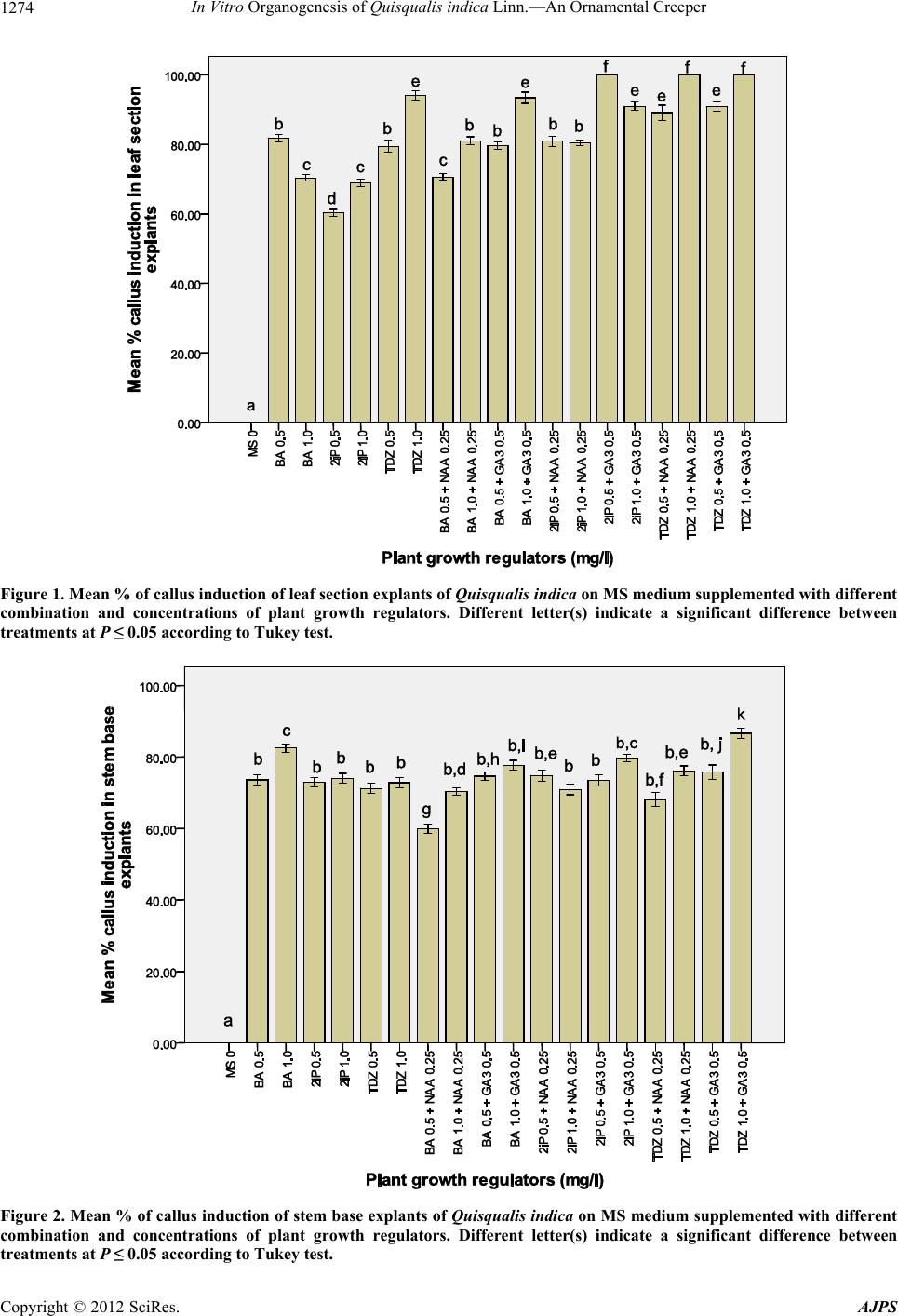 In Vitro Organogenesis of Quisqualis indica Linn.—An Ornamental Creeper Copyright © 2012 SciRes. AJPS 1274 Figure 1. Mean % of callus induction of leaf section explants of Quisqualis indica on MS medium supplemented with different combination and concentrations of plant growth regulators. Different letter(s) indicate a significant difference between treatments at P ≤ 0.05 according to Tukey test. Figure 2. Mean % of callus induction of stem base explants of Quisqualis indica on MS medium supplemented with different combination and concentrations of plant growth regulators. Different letter(s) indicate a significant difference between treatments at P ≤ 0.05 according to Tukey test. 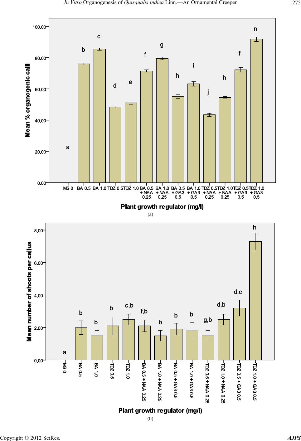 In Vitro Organogenesis of Quisqualis indica Linn.—An Ornamental Creeper 1275 (a) (b) Copyright © 2012 SciRes. AJPS 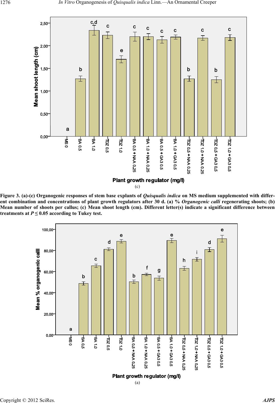 In Vitro Organogenesis of Quisqualis indica Linn.—An Ornamental Creeper 1276 (c) Figure 3. (a)-(c) Organogenic responses of stem base explants of Quisqualis indica on MS medium supplemented with differ- ent combination and concentrations of plant growth regulators after 30 d. (a) % Organogenic calli regenerating shoots; (b) Mean number of shoots per callus; (c) Mean shoot length (cm). Different letter(s) indicate a significant difference between treatments at P ≤ 0.05 according to Tukey test. (a) Copyright © 2012 SciRes. AJPS 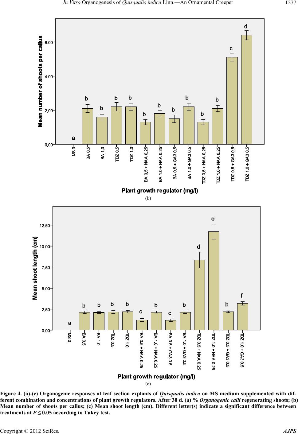 In Vitro Organogenesis of Quisqualis indica Linn.—An Ornamental Creeper 1277 (b) (c) Figure 4. (a)-(c) Organogenic responses of leaf section explants of Quisqualis indica on MS medium supplemented with dif- ferent combination and concentrations of plant growth regulators. After 30 d. (a) % Organogenic calli regenerating shoots; (b) Mean number of shoots per callus; (c) Mean shoot length (cm). Different letter(s) indicate a significant difference between treatments at P ≤ 0.05 according to Tukey test. Copyright © 2012 SciRes. AJPS 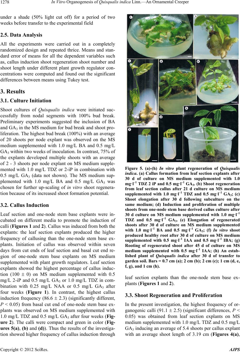 In Vitro Organogenesis of Quisqualis indica Linn.—An Ornamental Creeper Copyright © 2012 SciRes. AJPS 1278 under a shade (50% light cut off) for a period of two weeks before transfer to the experimental field 2.5. Data Analysis All the experiments were carried out in a completely randomized design and repeated thrice. Means and stan- dard error of means for all the dependent variables such as, callus induction shoot regener ation shoot number and shoot length under different plant growth regulator con- centrations were computed and found out the significant differences between means using Tukey test. 3. Results 3.1. Culture Initiation Shoot cultures of Quisqualis indica were initiated suc- cessfully from nodal segments with 100% bud break. Preliminary experiments suggested the inclusion of BA and GA3 in the MS medium for bud break an d shoot pro- liferation. The high est bud brea k (100%) w ith an av erage of 20 shoots per node explant was observed on the MS medium supplemented with 1.0 mg/L BA and 0.5 mg/L GA3 within two weeks of inocul at i on. I n contrast , 75% o f the explants developed multiple shoots with an average of 2 - 3 shoots per node explant on MS medium supple- mented with 1.0 mg/L TDZ or 2-iP in combination with 0.5 mg/L GA3 (data not shown). The MS medium sup- plemented with 1.0 mg/L BA and 0.5 mg/L GA3 was chosen for further up-scaling of in vitro shoot regenera- tion because of its increased shoot formation potential. Figure 5. (a)-(h) In vitro plant regeneration of Quisqualis indica. (a) Callus formation from leaf section explants after 30 d of culture on MS medium supplemented with 1.0 mg·l–1 TDZ 2 iP and 0.5 mg·l–1 GA3. (b) Shoot regeneration from leaf section callus after 21 d culture on MS medium supplemented with 1.0 mg·l–1 TDZ and 0.5 mg·l–1 GA3; (c) Shoot elongation after 30 d following subculture on the same medium; (d) Induction and proliferation of multiple shoots from one-node stem base derived callus culture after 30 d culture on MS medium supplemented with 1.0 mg·l–1 TDZ and 0.5 mg·l–1 GA3. (e) Elongation of regenerated shoots after 30 d of culture on MS medium supplemented with 1.0 mg·l–1 BA and 0.5 mg·l–1 GA3; (f) In vitro shoot produced healthy root after 30 d of culture on MS medium supplemented with 0.5 mg·l–1 IAA and 0.5 mg·l–1 IBA; (g) Rooting of regenerated shoot after 45 d of culture on MS medium supplemented with 0.5 mg·l–1 IAA; (h) An estab- lished plant of Quisqualis indica after 30 d of transfer to garden soil. Bars = 0.7 cm (a); 2 cm (b); 2 cm (c); 1 cm (d, e, f, g), and 1 cm (h). 3.2. Callus Induction Leaf section and one-node stem base explants were in- cubated on different media to promote the induction of calli (Figures 1 and 2). Callus was induced from both the explants: the leaf section explants produced the higher frequency of callusing than the one-node stem base ex- plants. Initiation of callus was observed within seven days from cut ends of leaf section and basal cut end re- gion of one-node stem base explants on MS medium supplemented with plant growth regulators. Leaf section explants showed the highest percentage of callus induc- tion (100 0) on MS medium supplemented with 0.5 mg/L 2-iP and 0.5 mg/L GA3 or 1.0 mg/L TDZ in com- bination with 0.25 mg/L NAA or 0.5 mg/L GA3 after four weeks (Figure 1). In contrast, the highest callus induction frequency (86.6 2.3) (significantly different, P < 0.05) from basal cut end of one-node stem base ex- plants was observed on MS medium supplemented with 1.0 mg/L TDZ and 0.5 mg/L GA3 after four weeks (Fig- ure 2). The calli were compact and green in color (Fig- ures 5(a), (b) and (d)). Thus the results of the investiga- tion showed higher fr equency of callus inductio n through leaf section explants than the one-node stem base ex- plants (Figures 1 and 2). 3.3. Shoot Regeneration and Proliferation In the present investigation, the highest frequency of or- ganogenic calli (91.1 2.5) (significant differences, P < 0.05) was obtained from leaf section explants on MS medium supplemented with 1.0 mg/L TDZ and 0.5 mg/L GA3 inducing an average of 5.4 shoots per callus explant with an average shoot length of 3.19 cm (Figures 4(a), 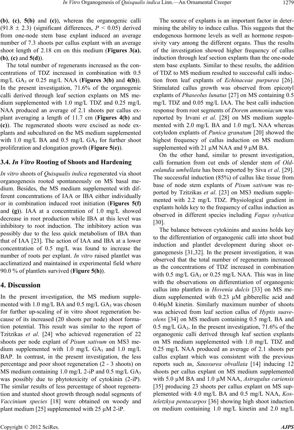 In Vitro Organogenesis of Quisqualis indica Linn.—An Ornamental Creeper 1279 (b), (c), 5(b) and (c)), whereas the organogenic calli (91.8 2.3) (significant differences, P < 0.05) derived from one-node stem base explant induced an average number of 7.3 shoots per callus explant with an average shoot length of 2.18 cm on this medium (Figures 3(a), (b), (c) and 5(d)). The total number of regenerants increased as the con- centrations of TDZ increased in combination with 0.5 mg/L GA3 or 0.25 mg/L NAA (Figures 3(b) and 4(b)). In the present investigation, 71.6% of the organogenic calli derived through leaf section explants on MS me- dium supplemented with 1.0 mg/L TDZ and 0.25 mg/L NAA produced an average of 2.1 shoots per callus ex- plant averaging a length of 11.7 cm (Figures 4(b) and (c)). The regenerated shoots were excised as node ex- plants and subcultured on the MS medium supplemented with 1.0 mg/L BA and 0.5 mg/L GA3 for further shoot proliferation and elongation growth (Figure 5(e)). 3.4. In Vitro Rooting of Shoots and Hardening In vitro shoots of Quisqualis indica regenerated via shoot organogenesis rooted spontaneously on MS basal me- dium. Besides, the MS medium supplemented with dif- ferent concentrations of IAA or IBA either individually or in combination induced root initiation (Figures 5(f) and (g)). IAA at a concentration of 1.0 mg/L showed decrease in root production while IBA at this level was inhibitory to root induction. The inhibitory action was possibly due to the less quick metabolism of IBA than that of IAA [23]. The action of IAA and IBA at a lower concentration of 0.5 mg/L was found to increase the number of roots per explant. In vitro raised plantlet was acclimatized and maintained in experimental field where 90.0 % of plantlets survived (Figure 5(h)). 4. Discussion In the present investigation, the MS medium supple- mented with 1.0 mg/L BA and 0.5 mg/L GA3 was chosen for further up-scaling of in vitro shoot regeneration be- cause of its increased (20 shoots per node) shoot forma- tion potential. This result was similar to the report of Tzitzikas et al. [24] who achieved regeneration of 22 shoots per node explant of Pisum sativum on MS3 me- dium supplemented with 1.0 mg/L GA3 and 1.0 mg/L BAP. In contrast, in the present investigation, the less percentage and poor shoot regeneration (2 - 3 shoots) on MS medium containing 1.0 mg/L 2-iP and 0.5 mg/L GA3 was possibly due to phytotoxicity of cytokinin (2-iP). The similar results of less percentage of shoot regenera- tion and stunted shoot growth through nodal segments of Vaccinium species [18] were obtained on woody and plant medium [25] supplemented with 25 µM 2-iP. The source of explants is an important factor in deter- mining the ability to induce callus. This suggests th at the endogenous hormone levels as well as hormone respon- sivity vary among the different organs. Thus the results of the investigation showed higher frequency of callus induction through leaf sectio n explants than the one-node stem base explants. Similar to these results, the addition of TDZ to MS medium resulted to successful calli induc- tion from leaf explants of Echinaceae purpurea [26]. Stimulated callus growth was observed from epicotyl explants of Phaseolus lunatus [27] on MS containing 0.5 mg/L TDZ and 0.05 mg/L IAA. The best calli induction response from root segments of Dorem ammoniacum was reported by Irvani et al. [28] on MS medium supple- mented with 2.0 mg/L BA and 1.0 mg/L NAA whereas cotyledon explants of Punica granatum [20] showed the highest frequency of callus induction on MS medium supplemented with 21 µM NAA and 9 µM BA. On the other hand, similar to present investigation, calli formation from cut ends of slender stem of Old- enlandia umbellata has been reported by Siva et al. [29]. The successful induction (85%) of callus like tissue from base of node stem explants of Pisum sativum was re- ported by Tzitzikas et al. [23] on MS3 medium supple- mented with 2.2 mg/L TDZ. Physiological gradient in explants hold s key to th e freque ncy of callu s indu ction as observed in different species including Fagus sylvatica [30]. The balance between cytokinins and auxins holds key to the differentiation of organogenic calli into shoot bud induction and plantlet development during shoot or- ganogenesis [31,32]. In the present investigation, it was observed that the total number of regenerants increased as the concentrations of TDZ increased in combination with 0.5 mg/L GA3 or 0.25 mg/L NAA. This was in line with the observations on differentiation of organogenic callus into plantlets in Hovenia dulcis [33] on MS me- dium supplemented with 0.23 µM gibberellic acid and 0.46µM kinetin. Similarly maximum number of shoots was achieved from leaf section callus of Hyptis suave- olens [34] on MS medium containing 0.5 mg/L BA and 0.5 mg/L GA3. In the present investigation, 71.6% of th e organogenic calli derived through leaf section explants on MS medium supplemented with 1.0 mg/L TDZ and 0.25 mg/L NAA produced an average of 2.1 shoots per callus explant which was consistent with the previous reports such as, Saussurea obvallata [14] inducing 12 shoots per callus explant on MS medium supplemented with 5.0 µM BA and 1.0 µM NAA, Astragalus cariensis [35] producing 23 shoots per callus explant on MS sup- plemented with 4.0 mg/L BA and 0.5 mg/L NAA, Kos- teletzkya pentacarpos [36] showing high shoot induction on medium containing 1.0 mg/L kinetin and 2.0 mg/L Copyright © 2012 SciRes. AJPS  In Vitro Organogenesis of Quisqualis indica Linn.—An Ornamental Creeper 1280 IAA. Similarly Tan et al. [37] could induce plantlet re- generation in Vanilla planifolia from leaf and node de- rived callus on MS medium supplemented with 1.0 mg/L BA and 0.5 mg/L NAA. There were reports of the best shoot regeneration response from leaf explants of Brun- felsia calycina [38] on MS medium supplemented with 2.85 µM IAA and 4.44 µM BA or 4.54 µM TDZ and hypocotyl derived organogenic calli of Dorem ammo- niacum [28] on MS medium with 2.0 mg/L BA and 0.2 mg/L IBA. Likewise, the MS medium supplemented with cytokinins and auxins (TDZ and NAA) induced adventi- tious shoot regeneration in many species such as, Prunus serotina [39], Aechmea fasciata [40] and Echinacea pur- purea [26]. Ghimire et al. [41] obtained highest mean number (10.65) of shoots per explant from in vitro leaves of Drymaria cordata on medium containing 1.0 mg/L BA and 0.1 mg/L NAA. Similar to the present results of indirect shoot organogenesis, successful callus initiation and proliferation and adventitious shoot production through nodal explants of silver maple and Acer species were achieved by Preece et al. [42] and Marks and Sim- pson [43] respectively. This was ascribed to the auxin accumulation at the excision sites by downward move- ment which in turn causes cell proliferation especially in the presence of cytokinins [43]. The Rooting efficiency of IAA or IBA has been docu- mented for many medicinal and ornamental plants [16, 20]. The inhibitory action of IBA at 1.0 mg/L compared to IAA was possibly due to the less quick metabolism of IBA than that of IAA [23]. The action of IAA and IBA at a lower concentration was found to increase the number of roots per explant. This result was consistent with the previous report on ro ot ing of Ailanthus triphysa [44]. 5. Conclusion In the present investigation, the TDZ treatment was su- perior to that of 2-iP and BA in combination with either GA3 or NAA in terms of inducing the formation of mul- tiple shoots from organogenic callus derived from leaf section and one-node stem base explants of this orna- mental shrubby climber Quisqualis indica. This was at- tributed to the special property of TDZ and NAA to ful- fill the capability of the ratios of cytokinin and auxin responses in plant species for achieving adventitious shoot production from callus tissues. The development of an efficient plant tissue culture system for the micro- propagation of Quisqualis indica through organogenic callus derived from leaf section and one-node stem base explants holds promise to facilitate conservation and the commercial production of this plant for its medicinal and ornamental use, besides its potential use in plant breeding and the production of transgenic plants through genetic engineering. REFERENCES [1] C. Limmatvapirat, T. Phaechamud and S. Keokitichai, “The Total Polyphenol Content of Some Edible Flowers of Thailand,” Proceeding of the 21st Congress of Federa- tion of Asian Pharmaceutical Associations, Yokohama, 18-21 November 2006, p. 288. [2] P. Wetwitayaklung, T. Phaechamud and S. Keokitichai, “The Study of Antioxidant Activites of Edible Flower,” Proceedings of International Workshop on Medicinal and aromatic Plants, Chiang Mai, 15-18 January 2007, p. 75. [3] P. Wetwitayaklung, T. Phaechamud, C. Limmatvapirat and S. Keokitichai, “The Study of Antioxidant Activities of Edible Flower Extracts,” ISHS Acta Horticulturae, Vol. 786, 2008, pp. 185-192. [4] A. Bose, S. Bose, S. Maji and P. Chakraborty, “Free Radical Scavenging Property of Quisqualis indica,” In- ternational Journal of Biomedical and Pharmaceutical Sciences, Vol. 3, No. 1, 2009, pp. 1-4. [5] B. Hazra, R. Sarkar, S. Biswas and N. Mandal, “Com- parative Study of the Antioxidant and Reactive Oxygen Species Scavenging Properties in the Extracts of the Fruits of Terminalia chebula, Terminalia belerica and Emblica officinalis,” BMC Complementary and Alternative Medicine, Vol. 10, 2010, p. 20. doi:10.1186/1472-6882-10-20 [6] H. S. Lee, N. H. Won, K. H. Kim, H. Lee, W. Jun and K. W. Lee, “Antioxidant Effects of Aqueous Extract of Ter- minalia chebula in Vivo and in Vitro,” Biological and Pharmaceutical Bulletin, Vol. 28, No. 9, 2005, pp. 1639- 1644. doi:10.1248/bpb.28.1639 [7] L. S. de Padua, et al., “Medicinal and Poisonous Plants 1,” In: I. F. Hanum and L. J. G. van der Maesen, Eds., Plant Resources of South-East Asia, Backhuys Publishers, Leiden, Vol. 12, No. 1, 1999, p. 711. [8] T. Ishizaki, K. Kato and M. Kumada, “Effect of Quis- qualic Acid upon Ascaris suum in Vitro in Comparison with Those of Kainic Acid, Alphar-Allokainic Acid and Pyrantel Palmoate,” Japanese Journal of Parasitology, Vol. 22, No. 4, 1973, pp. 181-186. [9] W. S. Kan, “Manual of Medicinal Plants in Taiwan,” National Resource Institute of Chinese Medicine, Vol. 3, 1985, p. 601. [10] L. Ta-Chen, M. Ying-Tsun, W. Jender and H. Feng-Lin, “Tannin and Related Compounds from Quisqualis in- dica,” Journal of the Chinese Chemical Society, Vol. 44, No. 2, 1997, pp. 151-155. [11] M. Lambardi and E. Rugini, “Micropropagation of Olive (Olea europaea L.),” In: S. M. Jain and K. Ishii, Eds., Micropropagation of Woody Trees and Fruits, Kluwer Academic Publishers, The Netherlands, 2003, pp. 621- 646. doi:10.1007/978-94-010-0125-0_21 [12] K. Nirmal Babu, A. Sajina, D. Minoo, C. Z. John, P. M. Mini, K. V. Tushar, J. Rema and P. N. Ravindran, “Mi- cropropagation of Camphor Tree (Cinnamomum cam- phora),” Plant Cell Tissue and Organ Culture, Vol. 74, No. 2, 2003, pp. 179-183. [13] B. Shyamkumar, C. Anjaneyulu and C. C. Giri, “Multiple Shoot Induction from Cotyledonary Node Explants of Copyright © 2012 SciRes. AJPS  In Vitro Organogenesis of Quisqualis indica Linn.—An Ornamental Creeper 1281 Terminalia chebula,” Journal of Plant Biology, Vol. 47, No. 4, 2003, pp. 585-588. [14] U. Dhar and M. Joshi, “Efficient Plant Regeneration Pro- tocol through Callus for Saussurea obvallata (DC) Edgew. (Asteraceae): Effect of Explant Type, Age, and Plant Growth Regulators,” Plant Cell Reports, Vol. 24, No. 4, 2005, pp. 195-200.doi:10.1007/s00299-005-0932-1 [15] A. Sadanandam, M. Ramesh, P. Umate and K. V. Rao, “Micropropagation of Terminalia bellirica Roxb.—A Sericulture and Medicinal Plant,” In Vitro Cellular & Developmental Biology—Plant, Vol. 41, No. 3, 2005, pp. 320-323. doi:10.1079/IVP2004626 [16] S. Pandey, M. Singh, U. Jaiswal and V. S. Jaiswal, “Shoot Initiation and Multiplication from a Mature Tree of Terminalia arjuna Roxb,” In Vitro Cellular & Devel- opmental Biology—Plant, Vol. 42, No. 5, 2006, pp. 389- 393. doi:10.1079/IVP2006790 [17] M. W. Bairu, W. A. Stirk, K. Dolezal and J. Van Staden, “Optimizing the Micropropagation Protocol for the En- dangered Aloe polyphylla: Can Meta-Topolin and Its De- rivatives Serve as Replacement for Benzyladenine and Zeatin?” Plant Cell, Tissue and Organ Culture, Vol. 90, No. 1, 2007, pp. 15-23. doi:10.1007/s11240-007-9233-4 [18] J. Meiners, M. Schwab and I. Szankowski, “Efficient in Vitro Regeneration Systems for Vaccinium species,” Plant Cell, Tissue and Organ Culture, Vol. 89, No. 4, 2007, pp. 169-176. doi:10.1007/s11240-007-9230-7 [19] K. Kwapata, R. Sabzikar, M. B. Sticklen and J. D. Kelly, “In Vitro Regeneration and Morphogenesis Studies in Common Bean,” Plant Cell, Tissue and Organ Culture, Vol. 100, No. 1, 2010, pp. 97-105. doi:10.1007/s11240-009-9624-9 [20] K. Kanwar, J. Joseph and R. Deepika, “Comparison of in Vitro Regeneration Pathways in Punica granatum L.,” Plant Cell, Tissue and Organ Culture, Vol. 100, No. 2, 2010, pp. 199-207. doi:10.1007/s11240-009-9637-4 [21] G. M. Abogadallah and W. P. Quick, “Fast Versatile Re- generation of Trifolium alexandrinum L.,” Plant Cell, Tissue and Organ Culture, Vol. 100, No. 1, 2010, pp. 39- 48. doi:10.1007/s11240-009-9614-y [22] T. Murashige and F. Skoog, “A Revised Medium for Rapid Growth and Bioassays with Tobacco Tissue Cul- tures,” Physiologia Plantarum, Vol. 15, No. 3, 1962, pp. 473-497. doi:10.1111/j.1399-3054.1962.tb08052.x [23] G. J. De Klerk, “Rooting of Micropropagules,” In: Y. Waisel, A. Eshel and U. Kafkafi, Eds., Plant Roots the Hidden Half, 3rd Edition, Marcel Dekker Inc., New York, 2002, pp. 349-357. doi:10.1201/9780203909423.ch21 [24] E. N. Tzitzikas, M. Bergervoet, K. Raemakers, J. P. Vincken, A. van Lammeren and R. G. F. Visser, “Regen- eration of Pea (Pisum sativum L.) by a Cyclic Organo- genic System,” Plant Cell Reports, Vol. 23, No. 7, 2004, pp. 453-460. doi:10.1007/s00299-004-0865-0 [25] G. Lloyd and B. McCown, “Commercially Feasible Mi- cropropagation of Mountain Laurel (Kalmia latifolia) by Use of Shoot Tip Culture,” Combined Proceedings of the International Plant Propagators’ Society, Vol. 30, 1980, pp. 421-427. [26] M. P. A. Jones, Z. Yi, S. J. Murch and P. K. Saxena, “Thidiazuron-Induced Regeneration of Echinacea pur- purea L.: Micropropagation in Solid and Liquid Culture Systems,” Plant Cell Reports, Vol. 26, No. 1, 2007, pp. 13-19. doi:10.1007/s00299-006-0209-3 [27] C. N. Kanchiswamy and M. Maffei, “Callus Induction and Shoot Regeneration of Phaseolus lunatus L. cv. Wonder Bush and cv. Pole Seiva,” Plant Cell, Tissue and Organ Culture, Vol. 92, No. 2, 2008, pp. 239-242. doi:10.1007/s11240-007-9322-4 [28] N. Irvani, M. Solouki, M. Omidi, A. R. Zare and S. Shabnazi, “Callus Induction and Plant Regeneration in Dorem ammoniacum D., an Endangered Medicinal Plant,” Plant Cell, Tissue and Organ Culture, Vol. 100, No. 3, 2010, pp. 293-299. doi:10.1007/s11240-009-9650-7 [29] R. Siva, C. Rajasekaran and G. Mudgal, “Induction of Somatic Embryogenesis and Organogenesis in Oldenlan- dia umbellata L., a Dye-Yielding Medicinal Plant,” Plant Cell, Tissue and Organ Culture, Vol. 98, No. 2, 2009, pp. 205-211. doi:10.1007/s11240-009-9553-7 [30] B. Cuenca, A. Ballester and A. M. Vieitez, “In Vitro Ad- ventitious Bud Regeneration from Internode Segments of Beech,” Plant Cell, Tissue and Organ Culture, Vol. 60, No. 3, 2000, pp. 213-220. doi:10.1023/A:1006428717309 [31] A. W. Woodward and B. Bartel, “Auxin. Regulation, Action and Interaction,” Annals of Botany, Vol. 95, No. 5, 2005, pp. 707-735. doi:10.1093/aob/mci083 [32] H. Sakakibara, “Cytokinins: Activity, Biosynthesis and Translocation,” Annual Review of Plant Biology, Vol. 57, 2006, pp. 431-449. doi:10.1146/annurev.arplant.57.032905.105231 [33] M. J. Jeong, H. J. Song, D. J. Park, J. Y. Min, J. S. Jo, B. M. Kim, H. G. Kim, Y. D. Ki m, R. M. Kim, C. S. Karigar and M. S. Choi, “High Frequency Plant Regeneration Following Abnormal Shoot Organogenesis in the Me- dicinal Tree Hovenia dulcis,” Plant Cell, Tissue and Or- gan Culture, Vol. 98, No. 1, 2009, pp. 59-65. doi:10.1007/s11240-009-9538-6 [34] J. Mandal, “Shoot Regeneration through Organogenesis of Leaf and Shoot Base Explants of Hyptis suaveolens (Linn.) Poit.,” Phytomorphology, Vol. 61, No. 3-4, 2011, pp. 85-92. [35] S. Erisen, M. Yorgancilar, E. Atalay and M. Babaoglu, “Prolific Shoot Regeneration of Astragalus cariensis Boiss,” Plant Cell, Tissue and Organ Culture, Vol. 100, No. 2, 2010, pp. 229-233. doi:10.1007/s11240-009-9638-3 [36] A. Piovan, R. Caniato, E. M. Cappelletti and R. Filippini, “Organogenesis from Shoot Segments and Via Callus of Endangered Kosteletzkya pentacarpos (L.) Ledeb,” Plant Cell, Tissue and Organ Culture, Vol. 100, No. 3, 2010, pp. 309-315. doi:10.1007/s11240-009-9652-5 [37] B. C. Tan, C. F. Chin and P. Alderson, “Optimization of Plantlet Regeneration from Leaf and Nodal Derived Cal- lus of Vanilla Planifolia Andrews,” Plant Cell, Tissue and Organ Culture, Vol. 105, No. 3, 2011, pp. 457-463. doi:10.1007/s11240-010-9866-6 [38] R. Liberman, L. Shahar, A. Nissim-Levi, D. Evenor, M. Reuveni and M. Oren-Shamir, “Shoot Regeneration from Copyright © 2012 SciRes. AJPS  In Vitro Organogenesis of Quisqualis indica Linn.—An Ornamental Creeper Copyright © 2012 SciRes. AJPS 1282 Leaf Explants of Brunfelsia calycina,” Plant Cell, Tissue and Organ Culture, Vol. 100, No. 3, 2010, pp. 345-348. doi:10.1007/s11240-009-9642-7 [39] X. Liu and P. M. Pijut, “Plant Regeneration from in Vitro Leaves of Mature Black Cherry (Prunus serotina),” Plant Cell, Tissue and Organ Culture, Vol. 94, No. 2, 2008, pp. 113-123. doi:10.1007/s11240-008-9393-x [40] P. L. Huang, L. J. Liao, C. C. Tsai and Z. H. Liu, “Mi- cropropagation of Bromeliad Aechmea fasciata via Floral Organ Segments and Effects of Acclimatization on Plant- let Growth,” Plant Cell, Tissue and Organ Culture, Vol. 105, No. 1, 2011, pp. 73-78. doi:10.1007/s11240-010-9843-0 [41] B. K. Ghimire, E. S. Seong, E. Goh, N. Y. Kim, W. H. Kang, E. H. Kim, C. Y. Yu and I. M. Chung, “High- Frequency Direct Shoot Regeneration from Drymaria cordata Willd. Leaves,” Plant Cell, Tissue and Organ Culture, Vol. 100, No. 2, 2010, pp. 209-217. doi:10.1007/s11240-009-9627-6 [42] J. E. Preece, C. A. Hutterman, W. C. Ashby and P. L. Roth, “Micropropagation and Cutting Propagation of Sil- ver Mapple. 1. Results with Adult and Juvenile Propa- gules,” Journal of the American Society for Horticultural Science, Vol.116, No. 1, 1991, pp. 142-148. [43] T. R. Marks and S. E. Simpson, “Factors Affecting Shoot Development in Apicaly Dominant Acer Cultivars in Vi- tro,” Journal of Horticultural Science, Vol. 69, No. 3, 1994, pp. 543-551. [44] S. R. Natesha and N. K. Vijayakumar, “In Vitro Propaga- tion of Ailanthus triphysa,” Journal of Tropical Forest Science, Vol. 16, 2004, pp. 402-412. |