

Paper Menu >>
Journal Menu >>
 American Journal of Plant Sciences, 2012, 3, 1225-1231 http://dx.doi.org/10.4236/ajps.2012.39148 Published Online September 2012 (http://www.SciRP.org/journal/ajps) 1225 Genetic Transformation Studies on Avocado Cultivar “Hass” (Persea americana) Muhammad F. Ahmed1, Arumugam S. Kantharajah2*, Paul Holford1 1University of Western Sydney, South Penrith DC, Australia; 2Department of Agricultural Sciences, American University of Beirut, Beirut, Lebanon. Email: *[email protected] Received March 16th, 2012; revised April 10th, 2012; accepted April 26th, 2012 ABSTRACT The use of traditional breeding for improvement of avocado cultivars is time consuming, hence other methods such as genetic transformation by Agrobacterium is indispensable to adop t. The str ain GV3850 /pBI12 1gav e best tran sfor mation outcome compared to five other binary vectors (AGL1/pCGP904; AGL1/pBI121; GV3850/pCGP904; LBA4404/pCG- P904 and LBA4404/pBI121) under different pH and acetosyringone concentrations. The optimal condition for reliable transformation was by using 200 µM acetosyringone and a pH of 5.2. Transformed embryonic shoots co-cultivated with GV3850/pBI121 were tested using th e histochemical x-gluc assay. Fur ther analysis was conducted by polymerase ch ain reaction using specific primers for the rep orter gene (GUS). Keywords: Avocado; Persea; Binary Vectors; GUS Reporter 1. Introduction The avocado is a major horticu ltural crop in tropical parts of the world. Although avocado has a high economic and nutritional importance, there are genetic problems asso- ciated with its production. The successful incorporation of transfer-DNA (T-DNA) from wild-type strains of Agrobacterium tumefaciens to avocado tissues has been observed. However the wild-type Ti-plasmids are not suitable as gene vectors as they produce disorganized growth of recipient plant cells owing to the effects of the oncogenes in the T-DNA. Consequently, such tumour cells have proven recalcitrant to attempts to induce re- generation into plantlets or normal tissues. In order to regenerate plants effectively, the T-DNA has to be dis- armed. This is achieved by deleting all of its oncogenic hormone biosynthesis genes without interfering with its ability to integrate into p lant chromosomes [1,2]. There are two types of disarmed tumor-inducing (Ti) plasmid vectors currently in use; these are co-integrative and binary vectors. The T-DNA and vir functions are maintained within the same Ti plasmid in co-integrative vectors. In contrast, binary vectors have the vir and T- DNA regions on separate replicons. In this latter system, the T-DNA borders are located on a replicon that will function in both E. coli and Agrobacterium, a feature that greatly facilitates construct formation. Although the vir and T-DNA regions are in trans, the inserted DNA be- tween the T-DNA borders is efficiently transferred to the plant’s genome [3]. pGV3850 is an example of a co-integrative vector [4]. Zambryski et al. [4] created a deletio n mutant of pTiC58 where mo st o f the DNA be tween th e righ t and lef t border sequences of the T-DNA had been lost, including the genes for hormone production. The nopaline synthase gene remained and acts as a T-DNA specific marker. In addition, the cloning vector, pBR322, was inserted in the T-DNA region. The pBR322 sequence can act as an ac- ceptor site for the insertion of genes to be transferred to the plant through a single recombination even t with plas- mids containing homologous sequences. Using this vec- tor, Zambryski et al. [4] were able to transform plant tissues and regenerate fertile adult p lants. Hoekema et al. [5] developed the binary vector strategy by creating the plasmid pAL1050. pAL1050 is a derivative of pTiAch5 that can replicate in both E. coli and A. tumefaciens and contains the T-DNA region. This plasmid was introduced into the cell line, LBA44 04, which harbours the plasmid, pAL4404. This latter plasmid was isolated as a sponta- neous deletion mutant of an octopine-type Ti plasmid that had lost its entire T-DNA but retained a complete comple ment of vir functions [6]. The combination of the two plasmids induced tumour formation on tomatoes, Kalanchoë, tobacco and peas despite the fact that the T-DNA and vir regions were on separate plasmids [5]. Since this time, several disarmed binary vector systems have been produced. *Corresponding author. Copyright © 2012 SciRes. AJPS 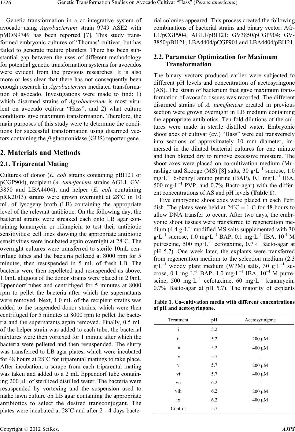 Genetic Transformation Studies on Avocado Cultivar “Hass” (Persea americana) 1226 Genetic transformation in a co-integrative system of avocado using Agrobacterium strain 9749 ASE2 with pMON9749 has been reported [7]. This study trans- formed embryonic cultures of ‘Thomas’ cultivar, but has failed to generate mature plantlets. There has been sub- stantial gap between the uses of different methodology for potential genetic transformation systems for avocado s were evident from the previous researches. It is also more or less clear that there has not consequently been enough research in Agrobacterium mediated transforma- tion of avocado. Investigations were made to find: 1) which disarmed strains of Agrobacterium is most viru- lent on avocado cultivar “Hass”; and 2) what culture conditions give maximum transformation. Therefore, the main purposes of this study were to d etermine the condi- tions for successful transformation using disarmed vec- tors containing the -glucuroni dase (GUS) reporter gene. 2. Materials and Methods 2.1. Triparental Mating Cultures of donor (E. coli strains containing pBI121 or pCGP904), recipient (A. tumefaciens strains AGL1, GV- 3850 and LBA4404), and helper (E. coli containing pRK2013) strains were grown overnight at 28˚C in 10 mL of lysogeny broth (LB) containing the appropriate level of the relevant antibio tic. On the following day, the bacterial strains were streaked each onto LB agar con- taining kanamycin or rifampicin to test their antibiotic sensitivities: cell lines showing the appropriate antibiotic sensitivities were incub ated again overnight at 28˚C. The overnight cultures were transferred to sterile 10mL cen- trifuge tubes and the bacteria pelleted at 8000 rpm for 5 minutes, then resuspended in 5 mL of fresh LB. The bacteria were then repelleted and resuspended as above. 1.0mL aliquots of the donor strains were placed in 2.0mL Eppendorf tubes and centrifuged for 5 minutes at 8000 rpm to pellet the bacteria after which the supernatants were removed. Next, 1.0 mL of the recipient strains was added to the suspended donor strains, which were then centrifuged for 5 minutes at 8000 rpm to pellet the bacte- ria and the supernatants again removed. Finally, 0.5 mL of the helper strain was added to each tube, the bacterial mixtures were then vortexed for 1 minute after which the bacteria were pelleted and then resuspended. The slurry was transferred to LB agar plates, which were incubated for 48 hours at 28˚C for triparental matings to take place. After incubation, a scrape from each triparental mating was taken and added to a 2 mL Eppendorf tube contain- ing 200 L of sterilized distilled water. The bacteria were resuspended by vortexing and the suspension used to make lawn culture on LB agar containing the appropriate antibiotics to select the desired transconjugant. The plates were incubated at 28˚C and after 2 - 4 days bacte- rial colonies appeared. This process created the following combinations of bacterial strains and binary vector: AG- L1/pCGP904; AGL1/pBI121; GV3850/pCGP904; GV- 3850/pBI121; LB A4404/pC GP904 and LB A4404/ pBI12 1. 2.2. Parameter Optimization for Maximum Transformation The binary vectors produced earlier were subjected to different pH levels and concentration of acetosyringone (AS). The strain of bacterium that gave maximum trans- formation of avocado tissues was recorded. The different disarmed strains of A. tumefaciens created in previous section were grown overnight in LB medium containing the appropriate antibiotics. Ten-fold dilutions of the cul- tures were made in sterile distilled water. Embryonic shoot axes of cultivar (cv.) “Hass” were cut transversely into sections of approximately 10 mm diameter, im- mersed in the diluted bacterial cultures for one minute and then blotted dry to remove excessive moisture. The shoot axes were placed on co-cultivation medium (Mu- rashige and Skooge (MS) [8] salts, 30 g·L–1 sucrose, 1.0 mg·L–1 6-benzyl amino purine (BAP), 0.1 mg·L–1 IBA, 500 mg·L–1 PVP, and 0.7% Bacto-agar) with the differ- ent concentrations of AS and pH levels (Table 1). Five embryonic shoot axes were placed in each Petri dish. The plates were held at 24˚C ± 1˚C for 48 hours to allow DNA transfer to occur. After two days, the embr- yonic shoot tissues were transferred to regeneration me- dium (4.4 g·L–1 modified MS salts supplemented with 30 g·L–1 sucrose, 1.0 mg·L–1 BAP, 0.1 mg·L–1 IBA, 10-4 M putrescine, 500 mg·L–1 cefotaxime, 0.7% Bacto-agar at pH 5.7). One week later, the explants were transferred from regeneration medium to the selection medium (2.3 g·L–1 woody plant medium (WPM) salts, 30 g·L–1 su- crose, 0.1 mg·L–1 BAP, 1.0 mg·L–1 IBA, 10–4 M putre- scine, 500 mg·L–1 cefotaxime, 60 mg·L–1 kanamycin, 0.7% Bacto-agar at pH 5.7). The majority of explants Table 1. Co-cultivation media with different concentrations of pH and acetosyringone. Treatment pH Acetosyringone i 5.2 - ii 5.2 200 M iii 5.2 400 M iv 5.7 - v 5.7 200 M vi 5.7 400 M vii 6.2 - viii 6.2 200 M ix 6.2 400 M Control 5.7 - Copyright © 2012 SciRes. AJPS 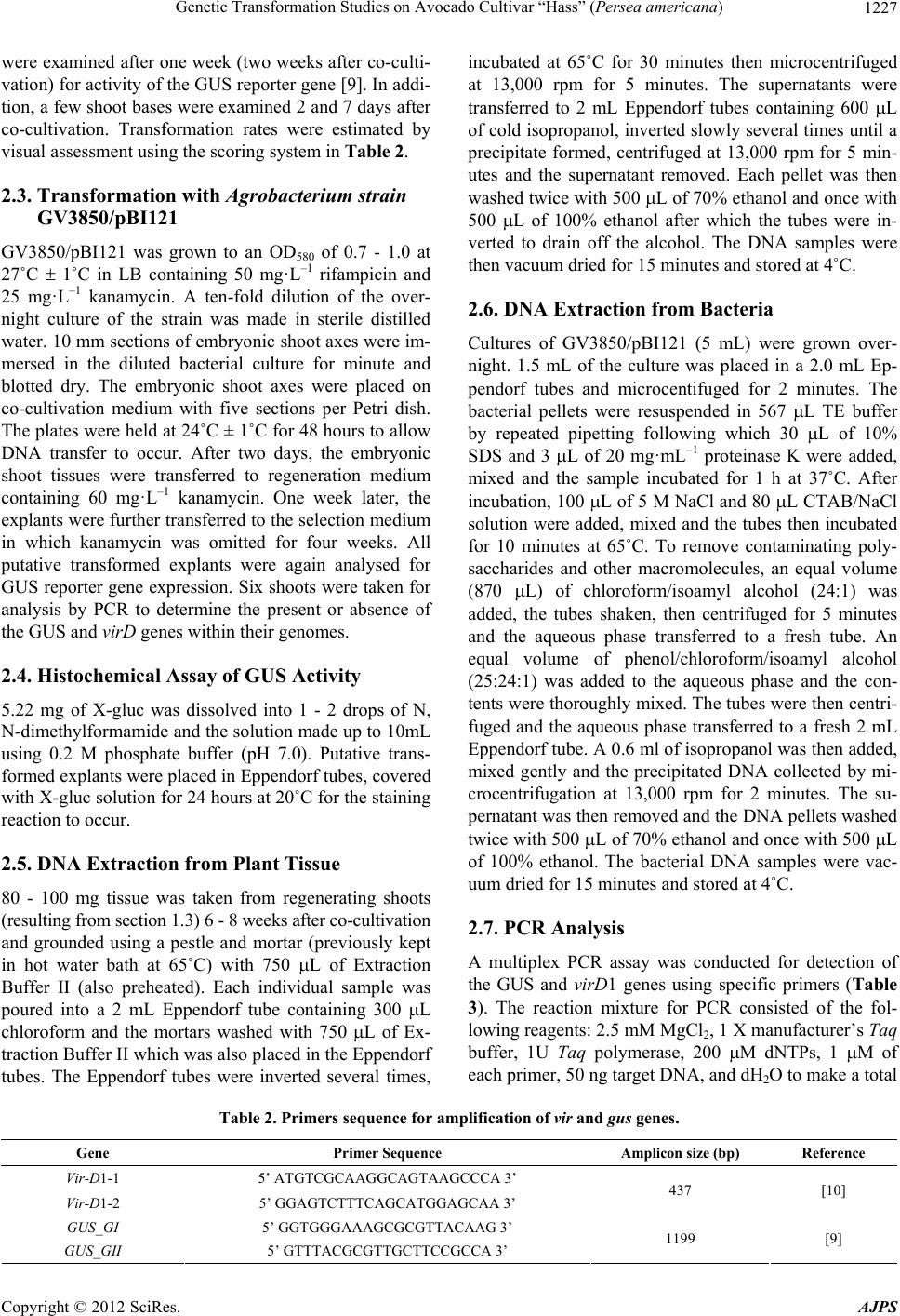 Genetic Transformation Studies on Avocado Cultivar “Hass” (Persea americana) Copyright © 2012 SciRes. AJPS 1227 were examined after one week (two weeks after co-culti- vation) for activity of the GUS reporter gene [9]. In addi- tion, a few shoot bases were examined 2 and 7 days after co-cultivation. Transformation rates were estimated by visual assessment using the scoring system in Table 2. 2.3. Transformation with Agrobacterium strain GV3850/pBI121 GV3850/pBI121 was grown to an OD580 of 0.7 - 1.0 at 27˚C 1˚C in LB containing 50 mg·L–1 rifampicin and 25 mg·L–1 kanamycin. A ten-fold dilution of the over- night culture of the strain was made in sterile distilled water. 10 mm sections of embryonic shoot axes were im- mersed in the diluted bacterial culture for minute and blotted dry. The embryonic shoot axes were placed on co-cultivation medium with five sections per Petri dish. The plates were held at 24˚C ± 1˚C for 48 hours to allow DNA transfer to occur. After two days, the embryonic shoot tissues were transferred to regeneration medium containing 60 mg·L–1 kanamycin. One week later, the explants were further transferred to the selection medium in which kanamycin was omitted for four weeks. All putative transformed explants were again analysed for GUS reporter gene expression. Six shoots were taken for analysis by PCR to determine the present or absence of the GUS and virD genes within their genomes. 2.4. Histochemical Assay of GUS Activity 5.22 mg of X-gluc was dissolved into 1 - 2 drops of N, N-dimethylformamide and the solution made up to 10 mL using 0.2 M phosphate buffer (pH 7.0). Putative trans- formed explants were placed in Eppendorf tubes, covered with X-gluc solu tion for 24 hours at 20 ˚C for the staining reaction to occur. 2.5. DNA Extraction from Plant Tissue 80 - 100 mg tissue was taken from regenerating shoots (resulting from section 1.3) 6 - 8 weeks after co-cultivation and grounded using a pestle and mortar (previously kept in hot water bath at 65˚C) with 750 L of Extraction Buffer II (also preheated). Each individual sample was poured into a 2 mL Eppendorf tube containing 300 L chloroform and the mortars washed with 750 L of Ex- traction Buffer II which was also placed in the Eppendorf tubes. The Eppendorf tubes were inverted several times, incubated at 65˚C for 30 minutes then microcentrifuged at 13,000 rpm for 5 minutes. The supernatants were transferred to 2 mL Eppendorf tubes containing 600 L of cold isoprop anol, inverted slowly several times until a precipitate formed, centrifuged at 13,000 rpm for 5 min- utes and the supernatant removed. Each pellet was then washed twice with 500 L of 70% ethanol and once with 500 L of 100% ethanol after which the tubes were in- verted to drain off the alcohol. The DNA samples were then vacuum dried for 15 minutes and stored at 4˚C. 2.6. DNA Extraction from Bacteria Cultures of GV3850/pBI121 (5 mL) were grown over- night. 1.5 mL of the culture was placed in a 2.0 mL Ep- pendorf tubes and microcentifuged for 2 minutes. The bacterial pellets were resuspended in 567 L TE buffer by repeated pipetting following which 30 L of 10% SDS and 3 L of 20 mg·mL–1 proteinase K were added, mixed and the sample incubated for 1 h at 37˚C. After incubation, 100 L of 5 M NaCl and 80 L CTAB/NaCl solution were added, mixed and the tubes then incubated for 10 minutes at 65˚C. To remove contaminating poly- saccharides and other macromolecules, an equal volume (870 L) of chloroform/isoamyl alcohol (24:1) was added, the tubes shaken, then centrifuged for 5 minutes and the aqueous phase transferred to a fresh tube. An equal volume of phenol/chloroform/isoamyl alcohol (25:24:1) was added to the aqueous phase and the con- tents were thoroughly mixed. The tubes were then centri- fuged and the aqueous phase transferred to a fresh 2 mL Eppendorf tube. A 0.6 ml of isopropanol was then added, mixed gently and the precipitated DNA collected by mi- crocentrifugation at 13,000 rpm for 2 minutes. The su- pernatant was then removed and the DNA pellets washed twice with 500 L of 70% ethanol and once w ith 500 L of 100% ethanol. The bacterial DNA samples were vac- uum dried for 15 minutes and stored at 4˚C. 2.7. PCR Analysis A multiplex PCR assay was conducted for detection of the GUS and vir D1 genes using specific primers (Table 3). The reaction mixture for PCR consisted of the fol- lowing reagents: 2.5 mM MgCl2, 1 X manufacturer’s Taq buffer, 1U Taq polymerase, 200 M dNTPs, 1 M of each primer, 50 ng target DNA, and dH2O to make a total Table 2. Primers sequence for amplification of vir and gus genes. Gene Primer Sequence Amplicon size (bp) Reference Vir-D1-1 5’ ATGTCGCAAGGCAGTAAGCCCA 3’ Vir-D1-2 5’ GGAGTCTTTCAGCATGGAGCAA 3’ 437 [10] GUS_GI 5’ GGTGGGAAAGCGCGTTACAAG 3’ GUS_GII 5’ GTTTACGCGTTGCTTCCGCCA 3’ 1199 [9] 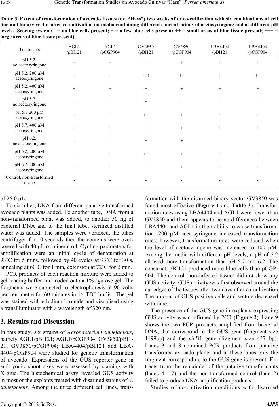 Genetic Transformation Studies on Avocado Cultivar “Hass” (Persea americana) 1228 Table 3. Extent of transformation of avocado tissues (cv. “Hass”) two weeks after co-cultivation with six combinations of cell line and binary vector after co-cultivation on media containing different concentrations of acetosyringone and at different pH levels. (Scoring system: - = no blue cells present; + = a few blue cells present; ++ = small areas of blue tissue present; +++ = large areas of blue tissue present). Treatments AGL1 /pBI121 AGL1 /pCGP904 GV3850 /pBI121 GV3850 /pCGP904 LBA4404 /pBI121 LBA4404 /pCGP904 pH 5.2, no acetosyringone - - + + + + pH 5.2, 200 M acetosyringone + + +++ ++ + ++ pH 5.2, 400 M acetosyringone + + + + + + pH 5.7, no acetosyringone - - + - - - pH 5.7 200 M acetosyringone + + ++ + + + pH 5.7, 400 M acetosyringone + + + + + + pH 6.2, no acetosyringone - - + + - - pH 6.2, 200 M acetosyringone + + ++ + + + pH 6.2, 400 M acetosyringone + + + + + + Control, non-tra ns formed tissue - - - - - - of 25.0 L. To six tubes, DNA from different putative transformed avocado plants was added. To another tube, DNA from a non-transformed plant was added, to another 50 ng of bacterial DNA and to the final tube, sterilized distilled water was added. The samples were vortexed, the tubes centrifuged for 10 seconds then the contents were over- layered with 40 L of mineral oil. Cycling parameters for amplification were an initial cycle of denaturation at 93˚C for 5 mins, followed by 40 cycles at 93˚C for 30 s, annealing at 60˚C for 1 min, extension at 72˚C for 2 min. PCR products of each reaction mixture were added to gel loading buffer and loaded onto a 1% agarose gel. The fragments were subjected to electrophoresis at 90 volts per centimetre for 60 minutes in 1× TBE buffer. The gel was stained with ethidium bromide and visualised using a transilluminator with a wavelength of 320 nm. 3. Results and Discussion In this study, six strains of Agrobacterium tumefaciens, namely AGL1/pBI121; AGL1/pCGP904; GV3850/pBI1- 21; GV3850/pCGP904; LBA4404/pBI121 and LBA- 4404/pCGP904 were studied for genetic transformation of avocado. Expressions of the GUS reporter gene in embryonic shoot axes were assessed by staining with X-gluc. The histochemical assay revealed GUS activity in most of the explants treated with disarmed strains of A. tumefaciens. Among the three different cell lines, trans- formation with the disarmed binary vector GV3850 was found most effective (Figure 1 and Table 3). Transfor- mation rates usin g LBA4404 and AGL1 were lower than GV3850 and there appears to be no differences between LBA4404 and AGL1 in their ability to cause transfo rma- tion. 200 M acetosyringone increased transformation rates; however, transformation rates were reduced when the level of acetosyringone was increased to 400 M. Among the media with different pH levels, a pH of 5.2 allowed more transformation than pH 5.7 and 6.2. The construct, pBI121 produced more blue cells than pCGP- 904. The control (non-infected tissue) did not show any GUS activity. GUS activity was first observed aroun d the cut edges of the tissues after two days after co-cultiv ation. The amount of GUS positive cells and sectors decreased with time. The presence of the GUS gene in explants expressing GUS activity was confirmed by PCR (Figure 2). Lane 9 shows the two PCR products, amplified from bacterial DNA, that correspond to the GUS gene (fragment size 1199bp) and the virD1 gene (fragment size 437 bp). Lanes 3 and 8 contained PCR products from putative transformed avocado plants and in these lanes only the fragment corresponding to the GUS gene is present. Ex- tracts from the remainder of the putative transformants (lanes 4 - 7) and the non-transformed control (lane 2) failed to produce DNA amplification products. Studies of co-cultivation conditions with disarmed Copyright © 2012 SciRes. AJPS 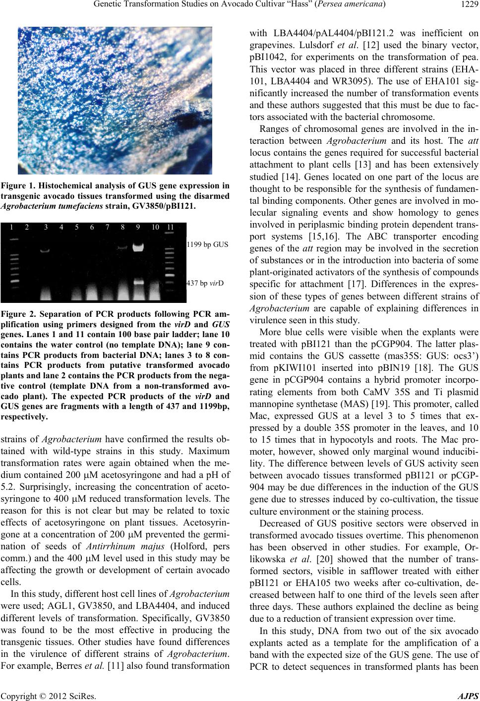 Genetic Transformation Studies on Avocado Cultivar “Hass” (Persea americana) 1229 Figure 1. Histochemical analysis of GUS gene expression in transgenic avocado tissues transformed using the disarmed Agrobacterium tumefaciens strain, GV3850/pBI121. 1 2 3 4 5 6 7 8 9 10 11 1199 bp GUS 437 bp virD Figure 2. Separation of PCR products following PCR am- plification using primers designed from the virD and GUS genes. Lanes 1 and 11 contain 100 base pair ladder; lane 10 contains the water control (no template DNA); lane 9 con- tains PCR products from bacterial DNA; lanes 3 to 8 con- tains PCR products from putative transformed avocado plants and lane 2 contains the PCR produc ts from the nega- tive control (template DNA from a non-transformed avo- cado plant). The expected PCR products of the virD and GUS genes are fragments with a length of 437 and 1199bp, respectively. strains of Agrobacterium have confirmed the results ob- tained with wild-type strains in this study. Maximum transformation rates were again obtained when the me- dium contained 200 M acetosyringone and had a pH of 5.2. Surprisingly, increasing the concentration of aceto- syringone to 400 M reduced transformation levels. The reason for this is not clear but may be related to toxic effects of acetosyringone on plant tissues. Acetosyrin- gone at a concentration of 200 M prevented the germi- nation of seeds of Antirrhinum majus (Holford, pers comm.) and the 400 M level used in this study may be affecting the growth or development of certain avocado cells. In this study, different host cell lines of Agrobacterium were used; AGL1, GV3850, and LBA4404, and induced different levels of transformation. Specifically, GV3850 was found to be the most effective in producing the transgenic tissues. Other studies have found differences in the virulence of different strains of Agrobacterium. For example, Berres et al. [11] also found transfor mation with LBA4404/pAL4404/pBI121.2 was inefficient on grapevines. Lulsdorf et al. [12] used the binary vector, pBI1042, for experiments on the transformation of pea. This vector was placed in three different strains (EHA- 101, LBA4404 and WR3095). The use of EHA101 sig- nificantly increased the number of transformation events and these authors suggested that this must be due to fac- tors associated with the bacterial chromosome. Ranges of chromosomal genes are involved in the in- teraction between Agrobacterium and its host. The att locus contains the genes required for successful bacterial attachment to plant cells [13] and has been extensively studied [14]. Genes located on one part of the locus are thought to be responsible for the synthesis of fundamen- tal binding components. Other genes are involved in mo- lecular signaling events and show homology to genes involved in periplasmic binding protein dependent trans- port systems [15,16]. The ABC transporter encoding genes of the att region may be involved in the secretion of substances or in the introduction into bacteria of some plant-originated activators of the synthesis of compounds specific for attachment [17]. Differences in the expres- sion of these types of genes between different strains of Agrobacterium are capable of explaining differences in virulence seen in this study. More blue cells were visible when the explants were treated with pBI121 than the pCGP904. The latter plas- mid contains the GUS cassette (mas35S: GUS: ocs3’) from pKIWI101 inserted into pBIN19 [18]. The GUS gene in pCGP904 contains a hybrid promoter incorpo- rating elements from both CaMV 35S and Ti plasmid mannopine synthetase (MAS) [19]. This promoter, called Mac, expressed GUS at a level 3 to 5 times that ex- pressed by a double 35S promoter in the leaves, and 10 to 15 times that in hypocotyls and roots. The Mac pro- moter, however, showed only marginal wound inducibi- lity. The difference between levels of GUS activity seen between avocado tissues transformed pBI121 or pCGP- 904 may be due differences in the induction of the GUS gene due to stresses induced by co-cultivation, the tissue culture environment or the staining process. Decreased of GUS positive sectors were observed in transformed avocado tissues overtime. This phenomenon has been observed in other studies. For example, Or- likowska et al. [20] showed that the number of trans- formed sectors, visible in safflower treated with either pBI121 or EHA105 two weeks after co-cultivation, de- creased between half to one third of the levels seen after three days. These authors explained the decline as being due to a reduction of transient expression over time. In this study, DNA from two out of the six avocado explants acted as a template for the amplification of a band with the expected size of the GUS gene. The use of PCR to detect sequences in transformed plants has been Copyright © 2012 SciRes. AJPS 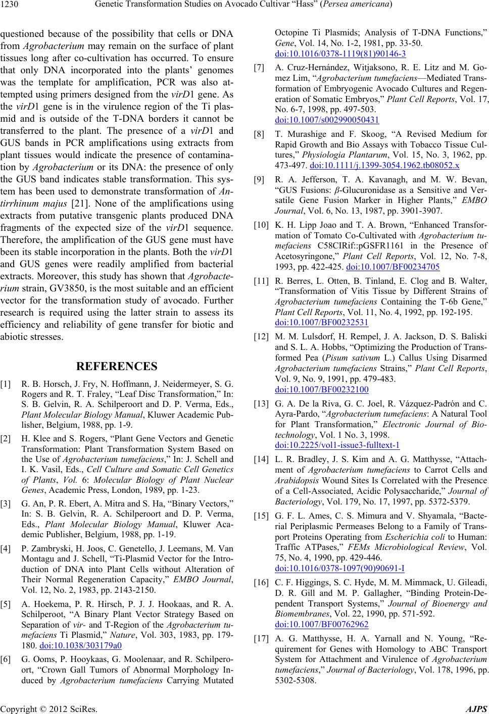 Genetic Transformation Studies on Avocado Cultivar “Hass” (Persea americana) 1230 questioned because of the possibility that cells or DNA from Agrobacterium may remain on the surface of plant tissues long after co-cultivation has occurred. To ensure that only DNA incorporated into the plants’ genomes was the template for amplification, PCR was also at- tempted using primers designed from the virD1 gene. As the virD1 gene is in the virulence region of the Ti plas- mid and is outside of the T-DNA borders it cannot be transferred to the plant. The presence of a virD1 and GUS bands in PCR amplifications using extracts from plant tissues would indicate the presence of contamina- tion by Agrobacterium or its DNA: the presence of only the GUS band indicates stable transformation. This sys- tem has been used to demonstrate transformation of An- tirrhinum majus [21]. None of the amplifications using extracts from putative transgenic plants produced DNA fragments of the expected size of the virD1 sequence. Therefore, the amplification of the GUS gene must have been its stable incorporation in the plan ts. Both the virD1 and GUS genes were readily amplified from bacterial extracts. Moreover , this study has shown that Agrobacte- rium strain, GV3850, is the most suitable and an efficient vector for the transformation study of avocado. Further research is required using the latter strain to assess its efficiency and reliability of gene transfer for biotic and abiotic stresses. REFERENCES [1] R. B. Horsch, J. Fry, N. Hoffmann, J. Neidermeyer, S. G. Rogers and R. T. Fraley, “Leaf Disc Transformation,” In: S. B. Gelvin, R. A. Schilperoort and D. P. Verma, Eds., Plant Molecular Biology Manual, Kluwer Academic Pub- lisher, Belgium, 1988, pp. 1-9. [2] H. Klee and S. Rogers, “Plant Gene Vectors and Genetic Transformation: Plant Transformation System Based on the Use of Agrobacterium tumefaciens,” In: J. Schell and I. K. Vasil, Eds., Cell Culture and Somatic Cell Genetics of Plants, Vol. 6: Molecular Biology of Plant Nuclear Genes, Academic Press, London, 1989, pp. 1-23. [3] G. An, P. R. Ebert, A. Mitra and S. Ha, “Binary Vectors,” In: S. B. Gelvin, R. A. Schilperoort and D. P. Verma, Eds., Plant Molecular Biology Manual, Kluwer Aca- demic Publisher, Belgium, 1988, pp. 1-19. [4] P. Zambry ski, H. Joos, C. Genetello, J. Leemans, M. Van Montagu and J. Schell, “Ti-Plasmid Vector for the Intro- duction of DNA into Plant Cells without Alteration of Their Normal Regeneration Capacity,” EMBO Journal, Vol. 12, No. 2, 1983, pp. 2143-2150. [5] A. Hoekema, P. R. Hirsch, P. J. J. Hookaas, and R. A. Schilperoot, “A Binary Plant Vector Strategy Based on Separation of vir- and T-Region of the Agrobacterium tu- mefaciens Ti Plasmid,” Nature, Vol. 303, 1983, pp. 179- 180. doi:10.1038/303179a0 [6] G. Ooms, P. Hooykaas, G. Moolenaar, and R. Schilpero- ort, “Crown Gall Tumors of Abnormal Morphology In- duced by Agrobacterium tumefaciens Carrying Mutated Octopine Ti Plasmids; Analysis of T-DNA Functions,” Gene, Vol. 14, No. 1-2, 1981, pp. 33-50. doi:10.1016/0378-1119(81)90146-3 [7] A. Cruz-Hernández, Witjaksono, R. E. Litz and M. Go- mez Lim, “Agrobacterium tumefaciens—Mediated Trans- formation of Embryogenic Avocado Cultures and Regen- eration of Somatic Embryos,” Plant Cell Reports, Vol. 17, No. 6-7, 1998, pp. 497-503. doi:10.1007/s002990050431 [8] T. Murashige and F. Skoog, “A Revised Medium for Rapid Growth and Bio Assays with Tobacco Tissue Cul- tures,” Physiologia Plantarum, Vol. 15, No. 3, 1962, pp. 473-497. doi:10.1111/j.1399-3054.1962.tb08052.x [9] R. A. Jefferson, T. A. Kavanagh, and M. W. Bevan, “GUS Fusions: β-Glucuronidase as a Sensitive and Ver- satile Gene Fusion Marker in Higher Plants,” EMBO Journal, Vol. 6, No. 13, 1987, pp. 3901-3907. [10] K. H. Lipp Joao and T. A. Brown, “Enhanced Transfor- mation of Tomato Co-Cultivated with Agrobacterium tu- mefaciens C58CIRif::pGSFR1161 in the Presence of Acetosyringone,” Plant Cell Reports, Vol. 12, No. 7-8, 1993, pp. 422-425. doi:10.1007/BF00234705 [11] R. Berres, L. Otten, B. Tinland, E. Clog and B. Walter, “Transformation of Vitis Tissue by Different Strains of Agrobacterium tumefaciens Containing the T-6b Gene,” Plant Cell Reports, Vol. 11, No. 4, 1992, pp. 192-195. doi:10.1007/BF00232531 [12] M. M. Lulsdorf, H. Rempel, J. A. Jackson, D. S. Baliski and S. L. A. Hobbs, “Optimizing the Production of Trans- formed Pea (Pisum sativum L.) Callus Using Disarmed Agrobacterium tumefaciens Strains,” Plant Cell Reports, Vol. 9, No. 9, 1991, pp. 479-483. doi:10.1007/BF00232100 [13] G. A. De la Riva, G. C. Joel, R. Vázquez-Padrón and C. Ayra-Pardo, “Agrobacterium tumefaciens: A Natural Tool for Plant Transformation,” Electronic Journal of Bio- technology, Vol. 1 No. 3, 1998. doi:10.2225/vol1-issue3-fulltext-1 [14] L. R. Bradley, J. S. Kim and A. G. Matthysse, “Attach- ment of Agrobacterium tumefaciens to Carrot Cells and Arabidopsis Wound Sites Is Correlated with the Presence of a Cell-Associated, Acidic Polysaccharide,” Journal of Bacteriology, Vol. 179, No. 17, 1997, pp. 5372-5379. [15] G. F. L. Ames, C. S. Mimura and V. Shyamala, “Bacte- rial Periplasmic Permeases Belong to a Family of Trans- port Proteins Operating from Escherichia coli to Human: Traffic ATPases,” FEMs Microbiological Review, Vol. 75, No. 4, 1990, pp. 429-446. doi:10.1016/0378-1097(90)90691-I [16] C. F. Higgings, S. C. Hyde, M. M. Mimmack, U. Gileadi, D. R. Gill and M. P. Gallagher, “Binding Protein-De- pendent Transport Systems,” Journal of Bioenergy and Biomembranes, Vol. 22, 1990, pp. 571-592. doi:10.1007/BF00762962 [17] A. G. Matthysse, H. A. Yarnall and N. Young, “Re- quirement for Genes with Homology to ABC Transport System for Attachment and Virulence of Agrobacterium tumefaciens,” Journal of Bacteriology, Vol. 178, 1996, pp. 5302-5308. Copyright © 2012 SciRes. AJPS  Genetic Transformation Studies on Avocado Cultivar “Hass” (Persea americana) Copyright © 2012 SciRes. AJPS 1231 [18] M. Bevan, “Binary Agrobacterium Vectors for Plant Transformation,” Nucleic Acids Research, Vol. 12, No. 22, 1984, pp. 8711-8721. doi:10.1093/nar/12.22.8711 [19] L. Comai, P. Moran and D. Maslyar, “Novel and Useful Properties of a Chimeric Plant Promoter Combining CaMV 35S and MAS Elements,” Plant Molecular Biol- ogy, Vol. 15, No. 3, 1990, pp. 373-381. doi:10.1007/BF00019155 [20] T. K. Orlikowska, H. J. Cranston and W. E. Dyer, “Fac- tors Influencing Agrobacterium tumefaciens—Mediated Transformation and Regeneration of the Safflower Culti- var ‘Centennial’,” Plant Cell, Tissue and Organ Culture, Vol. 40, 1995, pp. 85-91. doi:10.1007/BF00041122 [21] I. Senior, P. Holford, R. N. Cooley and H. J. Newbury, “Transformation of Antirrhinum majus Using Agrobacte- rium rhizogenese,” Journal of Experimental Botany, Vol. 46, No. 9, 1995, pp. 1233-1239. doi:10.1093/jxb/46.9.1233 |