

Paper Menu >>
Journal Menu >>
 American Journal of Plant Sciences, 2012, 3, 1187-1192 http://dx.doi.org/10.4236/ajps.2012.39144 Published Online September 2012 (http://www.SciRP.org/journal/ajps) 1187 Impact of Germination on Biochemical and Antioxidant Enzymes of Ceiba pentandra (Kapok) Seeds Chekuboyina Ravi Kiran1*, Dadi Bhaskar Rao1, Nagala Sirisha1, Tamanam Raghava Rao1 1Department of Biochemistry, College of Science and Technology, Andhra University, Visakhapatnam, India. Email: *[email protected] Received March 29th, 2012; revised April 27th, 2012; accepted May 6th, 2012 ABSTRACT Changes in biochemical components and the activities of superoxide dismutase (SOD), catalase (CAT), peroxidase (POD), glutathione peroxidase (GPx) and ascorbate oxidase (AO) in germinating and non-germinating seeds of Ceiba pentandra were evaluated in the present study. Results show that the levels of proteins and total soluble sugars are high and reducing sugars and free amino acids are low in non-germinating seeds whereas the contrary was observed in 4 days old germinating seeds. Enzymatic antioxidants like SOD, CAT, POD, GPx and AO showed enhanced activities during seed germination. Our findings indicate that three days after germination of Ceiba pentandra seeds, their palat- ability significantly improve. The nutritive utilization of protein and carbohydrates along with efficient participation of antioxidant mechanisms, including the synergistic activities of the different types of SOD, CAT, POD, GPx and AO, might play an important role during seed germination. The conclusion of this inquiry is that the germinating seeds can serve as natural antioxidant agents, setting ahead the possibility of employing them for therapeutic purposes. Keywords: Amino Acids; Ascorbate Oxidase; Catalase; Germination; Glutathione Peroxidase; Peroxidase; Reducing Sugars; Superoxide Dismutase 1. Introduction Seed germination and post-germination seedling deve- lopment are well-regulated processes in plant physiology involving high metabolic activity and generation of reac- tive oxygen species (ROS) in the cell [1]. ROS affect various aspects of seed physiology, displaying two major functions: as a kind of cytotoxin and as a special role in seed development, dormancy breakage, and in defence against biotic and abiotic stresses [2]. A series of new roles for ROS has recently been identified: the control and regulation of biological processes, such as cell cycle, programmed cell death and hormone signaling [3]. These studies extend our understanding of ROSs and suggest a dual role for ROS in plant biology as both toxic bypro- ducts of aerobic metabolism and key regulators of growth, development and defence pathways. ROS are produced in aerobic organisms within the cell and are normally in balance with antioxidant molecules. Oxida- tive stress arises from an imbalance between generation and elimination of ROS. These cytotoxic activated ROS can seriously disrupt normal metabolism through oxida- tive damage to lipids, protein and nucleic acids, selective permeability of bio-membranes, causing membrane leak- age and changes in the activity of membrane-bound en- zymes [2]. However, an elaborate and highly redundant plant ROS defence network, composed of antioxidant enzymes, antioxidants and ROS-producing enzymes, is responsible for maintaining ROS levels under tight con- trol. In plant cells, antioxidant enzymes, such as super- oxide dismutase (SOD), glutathione peroxidase (GPx), peroxidase (POD), catalase (CAT) and ascorbate oxidase (AO), are considered to form a defensive team, whose combined purpose is to protect cells from oxidative da- mage during growth, development and senescence [4]. Thus the key objective of this study was to determine the impact of germination on biochemical and enzymatic antioxidant activities of Ceiba pentandra seeds belong- ing to the order Malvalea and the family Malvaceae. 2. Materials and Methods 2.1. Chemicals Chemicals and reagents used for antioxidant estimations were purchased from Merck. All additional chemicals used were analytical grade. Altogether the experiments were performed at room temperature unless otherwise stated. 2.2. Collection of Seeds Mature Dried fruits of Ceiba pentandra were obtained *Corresponding author. Copyright © 2012 SciRes. AJPS  Impact of Germination on Biochemical and Antioxidant Enzymes of Ceiba pentandra (Kapok) Seeds 1188 from in and around Andhra University area, Visakhapat- nam, India. Healthy seeds were selected and washed thoroughly with running tap water and with 5% (w/v) Teepol for 10 minutes followed by treatment with ba- vistin, a commercial fungicide for 5 minutes. Then the seeds were subsequently surface sterilized with 0.1% (w/v) HgCl2 for 5 minutes and then washed with sterile dis- tilled water. The seeds were soaked for 24 hours before they were kept for germination [5] in sterile Petri-plates with double layered moistened filter paper. The germina- tion was carried out at 30˚C with 16 hours light and 8 hours dark [6]. Radicle emergence of 1 cm was used as a reference to consider seed germination. The 4 days ger- minated seeds were used for biochemical and antioxidant analysis. 2.3. Determination of Proteins, Amino Acids, Total Soluble Sugars and Reducing Sugars One gram of non-germinating (soaked overnight in 0.2 M Tris HCl Buffer, pH 7.2) and germinating Ceiba pentan- dra seeds were homogenized separately with 20 ml of pre-chilled 0.2 M Tris HCl Buffer, pH 7.2, containing 0.1 mM EDTA in chilled pestle and mortar. The homoge- nates were squeezed through double layered cheese cloth and centrifuged (Sorvall Instrument RC5C, Rotor SS-34) at 16,000 rpm for 15 minute at 4˚C. One ml of above ex- tract was taken and 1 ml of ice cold 20% TCA was added [7]. The pellet was washed twice with acetone and again centrifuged at 8000 rpm. Supernatant was discarded and pellet was dissolved in 5 mL of 0.1 N NaOH. This was used for protein estimation. Total proteins were esti- mated by the method of Lowry et al. [8] using BSA as standard. Soluble sugars and free amino acids were de- termined by phenol sulphuric acid method using glucose as standard [9] and ninhydrin method using leucine as standard [10]. 2.4. Assay of Superoxide Dismutase The assay of superoxide dismutase was carried out based on the reduction of Nitroblue tetrazolium (NBT) [11]. To 0.5 ml of seed extract, 1 ml 125 mM of Sodium Carbon- ate, 0.4 ml of 24 µM NBT and 0.2 ml of 0.1 mM EDTA were added. The reaction was initiated by adding 0.4 ml of 1mM Hydroxylamine hydrochloride. Zero time absor- bance was taken at 560 nm using spectrophotometer, followed by recording the absorbance after 5 min at 25˚C. The control was simultaneously run without seed extract. Units of SOD were expressed as amount of enzyme re- quired for inhibiting the reduction of NBT by 50%. The specific activity was expressed in terms of Units per mg of protein. 2.5. Assay of Catalase Catalase activity was determined by the titrimetric me- thod [12]. To 1 ml plant extract, 5 ml of 300 μM phos- phate buffer (pH 6.8) containing 100 μM hydrogen per- oxide (H2O2) was added and left at 25˚C for 1 min. The reaction was arrested by adding 10 ml of 2% sulphuric acid, and residual H2O2 was titrated with potassium per- manganate (0.01 N) till pink colour was obtained. Units of enzyme activity were expressed as ml of 0.1 N potas- sium permanganate equivalents of H2O2 decomposed per mg protein per min. 2.6. Assay of Peroxidase Assay of Peroxidase activity is carried out according to the procedure of Malik and Singh [13]. 3.5 ml of phos- phate buffer (pH 6.5) was taken in a clean dry cuvette, 0.2 ml seed extract and 0.1 ml of freshly prepared o-dia- nisidine solution was added. The temperature of assay mixture was brought to 28˚C - 30˚C and then placed the cuvett in the spectrophotometer set at 430 nm. Then, 0.2 ml of 0.2 M H2O2 was added and mixed. The initial ab- sorbance was read and then, at every 30 sec intervals up to 3 min. A graph was plotted with the increase in absor- bance against time. From the linear phase, the change in absorbance per min was read. Water blank was used in the assay. The enzyme activity was expressed in units per mg of protein per min. 2.7. Assay of Glutathione Peroxidase Glutathione Peroxidase was assayed by the method of Rotruck et al. [14]. 0.2 ml each of 0.8 mM EDTA, 10 mM sodium azide, 1 mM GSH, 2.5 mM H2O2, 0.32 M phosphate buffer (pH 7.0) and seed extract were mixed and incubated at 37˚C for 10 min. The reaction was ar- rested by the addition of 0.5 ml of 10% TCA and the tubes were centrifuged. To 0.5 ml of supernatant, 3.0 ml of 0.33 mM phosphate solution and 1.0 ml 0.6 mM DTNB reagent were added and the colour developed was read at 420 nm immediately. Graded amount of standards were also treated similarly. Glutathione peroxidase acti- vity was expressed as µg of glutathione utilized per mg of protein. 2.8. Assay of Ascorbate Oxidase To 3.0 ml of ascorbate solution (0.003%), 0.1 ml of plant extracts were added and change in absorbance at 265 nm was measured at an interval of 30 s for a period of 5 min. One unit of enzyme activity was expressed as 0.01 OD change per mg of protein [15]. 3. Statistical Analysis The results of in vitro study were given as Mean ± Stan- Copyright © 2012 SciRes. AJPS  Impact of Germination on Biochemical and Antioxidant Enzymes of Ceiba pentandra (Kapok) Seeds 1189 dard Deviation (SD) obtained from three independent ex- periments, and analyzed with Student’s t-test for paired data and a “p” value less than 0.05 was considered as significant difference in the analysis. 4. Results and Discussion 4.1. Biochemical Components The total protein content decreased during seed germina- tion in ceiba pentandra seeds. The total protein content in non-germinating seeds was 27 mg/gram tissue and decreased to 11.6 mg/g tissue in four days germinating seeds. On the contrary it was observed that there was an increase in the free amino acids from 2.20 mg/g of tissue in germinating seedlings to 0.78 mg/g of tissue in non- germinating seeds. It may be due to rapid hydrolysis of proteins, which would result in the release of free amino acids. Similar fallouts were reported by Ali Al-Heal [16] in Cassia senna seedlings. Considerable decrease in the protein content was observed in germinating Lupinus luteus and L. angustifolius [17] soybeans [18], Bambara groundnuts (Voandzeia subterranea L. Thouans) [19], fluted pumpkin (Telfairia occidentalis Hook) [20], and sunflower seeds (Helianthus annuus) [21]. The total soluble sugars content varied from 4 mg/g of tissue to 1.4 mg/g of tissue and reducing sugars varied from 0.56 mg/g of tissue and 1.26 mg/g of tissue in non-germina- ting and germinating seeds respectively. During germi- nation, there was a decrease in storage carbohydrates and an increase of reducing sugars. This might be due to re- quirement of energy by growing plant at initial stages of seed germination. The outcomes acquired were presented in Figure 1(a). These results agree well with the results of Jaya and Venkataraman [22] in chickpea and green gram and also in white beans by Kon et al. [23]. 4.2. Antioxidant Activity The germination process modified the antioxidant acti- vity; after a germination period of four days, Ceiba pen- tandra seeds showed higher antioxidant activity than raw seeds. Capacity measured by the constraints of enzymatic antioxidants includes SOD, CAT, POD, GPx and AO, all the activities increased with increasing concentrations ranging from 25 to 100 mg/ml. In germinating seed ex- tract higher enzymatic antioxidants at 100 mg/ml like SOD by 6.3 ± 0.089, CAT with 21.1 ± 0.089, peroxidase 69.2 ± 0.15, ascorbate oxidase through 0.84 ± 0.0009 and glutathione peroxidase with 836 ± 0.89 were detected against non-germinating seed extracts specifically SOD with 1.99 ± 0.005, CAT with 6.77 ± 0.05, peroxidase with 0.127 ± 0.0009, ascorbate oxidase with 0.29 ± 0.0005 and glutathione peroxidase with 84 ± 0.36. The control of steady-state ROS levels by SOD is an important protective mechanism against cellular oxida- tive damage, since 2 O acts as a precursor of more cyto- toxic or highly relative ROS [24]. SOD has been estab- lished to work in collaboration with POD and CAT which act in tandem to remove 2 and H2O2, respec- tively [4]. Early reports illustrate that increased SOD activities and cellular ROS levels were involved in the life of many plants including developmental course such as seed germination [25]. Enhanced SOD activity can be triggered by increased production of ROS or it might be a protective measure adopted by C. pentandra seeds against oxidative damage. Moreover, the changes of SOD activity in the degrading endosperms and develop- ing cotyledons were correlated to those of POD and CAT activities. Our findings, shown in Figure 1(b) were also in line with previous reports symptomatic of the partici- pation of SOD in the defense mechanism during germi- nation and early seedlings development [26]. O In oily seeds, CAT is particularly important in the early events of seedling growth, because it removes H2O2 produced during β-oxidation of the fatty acids [1]. In the present study, CAT activity was also examined, and a trend parallel to above was recorded; results are illus- trated in Figure 1 (c). Increased CAT activity could be an indication of the cellular evaluated ROS, since the amount of CAT present in aerobic cells is directly pro- portional to the oxidative state of the cells [2]. In plants, GPx and POD were considered to be associ- ated with a number of essential metabolic processes, such as cell elongation, lignification, phenolic oxidation, patho- gen defense and defense against stress [27,28]. Moreover, PODs probably play an important role in seed germina- tion, growth, morphogenesis, and even in the final stage of senescence and death [29,30]. Changes in PODs ac- tivities occur during developmental process in tissue spe- cific manner and differential regulation in response to germination process and plant species has been reported [31,32]. The present results indicate that PODs activities in the degrading endosperms and developing cotyledons were considerably greater than those of the raw seeds. Outcomes were laid on sight in Figures 1(d) and (e). Thus, increased PODs activities might be involved in the defence system during seed germination and early seed- lings development. AO catalyses the oxidation of L-AA (L-Ascobic acid) to MDHA (Monodehydroascorbate reductase) with the related reduction of molecular oxygen to water [33]. In- creasing evidence links this enzyme to the modulation of cell expansion and/or cell division, possibly via control of the oxidation status of the L-AA/DHA redox pair [34, 35]. It is highly expressed in fast-growing tissues [36] and germinating pea seeds similar to that of above anti- oxidant enzymes. AO is also found localised mainly in the cell wall and AO mRNA and proteins are highly ex- pressed in flowers, ovaries and very young fruits as well Copyright © 2012 SciRes. AJPS 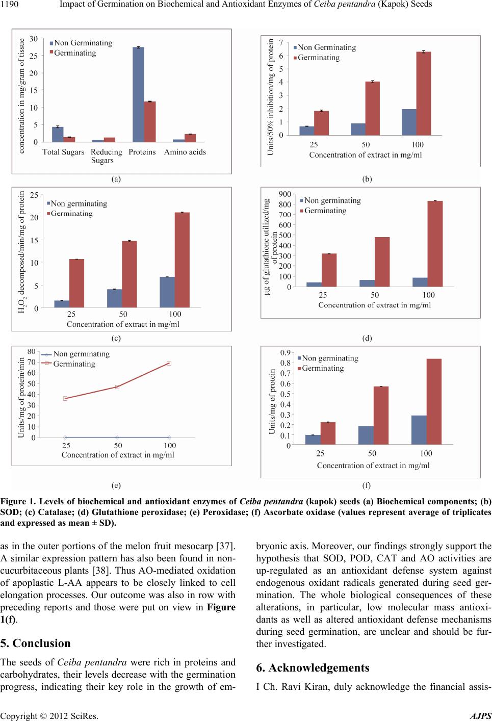 Impact of Germination on Biochemical and Antioxidant Enzymes of Ceiba pentandra (Kapok) Seeds Copyright © 2012 SciRes. AJPS 1190 Figure 1. Levels of biochemical and antioxidant enzymes of Ceiba pentandra (kapok) seeds (a) Biochemical components; (b) SOD; (c) Catalase; (d) Glutathione peroxidase; (e) Peroxidase; (f) Ascorbate oxidase (values represent average of triplicates and expressed as mean ± SD). as in the outer portions of the melon fruit mesocarp [37]. A similar expression pattern has also been found in non- cucurbitaceous plants [38]. Thus AO-mediated oxidation of apoplastic L-AA appears to be closely linked to cell elongation processes. Our outcome was also in row with preceding reports and those were put on view in Figure 1(f). 5. Conclusion The seeds of Ceiba pentandra were rich in proteins and carbohydrates, their levels decrease with the germination progress, indicating their key role in the growth of em- bryonic axis. Moreover, our findings strongly support the hypothesis that SOD, POD, CAT and AO activities are up-regulated as an antioxidant defense system against endogenous oxidant radicals generated during seed ger- mination. The whole biological consequences of these alterations, in particular, low molecular mass antioxi- dants as well as altered antioxidant defense mechanisms during seed germination, are unclear and should be fur- ther investigated. 6. Acknowledgements I Ch. Ravi Kiran, duly acknowledge the financial assis- 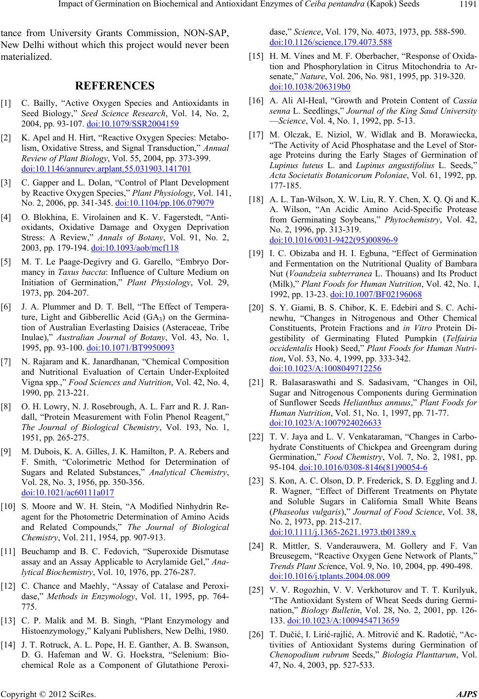 Impact of Germination on Biochemical and Antioxidant Enzymes of Ceiba pentandra (Kapok) Seeds 1191 tance from University Grants Commission, NON-SAP, New Delhi without which this project would never been materialized. REFERENCES [1] C. Bailly, “Active Oxygen Species and Antioxidants in Seed Biology,” Seed Science Research, Vol. 14, No. 2, 2004, pp. 93-107. doi:10.1079/SSR2004159 [2] K. Apel and H. Hirt, “Reactive Oxygen Species: Metabo- lism, Oxidative Stress, and Signal Transduction,” Annual Review of Plant Biology, Vol. 55, 2004, pp. 373-399. doi:10.1146/annurev.arplant.55.031903.141701 [3] C. Gapper and L. Dolan, “Control of Plant Development by Reactive Oxygen Species,” Plant Physiology, Vol. 141, No. 2, 2006, pp. 341-345. doi:10.1104/pp.106.079079 [4] O. Blokhina, E. Virolainen and K. V. Fagerstedt, “Anti- oxidants, Oxidative Damage and Oxygen Deprivation Stress: A Review,” Annals of Botany, Vol. 91, No. 2, 2003, pp. 179-194. doi:10.1093/aob/mcf118 [5] M. T. Le Paage-Degivry and G. Garello, “Embryo Dor- mancy in Taxus baccta: Influence of Culture Medium on Initiation of Germination,” Plant Physiology, Vol. 29, 1973, pp. 204-207. [6] J. A. Plummer and D. T. Bell, “The Effect of Tempera- ture, Light and Gibberellic Acid (GA3) on the Germina- tion of Australian Everlasting Daisics (Asteraceae, Tribe Inulae),” Australian Journal of Botany, Vol. 43, No. 1, 1995, pp. 93-100. doi:10.1071/BT9950093 [7] N. Rajaram and K. Janardhanan, “Chemical Composition and Nutritional Evaluation of Certain Under-Exploited Vigna spp.,” Food Sciences and Nutrition, Vol. 42, No. 4, 1990, pp. 213-221. [8] O. H. Lowry, N. J. Rosebrough, A. L. Farr and R. J. Ran- dall, “Protein Measurement with Folin Phenol Reagent,” The Journal of Biological Chemistry, Vol. 193, No. 1, 1951, pp. 265-275. [9] M. Dubois, K. A. Gilles, J. K. Hamilton, P. A. Rebers and F. Smith, “Colorimetric Method for Determination of Sugars and Related Substances,” Analytical Chemistry, Vol. 28, No. 3, 1956, pp. 350-356. doi:10.1021/ac60111a017 [10] S. Moore and W. H. Stein, “A Modified Ninhydrin Re- agent for the Photometric Determination of Amino Acids and Related Compounds,” The Journal of Biological Chemistry, Vol. 211, 1954, pp. 907-913. [11] Beuchamp and B. C. Fedovich, “Superoxide Dismutase assay and an Assay Applicable to Acrylamide Gel,” Ana- lytical Biochemistry, Vol. 10, 1976, pp. 276-287. [12] C. Chance and Maehly, “Assay of Catalase and Peroxi- dase,” Methods in Enzymology, Vol. 11, 1995, pp. 764- 775. [13] C. P. Malik and M. B. Singh, “Plant Enzymology and Histoenzymology,” Kalyani Publishers, New Delhi, 1980. [14] J. T. Rotruck, A. L. Pope, H. E. Ganther, A. B. Swanson, D. G. Hafeman and W. G. Hoekstra, “Selenium: Bio- chemical Role as a Component of Glutathione Peroxi- dase,” Science, Vol. 179, No. 4073, 1973, pp. 588-590. doi:10.1126/science.179.4073.588 [15] H. M. Vines and M. F. Oberbacher, “Response of Oxida- tion and Phosphorylation in Citrus Mitochondria to Ar- senate,” Nature, Vol. 206, No. 981, 1995, pp. 319-320. doi:10.1038/206319b0 [16] A. Ali Al-Heal, “Growth and Protein Content of Cassia senna L. Seedlings,” Journal of the King Saud University —Science, Vol. 4, No. 1, 1992, pp. 5-13. [17] M. Olczak, E. Niziol, W. Widlak and B. Morawiecka, “The Activity of Acid Phosphatase and the Level of Stor- age Proteins during the Early Stages of Germination of Lupinus luteus L. and Lupinus angustifolius L. Seeds,” Acta Societatis Botanicorum Poloniae, Vol. 61, 1992, pp. 177-185. [18] A. L. Tan-Wilson, X. W. Liu, R. Y. Chen, X. Q. Qi and K. A. Wilson, “An Acidic Amino Acid-Specific Protease from Germinating Soybeans,” Phytochemistry, Vol. 42, No. 2, 1996, pp. 313-319. doi:10.1016/0031-9422(95)00896-9 [19] I. C. Obizaba and H. I. Egbuna, “Effect of Germination and Fermentation on the Nutritional Quality of Bambara Nut (Voandzeia subterranea L. Thouans) and Its Product (Milk),” Plant Foods for Human Nutrition, Vol. 42, No. 1, 1992, pp. 13-23. doi:10.1007/BF02196068 [20] S. Y. Giami, B. S. Chibor, K. E. Edebiri and S. C. Achi- newhu, “Changes in Nitrogenous and Other Chemical Constituents, Protein Fractions and in Vitro Protein Di- gestibility of Germinating Fluted Pumpkin (Telfairia occidentalis Hook) Seed,” Plant Foods for Human Nutri- tion, Vol. 53, No. 4, 1999, pp. 333-342. doi:10.1023/A:1008049712256 [21] R. Balasaraswathi and S. Sadasivam, “Changes in Oil, Sugar and Nitrogenous Components during Germination of Sunflower Seeds Helianthus annuus,” Plant Foods for Human Nutrition, Vol. 51, No. 1, 1997, pp. 71-77. doi:10.1023/A:1007924026633 [22] T. V. Jaya and L. V. Venkataraman, “Changes in Carbo- hydrate Constituents of Chickpea and Greengram during Germination,” Food Chemistry, Vol. 7, No. 2, 1981, pp. 95-104. doi:10.1016/0308-8146(81)90054-6 [23] S. Kon, A. C. Olson, D. P. Frederick, S. D. Eggling and J. R. Wagner, “Effect of Different Treatments on Phytate and Soluble Sugars in California Small White Beans (Phaseolus vulgaris),” Journal of Food Science, Vol. 38, No. 2, 1973, pp. 215-217. doi:10.1111/j.1365-2621.1973.tb01389.x [24] R. Mittler, S. Vanderauwera, M. Gollery and F. Van Breusegem, “Reactive Oxygen Gene Network of Plants,” Trends Plant Science, Vol. 9, No. 10, 2004, pp. 490-498. doi:10.1016/j.tplants.2004.08.009 [25] V. V. Rogozhin, V. V. Verkhoturov and T. T. Kurilyuk, “The Antioxidant System of Wheat Seeds during Germi- nation,” Biology Bulletin, Vol. 28, No. 2, 2001, pp. 126- 133. doi:10.1023/A:1009454713659 [26] T. Dučić, I. Lirić-rajlić, A. Mitrović and K. Radotić, “Ac- tivities of Antioxidant Systems during Germination of Chenopodium rubrum Seeds,” Biologia Planttarum, Vol. 47, No. 4, 2003, pp. 527-533. Copyright © 2012 SciRes. AJPS  Impact of Germination on Biochemical and Antioxidant Enzymes of Ceiba pentandra (Kapok) Seeds Copyright © 2012 SciRes. AJPS 1192 [27] N. G. Lewis and E. Yamamoto, “Lignin: Occurrence, Biosynthesis and Biodegradation,” Annual Review of Plant Physiology and Plant Molecular Biology, Vol. 41, 1990, pp. 455-496. [28] F. Passardi, C. Cosio, C. Penel and C. Dunand, “Peroxi- dases Have More Functions than a Swiss Army Knife,” Plant Cell Reports, Vol. 24, No. 5, 2005, pp. 255-265. doi:10.1007/s00299-005-0972-6 [29] H. Laloue, F. Weber-Lofti, A. Lucau-Danila and P. Guille- mat, “Identification of Ascorbate and Guaiacol Peroxi- dase in Needle Chloroplasts of Spruce Trees,” Plant Physiology and Biochemistry, Vol. 35, No. 5, 1997, pp. 341-346. [30] T. Kawano, “Roles of the Reactive Oxygen Species- Generating Peroxidase Reactions in Plant Defence and Growth Induction,” Plant Cell Reports, Vol. 21, No. 9, 2003, pp. 829-837. [31] O. Omidiji, J. Okpuzor and O. Otubu, “Peroxidase Activ- ity of Germin Ating Sorghum bicolor Grains: Effect of Some Cations,” Journal of the Science of Food and Ag- riculture, Vol. 82, No. 15, 2003, pp. 1881-1885. doi:10.1002/jsfa.1144 [32] Ł. Wojtyla, M. Garnczarska, T. Zalewski, W. Bednarski, L. Ratajczak and S. A. Jurga, “Comparative Study of Water Distribution, Free Radical Production and Activa- tion of Antioxidative Metabolism in Germinating Pea Seeds,” Journal of Plant Physiology, Vol. 163, No. 12, 2006, pp. 1207-1220. doi:10.1016/j.jplph.2006.06.014 [33] F. A. Loewus, “L-Ascorbic Acid: Metabolism, Biosyn- thesis, Function,” In: P. K. Stumpf and E. E. Conn, Eds., The Biochemistry of Plants: A Comprehensive Treatise, Academic Press, New York, 1980, pp. 77-101. [34] O. Arrigoni, “Ascorbate System in Plant Development,” Journal of Bioenergetics and Biomembranes, Vol. 26, No. 4, 1994, pp. 407-419. [35] N. Kato and M. Esaka, “Changes in Ascorbate Oxidase Gene Expression and Ascorbate Levels in Cell Division and Cell Elongation in Tobacco Cells,” Physiologia Plantarum, Vol. 105, No. 2, 1999, pp. 321-329. doi:10.1034/j.1399-3054.1999.105218.x [36] L. S. Lin and J. E. Varner, “Expression of Ascorbic Acid Oxidase in Zucchini Squash (Curcubito pepo L.),” Plant Physiology, Vol. 96, No. 1, 1991, pp. 696-976. doi:10.1104/pp.96.1.159 [37] G. Diallinas, I. Pateraki, M. Sanmartin, A. Scossa, E. Stilanou, N. J. Panopoulos and A. K. Kanellis, “Melon Ascorbate Oxidase: Cloning of a Multigene Family, In- duction during Fruit Development and Repression by Wounding,” Plant Molecular Biology, Vol. 34, No. 5, 1997, pp. 759-770. doi:10.1023/A:1005851527227 [38] N. Kato and M. Esaka, “cDNA Cloning and Gene Ex- pression of Ascorbate Oxidase in Tobacco,” Plant Mo- lecular Biology, Vol. 30, No. 4, 1996, pp. 833-837. doi:10.1007/BF00019015 |