

Paper Menu >>
Journal Menu >>
 Open Journal of Nephrology, 2012, 2, 29-34 http://dx.doi.org/10.4236/ojneph.2012.23005 Published Online September 2012 (http://www.SciRP.org/journal/ojneph) Do We Have a Biocompatible Peritoneal Dialysis Fluid? Shadi Hassan1, Batya Kristal2,3, Khalid Khazim3, Fadi Hassan4, Dunia Hassan5, Kamal Hassan2,3 1Internal Medicine Department, Carmel Medical Center, Haifa, Israel 2Faculty of Medicine in the Galilee, Bar-Ilan University, Safed, Israel 3Nephrology and Hypertension Department, Western Galilee Hospital, Nahariya, Israel 4Sackler Faculty of Medicine, Tel Aviv University, Tel Aviv, Israel 5The Ruth and Bruce Faculty of Medicine, Technion Israel Institute of Technology, Haifa, Israel Email: [email protected], [email protected] Received April 21, 2012; revised June 7, 2012; accepted June 21, 2012 ABSTRACT Objective: Cardiovascular disease remains the leading cause of morbidity and mortality in patients on maintenance dialysis. Diabetes mellitus, dyslipidemia, hypertension, inflammation and hyperhomocyteinemia are major cardiovas- cular risk factors. Aim: To evaluate the effects of Icodextrin and amino acid peritoneal dialysis fluid (AAPDF) on these major cardiovascular risk factors looking for a more biocompatible PDF formula. Methods: 20 adult stable peritoneal dialysis patients were included in the study. 10 patients received 2 L Icodextrin and other 10 patients received 2 L AAPDF in their dialysis prescription for 8 weeks. Results: Icodextrin decreased fasting plasma glucose (p < 0.001), LDL-C (p = 0.03), SBP (p < 0.01), DBP (p < 0.05) and plasma homocysteine (p = 0.002), and increased HDL-C (p = 0.009), CRP (p = 0.035) and fibrinogen (p = 0.009). AAPDF did not affect fasting plasma glucose, LDL-C, HDL-C, CRP and fibrinogen but increased serum albumin (p = 0.03), SBP (p < 0.01), DBP (p < 0.05) and PHcy (p = 0.03). Conclusions: A biocompatible PDF should provide not only adequate dialysis and ultrafiltration but should also improve nutritional and metabolic status, blood pressure control and reduce inflammation and plasma homocyteine. Keywords: Icodextrin; Amino Acids PDF; Homocyteine; Carbohydrates; Lipids; Inflammation 1. Introduction Cardiovascular disease (CVD) remains the leading cause of morbidity and mortality in end-stage renal disease patients on maintenance dialysis [1]. Diabetes mellitus, dyslipidemia, hypertension, inflammation and hyperho- mocyteinemia are major cardiovascular risk factors. Re- cent studies have suggested that novel risk factors, ure- mia or dialysis-related, are of great importance, as they act synergistically with the highly prevalent traditional risk factors for CVD in chronic kidney disease (CKD) patients [2]. Glucose based peritoneal dialysis fluids (PDFs) are usually associated with progressive loss of the osmotic gradient, concomitant reduction in ultrafil- tration (UF) and to the development of hyperglycemia and dyslipidemia. Dyslipidemia increases the risk of CVD and becomes worse in peritoneal dialysis (PD) pa- tients [3]. Up to 80% of peritoneal dialysis patients are hypertensive [4]. Hypertension plays an important role in the development of CVD in this population [4]. Elevated plasma inflammation markers associated with increased risk for CVD [5]. Almost all (97% - 98%) PD and hemo- dialysis patients have hyperhomocyteinemia that known to be associated with an increased risk of cardiovascular, cerebrovascular and venous thromboembolic diseases [6-8]. Icodextrin and Amino acid PDF (AAPDF) consid- ered more biocompatible PDFs. Icodextrin PDF is tar- geted to have more sustained oncotic effect, to reduce plasma glucose and glucose-induced lipid abnormalities, and to avoid the production of glucose degradation products (GDPs) [9,10]. Generally, there is some concern that Icodextrin treatment may induce a subclinical in- flammatory response, both intraperitoneally and sys- temically [10-12]. AAPDF was designed to correct nu- triational status by supplying extra nitrogen through the intraperitoneal route [13]. Although the effects of Ico- dextrin and AAPDF on metabolic status, blood pressure control, inflammation and plasma homocyteine were studied but some issues still controversial. Aim of this study was to evaluate the effects of Icodextrin and AAPDF on these major cardiovascular risk factors. 2. Patients and Methods Randomly, 20 adult stable PD patients on continuous ambulatory peritoneal dialysis (CAPD) or automated Peritoneal Dialysis (APD) for at least 3 months and with Kt/V > 1.8 were included in the study. Demographic and clinical characteristics of the enrolled subjects are listed in Table 1. The study protocol was approved by the local C opyright © 2012 SciRes. OJNeph 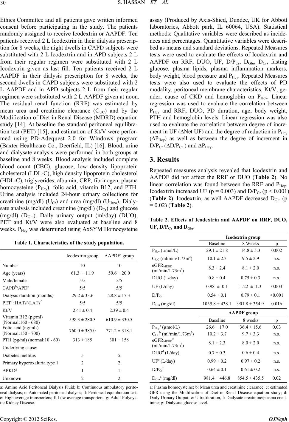 S. HASSAN ET AL. 30 Ethics Committee and all patients gave written informed consent before participating in the study. The patients randomly assigned to receive Icodextrin or AAPDF. Ten patients received 2 L Icodextrin in their dialysis prescrip- tion for 8 weeks, the night dwells in CAPD subjects were substituted with 2 L Icodextrin and in APD subjects 2 L from their regular regimen were substituted with 2 L Icodextrin given as last fill. Ten patients received 2 L AAPDF in their dialysis prescription for 8 weeks, the second dwells in CAPD subjects were substituted with 2 L AAPDF and in APD subjects 2 L from their regular regimen were substituted with 2 L AAPDF given at noon. The residual renal function (RRF) was estimated by mean urea and creatinine clearance (CUC) and by the Modification of Diet in Renal Disease (MDRD) equation study [14]. At baseline the standard peritoneal equilibra- tion test (PET) [15], and estimation of Kt/V were perfor- med using PD-Adequest 2.0 for Windows program (Baxter Healthcare Co., Deerfield, IL) [16]. Blood, urine and dialysate analysis were performed in both groups at baseline and 8 weeks. Blood analysis included complete blood count (CBC), glucose, low density lipoprotein cholesterol (LDL-C), high density lipoprotein cholesterol (HDL-C), triglycerides, albumin, CRP, fibrinogen, plasma homocysteine (PHcy), folic acid, vitamin B12, and PTH. Urine analysis included 24-hour urinary collections for creatinine (mg/dl) (UCr) and urea (mg/dl) (UUrea). Dialy- sate analysis included creatinine (mg/dl) (DCr) and glucose (mg/dl) (DGlu). Daily urinary output (ml/day) (DUO), PET and Kt/V were also evaluated at baseline and 8 weeks. PHcy was determined using AxSYM Homocysteine Table 1. Characteristics of the study population. Icodextrin group AAPDFa group Number 10 10 Age (years) 61.3 ± 11.9 59.6 ± 20.0 Male/female 5/5 5/5 CAPDb/APDc 5/5 5/5 Dialysis duration (months) 29.2 ± 33.6 28.8 ± 17.3 PETd: HATse/LATsf 5/5 5/5 Kt/V 2.41 ± 0.4 2.39 ± 0.4 Vitamin B12 (pg/ml) (Normal:160 - 680) 598.3 ± 280.3 610.9 ± 330.5 Folic acid (ng/mL) (Normal:150 - 700) 760.0 ± 385.0 771.2 ± 318.1 PTH (pg/ml) (normal:10 - 60) 313 ± 185 301 ± 158 Underlying cause: Diabetes mellitus 5 5 Primary hyperoxaluria type 1 2 2 APKDg 1 1 Unknown 2 2 a: Amino Acid Peritoneal Dialysis Fluid; b: Continuous ambulatory perito- neal dialysis; c: Automated peritoneal dialysis; d: Peritoneal equilibration test; e: High average transporters; f: Low average transporters; g: Adult Polycys- tic Kidney Disease. assay (Produced by Axis-Shied, Dundee, UK for Abbott laboratories, Abbott park, IL 60064, USA). Statistical methods: Qualitative variables were described as incide- nces and percentages. Quantitative variables were descri- bed as means and standard deviations. Repeated Measures tests were used to evaluate the effects of Icodextrin and AAPDF on RRF, DUO, UF, D/PCr, DGlu, DCr, fasting glucose, plasma lipids, plasma inflammation markers, body weight, blood pressure and PHcy. Repeated Measures tests were also used to evaluate the effects of PD modality, peritoneal membrane characteristics, Kt/V, ge- nder, cause of CKD and hemoglobin on PHcy. Linear regression was used to evaluate the correlation between PHcy and RRF, DUO, PD duration, age, body weight, PTH and hemoglobin levels. Linear regression was also used to evaluate the correlation between degree of incre- ment in UF (∆Net UF) and the degree of reduction in PHcy (∆PHcy) as well as between the degree of increment in D/PCr (∆D/PCr ) and ∆PHcy. 3. Results Repeated measures analysis revealed that Icodextrin and AAPDF did not affect the RRF or DUO (Table 2). No linear correlation was found between the RRF and PHcy. Icodextrin increased UF (p = 0.003) and D/PCr (p < 0.001) (Table 2). Icodextrin, as well AAPDF decreased DGlu (p = 0.02) (Table 2). Table 2. Effects of Icodextrin and AAPDF on RRF, DUO, UF, D/PCr and DGlu. Icodextrin group Baseline 8 Weeks p PHcy (µmol/L) 29.1 ± 21.8 14.8 ± 5.3 0.002 CUC (ml/min/1.73m2) 10.1 ± 2.3 9.5 ± 2.9 n.s. eGFRMDRD (ml/min/1.73m2) 8.3 ± 2.4 8.1 ± 2.0 n.s. DUO (L/day) 0.8 ± 0.4 0.75 ± 0.3 n.s. UF (L/day) 0.98 ± 0.1 1.22 ± 1.3 0.003 D/PCr 0.54 ± 0.1 0.79 ± 0.1 <0.001 DGlu (mg/dl) 1035.8 ± 438.1 901.8 ± 354.90.016 AAPDF group Baseline 8 weeks p PHcya (µmol/L) 26.6 ± 17.0 36.4 ± 15.6 0.03 CUCb (ml/min/1.73m2) 10.2 ± 3.7 9.7 ± 3.3 n.s. eGFRMDRDc (ml/min/1.73m2) 8.1 ± 2.3 8.0 ± 2.0 n.s. DUOd (L/day) 0.7 ± 0.3 0.6 ± 0.4 n.s. UFe (L/day) 0.99 ± 0.2 0.97 ± 0.2 n.s. D/PCrf 0.64 ± 0.1 0.61 ± 0.2 n.s. DGlug (mg/dl) 981.4 ± 446.8 854.5 ± 435.50.02 a: Plasma homocysteine; b: Mean urea and creatinine clearance; c: estimated GFR using the Modification of Diet in Renal Disease equation study; d: Daily Urinary Output; e: Ultrafiltration, f: Dialysate creatinine/plasma creat- inine; g: Dialysate glucose level. Copyright © 2012 SciRes. OJNeph 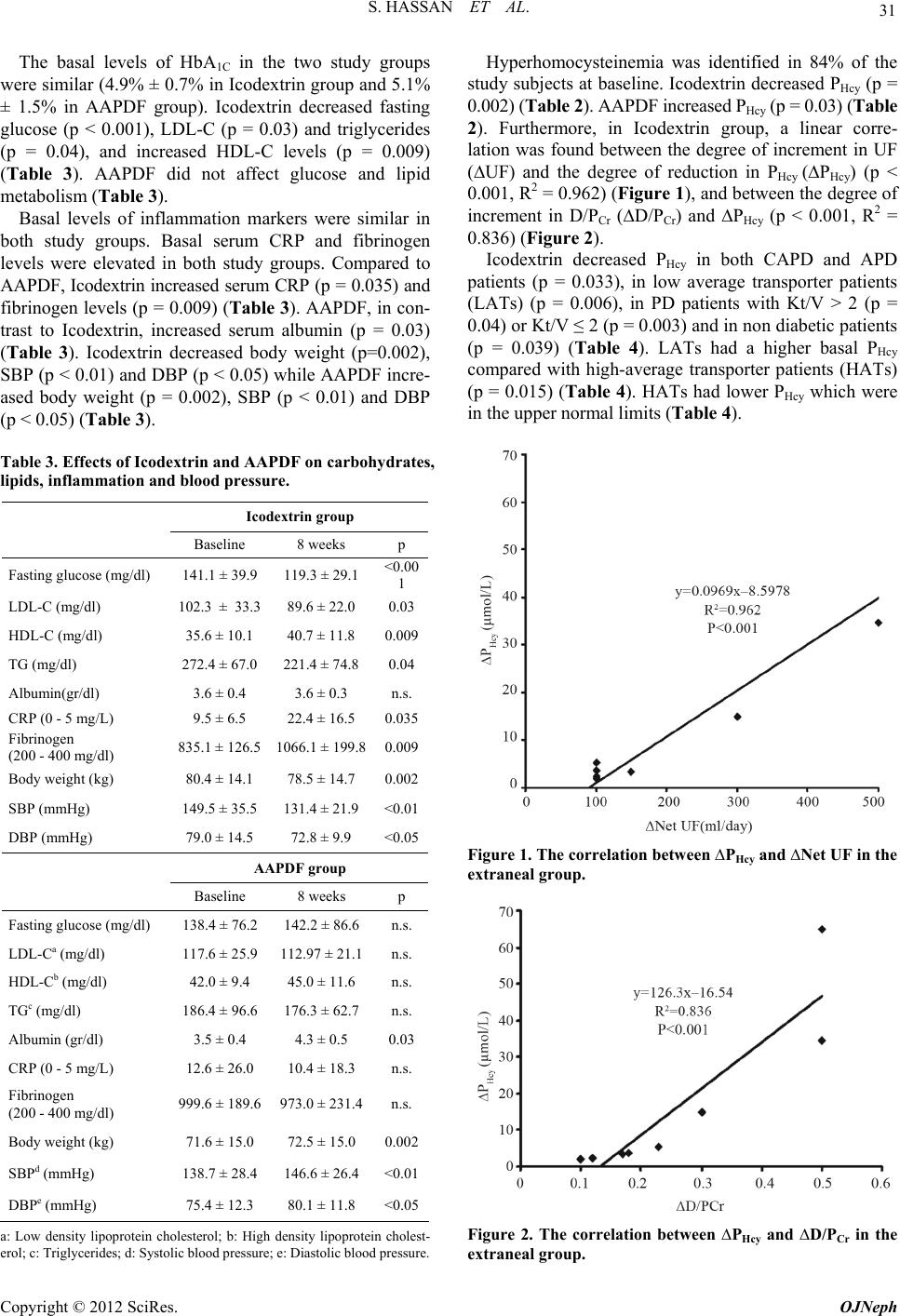 S. HASSAN ET AL. 31 The basal levels of HbA1C in the two study groups were similar (4.9% ± 0.7% in Icodextrin group and 5.1% ± 1.5% in AAPDF group). Icodextrin decreased fasting glucose (p < 0.001), LDL-C (p = 0.03) and triglycerides (p = 0.04), and increased HDL-C levels (p = 0.009) (Table 3). AAPDF did not affect glucose and lipid metabolism (Table 3). Basal levels of inflammation markers were similar in both study groups. Basal serum CRP and fibrinogen levels were elevated in both study groups. Compared to AAPDF, Icodextrin increased serum CRP (p = 0.035) and fibrinogen levels (p = 0.009) (Table 3). AAPDF, in con- trast to Icodextrin, increased serum albumin (p = 0.03) (Table 3). Icodextrin decreased body weight (p=0.002), SBP (p < 0.01) and DBP (p < 0.05) while AAPDF incre- ased body weight (p = 0.002), SBP (p < 0.01) and DBP (p < 0.05) (Table 3). Table 3. Effects of Icodextrin and AAPDF on carbohydrates, lipids, inflammation and blood pressure. Icodextrin group Baseline 8 weeks p Fasting glucose (mg/dl) 141.1 ± 39.9 119.3 ± 29.1 <0.00 1 LDL-C (mg/dl) 102.3 ± 33.3 89.6 ± 22.0 0.03 HDL-C (mg/dl) 35.6 ± 10.1 40.7 ± 11.8 0.009 TG (mg/dl) 272.4 ± 67.0 221.4 ± 74.8 0.04 Albumin(gr/dl) 3.6 ± 0.4 3.6 ± 0.3 n.s. CRP (0 - 5 mg/L) 9.5 ± 6.5 22.4 ± 16.5 0.035 Fibrinogen (200 - 400 mg/dl) 835.1 ± 126.5 1066.1 ± 199.80.009 Body weight (kg) 80.4 ± 14.1 78.5 ± 14.7 0.002 SBP (mmHg) 149.5 ± 35.5 131.4 ± 21.9 <0.01 DBP (mmHg) 79.0 ± 14.5 72.8 ± 9.9 <0.05 AAPDF group Baseline 8 weeks p Fasting glucose (mg/dl) 138.4 ± 76.2 142.2 ± 86.6 n.s. LDL-Ca (mg/dl) 117.6 ± 25.9 112.97 ± 21.1n.s. HDL-Cb (mg/dl) 42.0 ± 9.4 45.0 ± 11.6 n.s. TGc (mg/dl) 186.4 ± 96.6 176.3 ± 62.7 n.s. Albumin (gr/dl) 3.5 ± 0.4 4.3 ± 0.5 0.03 CRP (0 - 5 mg/L) 12.6 ± 26.0 10.4 ± 18.3 n.s. Fibrinogen (200 - 400 mg/dl) 999.6 ± 189.6 973.0 ± 231.4n.s. Body weight (kg) 71.6 ± 15.0 72.5 ± 15.0 0.002 SBPd (mmHg) 138.7 ± 28.4 146.6 ± 26.4 <0.01 DBPe (mmHg) 75.4 ± 12.3 80.1 ± 11.8 <0.05 a: Low density lipoprotein cholesterol; b: High density lipoprotein cholest- erol; c: Triglycerides; d: Systolic blood pressure; e: Diastolic blood pressure. Hyperhomocysteinemia was identified in 84% of the study subjects at baseline. Icodextrin decreased PHcy (p = 0.002) (Table 2). AAPDF increased PHcy (p = 0.03) (Table 2). Furthermore, in Icodextrin group, a linear corre- lation was found between the degree of increment in UF (∆UF) and the degree of reduction in PHcy (∆PHcy) (p < 0.001, R2 = 0.962) (Figure 1), and between the degree of increment in D/PCr (∆D/PCr) and ∆PHcy (p < 0.001, R2 = 0.836) (Figure 2). Icodextrin decreased PHcy in both CAPD and APD patients (p = 0.033), in low average transporter patients (LATs) (p = 0.006), in PD patients with Kt/V > 2 (p = 0.04) or Kt/V ≤ 2 (p = 0.003) and in non diabetic patients (p = 0.039) (Table 4). LATs had a higher basal PHcy compared with high-average transporter patients (HATs) (p = 0.015) (Table 4). HATs had lower PHcy which were in the upper normal limits (Table 4). Figure 1. The correlation between ∆PHcy and ∆Net UF in the extraneal group. Figure 2. The correlation between ∆PHcy and ∆D/PCr in the extraneal group. Copyright © 2012 SciRes. OJNeph 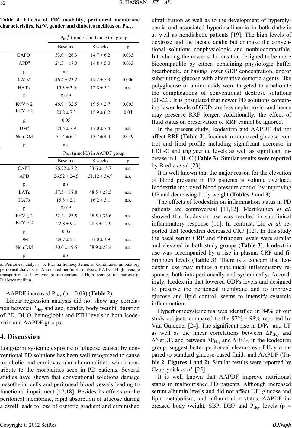 S. HASSAN ET AL. 32 Table 4. Effects of PDa modality, peritoneal membrane characteristics, Kt/V, gender and diabetes mellitus on PHcy. PHcyb (µmol/L) in Icodextrin group Baseline 8 weeks p CAPDc 33.0 ± 26.3 14.7 ± 6.2 0.033 APDd 24.3 ± 17.0 14.8 ± 5.0 0.033 p n.s. LATse 46.4 ± 23.2 17.2 ± 5.3 0.006 HATsf 15.3 ± 3.0 12.8 ± 5.1 n.s. P 0.015 Kt/V ≤ 2 46.9 ± 32.5 19.5 ± 2.7 0.003 Kt/V > 2 20.2 ± 7.3 15.9 ± 6.2 0.04 p 0.05 DMg 24.5 ± 7.9 17.0 ± 7.4 n.s. Non DM 31.4 ± 6.7 13.7 ± 4.4 0.039 p n.s. PHcy (µmol/L) in AAPDF group Baseline 8 weeks p CAPD 26.72 ± 7.2 33.6 ± 15.7 n.s. APD 26.52 ± 24.5 31.12 ± 34.9 n.s. p n.s. LATs 37.5 ± 18.8 48.5 ± 28.5 n.s. HATs 15.8 ± 2.1 16.2 ± 3.1 n.s. p 0.015 Kt/V ≤ 2 32.3 ± 25.5 38.5 ± 36.6 n.s. Kt/V > 2 22.8 ± 9.4 28.3 ± 17.9 n.s. p 0.05 DM 28.7 ± 5.1 37.0 ± 3.9 n.s. Non DM 30.0 ± 19.5 38.9 ± 28.4 n.s. p n.s. a: Peritoneal dialysis; b: Plasma homocysteine; c: Continuous ambulatory peritoneal dialysis; d: Automated peritoneal dialysis, HATs = High average transporters; e: Low average transporters; f: High average transporters; g: Diabetes mellitus. AAPDF increased PHcy (p = 0.03) (Table 2). Linear regression analysis did not show any correla- tion between PHcy and age, gender, body weight, duration of PD, DUO, hemoglobin and PTH levels in both Icode- xtrin and AAPDF groups. 4. Discussion Long-term systemic exposure of glucose caused by con- ventional PD solutions has been well recognized to cause metabolic and cardiovascular abnormalities, which con- tribute to the morbidities seen in PD patients. Several studies have shown that conventional solutions damage mesothelial cells and peritoneal blood vessels leading to functional impairment [17,18]. Besides its effects on the peritoneal membrane, rapid absorption of glucose during a dwell leads to loss of osmotic gradient and diminished ultrafiltration as well as to the development of hypergly- cemia and associated hyperinsulinemia in both diabetic as well as nondiabetic patients [19]. The high levels of dextrose and the lactate acidic buffer make the conven- tional solutions nonphysiologic and nonbiocompatible. Introducing the newer solutions that designed to be more biocompatible by either, containing physiologic buffer bicarbonate, or having lower GDP concentration, and/or substituting glucose with alternative osmotic agents, like polyglucose or amino acids were targeted to ameliorate the complications of conventional dextrose solutions [20-22]. It is postulated that newer PD solutions contain- ing lower levels of GDPs are less nephrotoxic, and hence may preserve RRF longer. Additionally, the effect of fluid status on preservation of RRF cannot be ignored. In the present study, Icodextrin and AAPDF did not affect RRF (Table 2). Icodextrin improved glucose con- trol and lipid profile including significant decrease in LDL-C and triglyceride levels as well as significant in- crease in HDL-C (Table 3). Similar results were reported by Bredie et al. [23]. It is well known that the major reason for the elevation of blood pressure in PD patients is volume overload. Icodextrin improved blood pressure control by improving UF and decreasing body weight (Tables 2 and 3). The effects of Icodextrin on inflammation status in PD patients are controversial [11,12]. Martikainen et al. showed that Icodextrin use was resulted in subclinical inflammatory response [11]. In contrast, Lin et al. re- ported that Icodextrin decreased CRP [12]. In this study the basal serum CRP and fibrinogen levels were similar and elevated in both study groups (Table 3). Icodextrin use was accompanied by a rise in plasma CRP and fi- brinogen levels (Table 3). There is a concern that Ico- dextrin use may induce a subclinical inflammatory re- sponse, both intraperitoneally and systemically. Accord- ingly, Icodextrin that lowered GDPs levels and designed to preserve the peritoneal membrane and to improve glucose and lipid control, seems to intensify systemic inflammation. Hyperhomocysteinemia was identified in 84% of our study subjects compared to the 97% - 98% reported by Van Guldener [24]. The significant rise in D/PCr and UF as well as the linear correlations between ∆PHcy and ∆NetUF, and between ∆PHcy and ∆D/PCr in the Icodextrin group, suggest better peritoneal clearances of Hcy com- pared to standard glucose-based fluids and AAPDF (Ta- ble 2, Figures 1 and 2). Similar results were reported by Czupryniak et al. [25]. It is well known that AAPDF improve nutritional status in malnourished PD patients. Although increased serum albumin levels and did not affect UF, glucose and lipid metabolism, and inflammation status, AAPDF in- creased body weight, SBP, DBP and PHcy levels (p = Copyright © 2012 SciRes. OJNeph  S. HASSAN ET AL. 33 0.002, p < 0.01, p < 0.05, p = 0.03—respectively) (Ta- bles 2 and 3). The increase in PHcy in the AAPDF group is, most likely, related to methionine, a precursor of Hcy, absorption as well as to the decrease in D/PCr and UF in the AAPDF group (Table 2). Then, AAPDF that intro- duced to improve nutritional status and survival may contribute to the unfavorable rise of PHcy. The rise in PHcy in PD patients treated with AAPDF described also in previous studies [26,27]. End stage renal disease patients are unable to excrete the daily acid load. The standard PDFs, Icodextrin and AAPDF use lactate as a buffer that, by its acidic pH, causes harmful effects on the peritoneal cells [17]. In contrast, bicarbonate-based PDFs include a physiologic buffer with neutral pH, cause less peritoneal damage [28]. The relationships between the PD modality, the peri- toneal membrane characteristics and Kt/V, and PHcy re- main a controversial issue [29,30]. Basal PHcy was elevated and not different in the two study groups and all sub- groups apart from HATs (Table 4). In the Icodextrin group PHcy decreased in both PD modalities (p = 0.033) (Table 3). On the other hand, AAPDF increase PHcy (p = 0.033) (Table 2). This may be due to its low osmotic drive and as a result of the absorption of methionine through the peritoneal membrane. These results suggest that the peritoneal membrane characteristics and the composition of the PDF have an important role in the peritoneal elimination of Hcy. Basal PHcy in HATs were near normal in both study groups (Table 4). It may be related to the higher solute clearances across the peritoneal membrane in HATs. Basal PHcy was marginally lower in subjects with Kt/V > 2 compared to those with Kt/V ≤ 2 (p = 0.05) (Table 4). Icodextrin, but not AAPDF, significantly decreased PHcy in PD patients with Kt/V > 2, in those with Kt/V ≤ 2 and in non diabetic patients (Table 4). Therefore, Icodextrin may influence the atherosclerotic outcomes through Hcy-lowering effects, as it was stated previously by Do- cloux et al [29]. No associations between age, gender, body weight, du- ration of PD, hemoglobin, PTH, RRF and DUO and PHcy were found. In summary: Higher UF, higher D/PCr, HATs, Kt/V > 2, non diabetic patients and Icodextrin use were associ- ated with decline in PHcy. Conversely, lower UF, lower D/PCr, LATs, Kt/V ≤ 2, diabetic patients and AAPDF use were associated with rise in PHcy. Although it exhibited favorable effects on metabolic status, blood pressure control and PHcy levels, Icodextrin seems to intensify systemic inflammation. On the other hand, although it did not seem to affect adversely the metabolic status and systemic inflammation, AAPDF increased PHcy and blood pressure. The results of the present study suggested that the use of Icodextrin and AAPDF was associated with beneficial effects as well as with considerable harmful consequences that may adversely affect the prognosis and survival of PD patients. 5. Conclusion A new PDF containing a mixture of a biocompatible sub- stance, amino acids without methionine, lower glucose and GDPs content, and neutral pH will provide sustained oncotic effect, adequate dialysis and ultrafiltration as well as will improve nutritional status and metabolic profile, reduce inflammation, plasma homocyteine and blood pressure. A PDF with those properties will preserve the peritoneal membrane and improve the prognosis and survival of peritoneal dialysis patients. 6. Acknowledgements The authors would like to thank Prof. Ben Ami Sela, Clinical Biochemistry Laboratory-Sheba Medical Center, Tel Hashomer, Israel for his assistance in determination of PHcy. We also would like to thank Tobie Kuritsky for her assistance in editing our manuscript. REFERENCES [1] P. S. Parfrey and J. D. Harnett, “Long Term Cardiac Morbidity and Mortality during Dialysis Therapy,” Ad- vanced Nephrology, Vol. 23, 1994, pp. 311-330. [2] S. H. Park, P. Stenvinkel and B. Lindholm, “Cardiovas- cular Biomarkers in Chronic Kidney Disease,” Journal of Renal Nutrition, Vol. 22, No. 1, 2012, pp. 120-127. doi:10.1053/j.jrn.2011.10.021 [3] A. H. Mitwalli, A. A. Alam, J. S. Al Wakeel and A. C. Isnani, “Dyslipidemia in Dialysis Patients,” Saudi Jour- nal of Kidney Disease and Transplantation, Vol. 22, No. 4, 2011, pp. 689-694. [4] L. M. Ortega and B. J. Materson, “Hypertension in Peri- toneal Dialysis Patients: Epidemiology, Pathogenesis, and Treatment,” Journal of the American Society of Hyper- tension, Vol. 5, No. 3, 2011, pp. 128-136. doi:10.1016/j.jash.2011.02.004 [5] M. Moriishi and H. Kawanishi, “Icodextrin and Intrap- eritoneal Inflammation,” Peritoneal Dialysis Interna- tional, Vol. 28, Suppl. 3, 2008, pp. S96-S100. [6] T. Huang, G. Yuan, Z. Zhang, Z. Zou and D. Li, “Car- diovascular Pathogenesis in Hyperhomocysteinemia,” Asia Pacific Journal of Clinical Nutrition, Vol. 17, No. 1, 2008, pp. 8-16. [7] C. van Guldener and F. Stam, “Stehouwer CDA. Homo- cysteine Metabolism in Renal Failure,” Kidney Interna- tional, Vol. 59, Suppl. 78, 2001, pp. S234-S237. doi:10.1046/j.1523-1755.2001.07859.x [8] M. M. Sagheb, M. A. Ostovan, Z. Sohrabi, E. Atabati, G. A. Raisjalai and J. Roozbeh, “Hyperhomocysteinemia and Cardiovascular Risks in Hemodialysis Patients,” Saudi Copyright © 2012 SciRes. OJNeph  S. HASSAN ET AL. Copyright © 2012 SciRes. OJNeph 34 Journal of Kidney Diseases and Transplantation, Vol. 21, No. 5, 2010, pp. 863-866. [9] L. A. Cooker, C. J. Holmes and C. M. Hoff, “Biocom- patibility of Icodextrin,” Kidney International, Vol. 62, Suppl. 81, 2002, pp. S34-S45. doi:10.1046/j.1523-1755.62.s81.6.x [10] M. Canbakan and G. M. Sahin, “Icodextrine and Insulin Resistance in Continuous Ambulatory Peritoneal Dialysis Patients,” Renal Failure, Vol. 29, No. 3, 2007, pp. 289- 293. doi:10.1080/08860220601166271 [11] T. A. Martikainen, A. M. Teppo, C. Grönhagen-Riska and A. V. Ekstrand, “Glucose-Free Dialysis Solutions: In- ductors of Inflammation or Preservers of Peritoneal Membrane?” Peritoneal Dialysis International, Vol. 25, No. 5, 2005, pp. 453-460. [12] W. Lin, Y. C. Chen, M. S. Wu, H. J. Hsu, C. Y. Sun, Y. K.Lin and I. W. Wu, “Icodextrin Dialysate Improves Nu- tritional and Inflammatory Profiles in Peritoneal Dialysis Patients,” Renal Failure, Vol. 31, No. 2, 2009, pp. 98-105. doi:10.1080/08860220802595500 [13] F. K. Li, L. Y.Chan , J. C. Woo, et al., “A 3-Year, Pro- spective, Randomized, Controlled, Study on Amino Acid Dialysate in Patients on CAPD,” American Journal of Kidney Diseases, Vol. 42, No. 1, 2003, pp. 173-183. doi:10.1016/S0272-6386(03)00421-9 [14] A. S. Levey, J. P. Bosch, J. B. Lewis, T. Greene, N. Rogers and D. Roth, “A More Accurate Method to Esti- mate Glomerular Filtration Rate from Serum Creatinine: A New Prediction Equation, Modification of Diet in Re- nal Disease Study Group,” Annals of Internal Medicine, Vol. 130, No. 6, 1999, pp. 461-470. [15] Z. J. Twardowski, “Clinical Value of Standardized Equilibration Tests in CAPD Patients,” Blood Purifica- tion, Vol. 7, No. 2-3, 1989, pp. 95-108. doi:10.1159/000169582 [16] G. Amici, S. Mastrosimone, G. Da Rin, C. Bocci and A. Bonadonna, “Clinical Validation of PD Adequest Soft- ware: Modeling Error Assessment,” Peritoneal Dialysis International, Vol. 18, No. 3, 1998, pp. 317-321. [17] H. Ha, M. R. Yu, H. N. Choi, M. K. Cha, H. S. Kang, M. H. Kim and H. B. Lee, “Effects of Conventional and New Peritoneal Dialysis Solutions on Human Peritoneal Meso- thelial Cell Viability and Proliferation,” Peritoneal Di- alysis International, Vol. 20, Suppl. 5, 2000, pp. S10- S18. [18] R. T. Krediet, M. M. Zweers, A. C. vander Wal and D. J. Struijk, “Neoangiogenesis in the Peritoneal Membrane,” Peritoneal Dialysis International, Vol. 20, Suppl. 2, 2000, pp. S19-S25. [19] J. Delarue and C. Maingourd, “Acute Metabolic Effects of Dialysis Fluids during CAPD,” American Journal of Kidney Diseases, Vol. 37, Suppl. 2, 2001, pp. S103-S107. doi:10.1053/ajkd.2001.20762 [20] N. Posthuma, P. M. ter Wee, A. J. Donker, P. L. Oe, E. M. Peers and H. A. Verbrugh, “Assesement of the Effective- ness, Safety, and Biocompatibility of Icodextrin in Auto- mated Peritoneal Dialysis. The Dextrin in APD in Am- sterdam (DIANA) Group,” Peritoneal Dialysis Interna- tional, Vol. 20, Suppl. 2, 2000, pp. S106-S113. [21] T. W. Kao, H. F. Chuang, K. Y. Hung, K. D. Wu and T. J. Tsai, “Rate of Decline of Residual Renal Function is As- sociated with All-Cause Mortality and Technique Failure in Patients on Long-Term Peritoneal Dialysis,” Nephrol- ogy Dialysis Transplantation, Vol. 24, No. 9, 2009, pp. 2909-2914. doi:10.1093/ndt/gfp056 [22] S. Kim, J. Oh, S. Kim, W. Chung, C. Ahn, S. G. Kim and K. H. Oh, “Benefits of Biocompatible PD Fluid for Pres- ervation of Residual Renal Function in Incident CAPD Patients: A 1-Year Study,” Nephrology Dialysis Trans- plantation, Vol. 24, No. 9, 2009, pp. 2899-2908. doi:10.1093/ndt/gfp054 [23] S. J. Bredie, F. H. Bosch, P. N. Demacker, A. F. Stalen- hoef and R. Van Leusen, “Effects of Peritoneal Dialysis with an Overnight Icodextrin Dwell on Parameters of Glucose and Lipid Metabolism,” Peritoneal Dialysis In- ternational, Vol. 21, No. 3, 2001, pp. 275-281. [24] C. Van Guldener, “Why is Homocysteine Elevated in Renal Failure and What Can Be Expected from Homo- cysteine-Lowering?” Peritoneal Dialysis International, Vol. 21, No. 5, 2006, pp. 1161-1166. doi:10.1093/ndt/gfl044 [25] A. Czupryniak, M. Nowicki, G. Chwatko, A. Jander and E. Bald, “Peritoneal Clearance of Homocysteine with Ico- dextrin or Standard Glucose Solution Exchange,” Ne- phrology (Carlton), Vol. 10, No. 6, 2005, pp. 571-575. [26] H. F. Brulez, C. van Guldener, A. J. Donker and P. M. Ter Wee, “The Impact of an Amino Acid-Based Perito- neal Dialysis Fluid on Plasma Total Homocysteine Levels, Lipid Profile and Body Fat Mass,” Nephrology Dialysis Transplantation, Vol. 14, No. 1, 1999, pp. 154-159. doi:10.1093/ndt/14.1.154 [27] S. Y. Yang, J. W. Huang, K. Y. Shih, et al., “Factors Associated with Increased Plasma Homocysteine in Pa- tients Using an Amino Acid Peritoneal Dialysis Fluid,” Nephrology Dialysis Transplantation, Vol. 20, No. 1, 2005, pp. 161-166. doi:10.1093/ndt/gfh554 [28] S. Haas, C. P. Schmitt, K. Arbeiter, K. E. Bonzel, M. Fischbach, U. John, A. K. Pieper, T. P. Schaub, J. Passlick-Deetjen and O. Mehls, “Schaefer FImproved Acidosis Correction and Recovery of Mesothelial Cell Mass with Neutral-pH Bicarbonate Dialysis Solution among Children Undergoing Automated Peritoneal Di- alysis,” Journal of the American Society of Hypertension, Vol. 14, No. 10, 2003, pp. 2632-2638. doi:10.1097/01.ASN.0000086475.83211.DF [29] D. Ducloux, L. Heuze-Lecornu, R. Gibey, C. Bresson- Vautrin, P. Vautrin and J. M. Chalopin, “Dialysis Ade- quacy and Homocyst(e)ine Concentrations in Peritoneal Dialysis Patients,” Nephrology Dialysis Transplantation, Vol. 14, No. 3, 1999, pp. 728-731. doi:10.1093/ndt/14.3.728 [30] A. Vychytil, M. Fodinger, M. Papagiannopoulos, et al., “Peritoneal Elimination of Homocysteine Moieties in Continuous Ambulatory Peritoneal Dialysis Patients,” Kidney International, Vol. 55, No. 5, 1999, pp. 2054- 2061. doi:10.1046/j.1523-1755.1999.00437.x |