

Paper Menu >>
Journal Menu >>
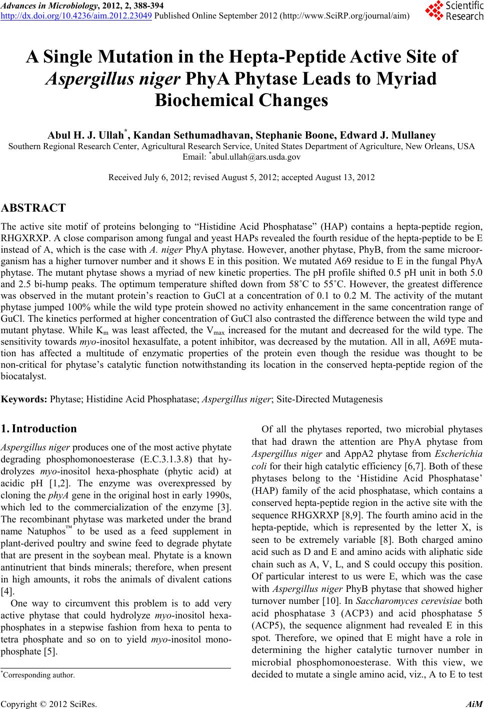 Advances in Microbiology, 2012, 2, 388-394 http://dx.doi.org/10.4236/aim.2012.23049 Published Online September 2012 (http://www.SciRP.org/journal/aim) A Single Mutation in the Hepta-Peptide Active Site of Aspergillus niger PhyA Phytase Leads to Myriad Biochemical Changes Abul H. J. Ullah*, Kandan Sethumadhavan, Stephanie Boone, Edward J. Mullaney Southern Regional Research Center, Agricultural Research Service, United States Department of Agriculture, New Orleans, USA Email: *[email protected] Received July 6, 2012; revised August 5, 2012; accepted August 13, 2012 ABSTRACT The active site motif of proteins belonging to “Histidine Acid Phosphatase” (HAP) contains a hepta-peptide region, RHGXRXP. A close comparison among fungal and yeast HAPs revealed the fourth residue of the hepta-peptide to be E instead of A, which is the case with A. niger PhyA phytase. However, another phytase, PhyB, from the same microor- ganism has a higher turnover number and it shows E in this position. We mutated A69 residue to E in the fungal PhyA phytase. The mutant phytase shows a myriad of new kinetic properties. The pH profile shifted 0.5 pH unit in both 5.0 and 2.5 bi-hump peaks. The optimum temperature shifted down from 58˚C to 55˚C. However, the greatest difference was observed in the mutant protein’s reaction to GuCl at a concentration of 0.1 to 0.2 M. The activity of the mutant phytase jumped 100% while the wild type protein showed no activity enhancement in the same concentration range of GuCl. The kinetics performed at higher concentration of GuCl also contrasted the difference between the wild type and mutant phytase. While Km was least affected, the Vmax increased for the mutant and decreased for the wild type. The sensitivity towards myo-inositol hexasulfate, a potent inhibitor, was decreased by the mutation. All in all, A69E muta- tion has affected a multitude of enzymatic properties of the protein even though the residue was thought to be non-critical for phytase’s catalytic function notwithstanding its location in the conserved hepta-peptide region of the biocatalyst. Keywords: Phytase; Histidine Acid Phosphatase; Aspergillus niger; Site-Directed Mutagenesis 1. Introduction Aspergillus niger produces one of the most active phytate degrading phosphomonoesterase (E.C.3.1.3.8) that hy- drolyzes myo-inositol hexa-phosphate (phytic acid) at acidic pH [1,2]. The enzyme was overexpressed by cloning the phyA gene in the original host in early 1990s, which led to the commercialization of the enzyme [3]. The recombinant phytase was marketed under the brand name Natuphos™ to be used as a feed supplement in plant-derived poultry and swine feed to degrade phytate that are present in the soybean meal. Phytate is a known antinutrient that binds minerals; therefore, when present in high amounts, it robs the animals of divalent cations [4]. One way to circumvent this problem is to add very active phytase that could hydrolyze myo-inositol hexa- phosphates in a stepwise fashion from hexa to penta to tetra phosphate and so on to yield myo-inositol mono- phosphate [5]. Of all the phytases reported, two microbial phytases that had drawn the attention are PhyA phytase from Aspergillus niger and AppA2 phytase from Escherichia coli for their high catalytic efficiency [6,7]. Both of these phytases belong to the ‘Histidine Acid Phosphatase’ (HAP) family of the acid phosphatase, which contains a conserved hepta-peptide region in the active site with the sequence RHGXRXP [8,9]. The fourth amino acid in the hepta-peptide, which is represented by the letter X, is seen to be extremely variable [8]. Both charged amino acid such as D and E and amino acids with aliphatic side chain such as A, V, L, and S could occupy this position. Of particular interest to us were E, which was the case with Aspergillus niger PhyB phytase that showed higher turnover number [10]. In Saccharomyces cerevisiae both acid phosphatase 3 (ACP3) and acid phosphatase 5 (ACP5), the sequence alignment had revealed E in this spot. Therefore, we opined that E might have a role in determining the higher catalytic turnover number in microbial phosphomonoesterase. With this view, we decided to mutate a single amino acid, viz., A to E to test *Corresponding author. C opyright © 2012 SciRes. AiM 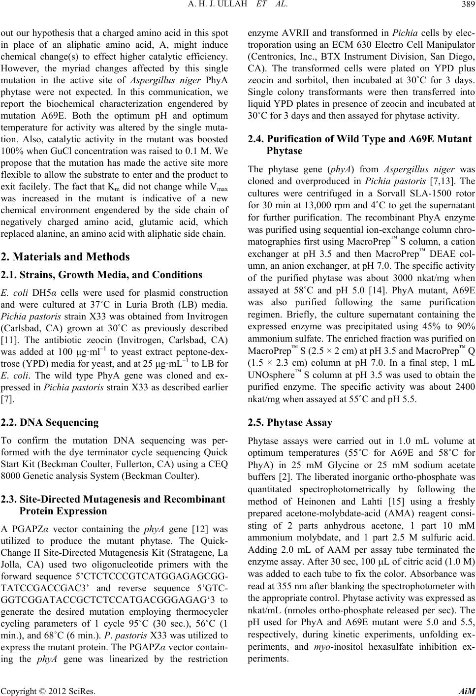 A. H. J. ULLAH ET AL. 389 out our hypothesis that a charged amino acid in this spot in place of an aliphatic amino acid, A, might induce chemical change(s) to effect higher catalytic efficiency. However, the myriad changes affected by this single mutation in the active site of Aspergillus niger PhyA phytase were not expected. In this communication, we report the biochemical characterization engendered by mutation A69E. Both the optimum pH and optimum temperature for activity was altered by the single muta- tion. Also, catalytic activity in the mutant was boosted 100% when GuCl concentration was raised to 0.1 M. We propose that the mutation has made the active site more flexible to allow the substrate to enter and the product to exit facilely. The fact that Km did not change while Vmax was increased in the mutant is indicative of a new chemical environment engendered by the side chain of negatively charged amino acid, glutamic acid, which replaced alanine, an amino acid with aliphatic side chain. 2. Materials and Methods 2.1. Strains, Growth Media, and Conditions E. coli DH5α cells were used for plasmid construction and were cultured at 37˚C in Luria Broth (LB) media. Pichia pastoris strain X33 was obtained from Invitrogen (Carlsbad, CA) grown at 30˚C as previously described [11]. The antibiotic zeocin (Invitrogen, Carlsbad, CA) was added at 100 μg·ml–1 to yeast extract peptone-dex- trose (YPD) media for yeast, and at 25 μg·mL–1 to LB for E. coli. The wild type PhyA gene was cloned and ex- pressed in Pichia pastoris strain X33 as described earlier [7]. 2.2. DNA Sequencing To confirm the mutation DNA sequencing was per- formed with the dye terminator cycle sequencing Quick Start Kit (Beckman Coulter, Fullerton, CA) using a CEQ 8000 Genetic analysis System (Beckman Coulter). 2.3. Site-Directed Mutagenesis and Recombinant Protein Expression A PGAPZα vector containing the phyA gene [12] was utilized to produce the mutant phytase. The Quick- Change II Site-Directed Mutagenesis Kit (Stratagene, La Jolla, CA) used two oligonucleotide primers with the forward sequence 5’CTCTCCCGTCATGGAGAGCGG- TATCCGACCGAC3’ and reverse sequence 5’GTC- GGTCGGATACCGCTCTCCATGACGGGAGAG‘3 to generate the desired mutation employing thermocycler cycling parameters of 1 cycle 95˚C (30 sec.), 56˚C (1 min.), and 68˚C (6 min.). P. pastoris X33 was utilized to express the mutant protein. The PGAPZα vector contain- ing the phyA gene was linearized by the restriction enzyme AVRII and transformed in Pichia cells by elec- troporation using an ECM 630 Electro Cell Manipulator (Centronics, Inc., BTX Instrument Division, San Diego, CA). The transformed cells were plated on YPD plus zeocin and sorbitol, then incubated at 30˚C for 3 days. Single colony transformants were then transferred into liquid YPD plates in presence of zeocin and incubated at 30˚C for 3 days and then assayed for phytase activity. 2.4. Purification of Wild Type and A69E Mutant Phytase The phytase gene (phyA) fro m Aspergillus niger was cloned and overproduced in Pichia pastoris [7,13]. The cultures were centrifuged in a Sorvall SLA-1500 rotor for 30 min at 13,000 rpm and 4˚C to get the supernatant for further purification. The recombinant PhyA enzyme was purified using sequential ion-exchange column chro- matographies first using MacroPrep™ S column, a cation exchanger at pH 3.5 and then MacroPrep™ DEAE col- umn, an anion exchanger, at pH 7.0. The specific activity of the purified phytase was about 3000 nkat/mg when assayed at 58˚C and pH 5.0 [14]. PhyA mutant, A69E was also purified following the same purification regimen. Briefly, the culture supernatant containing the expressed enzyme was precipitated using 45% to 90% ammonium sulfate. The enriched fraction was purified on MacroPrep™ S (2.5 × 2 cm) at pH 3.5 and MacroPrep™ Q (1.5 × 2.3 cm) column at pH 7.0. In a final step, 1 mL UNOsphere™ S column at pH 3.5 was used to obtain the purified enzyme. The specific activity was about 2400 nkat/mg when assayed at 55˚C and pH 5.5. 2.5. Phytase Assay Phytase assays were carried out in 1.0 mL volume at optimum temperatures (55˚C for A69E and 58˚C for PhyA) in 25 mM Glycine or 25 mM sodium acetate buffers [2]. The liberated inorganic ortho-phosphate was quantitated spectrophotometrically by following the method of Heinonen and Lahti [15] using a freshly prepared acetone-molybdate-acid (AMA) reagent consi- sting of 2 parts anhydrous acetone, 1 part 10 mM ammonium molybdate, and 1 part 2.5 M sulfuric acid. Adding 2.0 mL of AAM per assay tube terminated the enzyme assay. After 30 sec, 100 L of citric acid (1.0 M) was added to each tube to fix the color. Absorbance was read at 355 nm after blanking the spectrophotometer with the appropriate control. Phytase activity was expressed as nkat/mL (nmoles ortho-phosphate released per sec). The pH used for PhyA and A69E mutant were 5.0 and 5.5, respectively, during kinetic experiments, unfolding ex- periments, and myo-inositol hexasulfate inhibition ex- periments. Copyright © 2012 SciRes. AiM 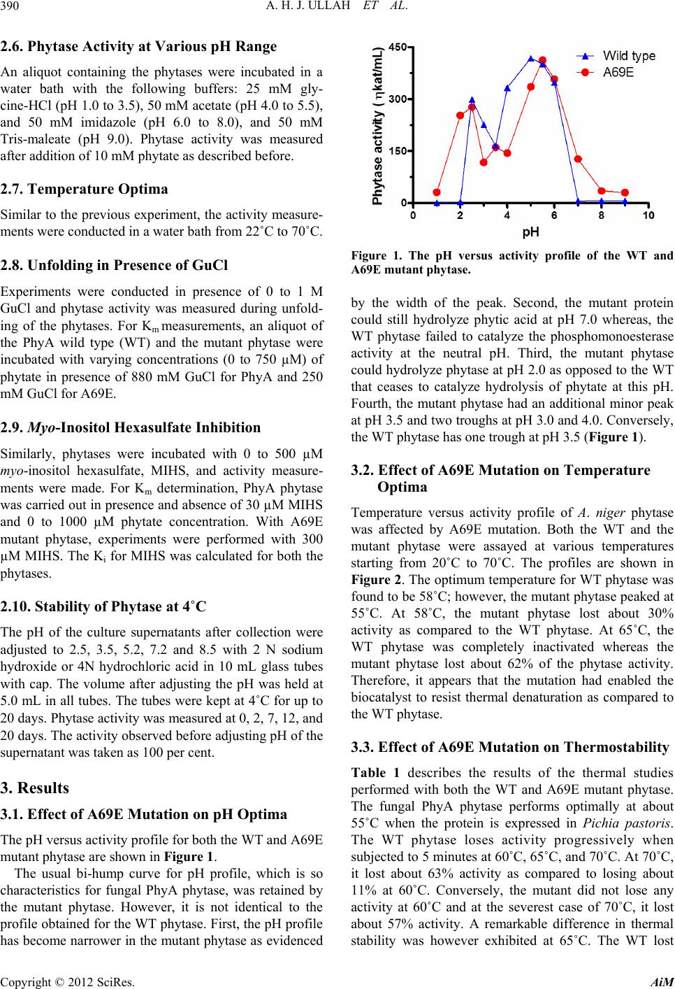 A. H. J. ULLAH ET AL. 390 2.6. Phytase Activity at Various pH Range An aliquot containing the phytases were incubated in a water bath with the following buffers: 25 mM gly- cine-HCl (pH 1.0 to 3.5), 50 mM acetate (pH 4.0 to 5.5), and 50 mM imidazole (pH 6.0 to 8.0), and 50 mM Tris-maleate (pH 9.0). Phytase activity was measured after addition of 10 mM phytate as described before. 2.7. Temperature Optima Similar to the previous experiment, the activity measure- ments were conducted in a water bath from 22˚C to 70˚C. 2.8. Unfolding in Presence of GuCl Experiments were conducted in presence of 0 to 1 M GuCl and phytase activity was measured during unfold- ing of the phytases. For Km measurements, an aliquot of the PhyA wild type (WT) and the mutant phytase were incubated with varying concentrations (0 to 750 µM) of phytate in presence of 880 mM GuCl for PhyA and 250 mM GuCl for A69E. 2.9. Myo-Inositol Hexasulfate Inhibition Similarly, phytases were incubated with 0 to 500 µM myo-inositol hexasulfate, MIHS, and activity measure- ments were made. For Km determination, PhyA phytase was carried out in presence and absence of 30 µM MIHS and 0 to 1000 µM phytate concentration. With A69E mutant phytase, experiments were performed with 300 µM MIHS. The Ki for MIHS was calculated for both the phytases. 2.10. Stability of Phytase at 4˚C The pH of the culture supernatants after collection were adjusted to 2.5, 3.5, 5.2, 7.2 and 8.5 with 2 N sodium hydroxide or 4N hydrochloric acid in 10 mL glass tubes with cap. The volume after adjusting the pH was held at 5.0 mL in all tubes. The tubes were kept at 4˚C for up to 20 days. Phytase activity was measured at 0, 2, 7, 12, and 20 days. The activity observed before adjusting pH of the supernatant was taken as 100 per cent. 3. Results 3.1. Effect of A69E Mutation on pH Optima The pH versus activity profile for both the WT and A69E mutant phytase are shown in Figure 1. The usual bi-hump curve for pH profile, which is so characteristics for fungal PhyA phytase, was retained by the mutant phytase. However, it is not identical to the profile obtained for the WT phytase. First, the pH profile has become narrower in the mutant phytase as evidenced Figure 1. The pH versus activity profile of the WT and A69E mutant phytase. by the width of the peak. Second, the mutant protein could still hydrolyze phytic acid at pH 7.0 whereas, the WT phytase failed to catalyze the phosphomonoesterase activity at the neutral pH. Third, the mutant phytase could hydrolyze phytase at pH 2.0 as opposed to the WT that ceases to catalyze hydrolysis of phytate at this pH. Fourth, the mutant phytase had an additional minor peak at pH 3.5 and two troughs at pH 3.0 and 4.0. Conversely, the WT phytase has one trough at pH 3.5 (Figure 1). 3.2. Effect of A69E Mutation on Temperature Optima Temperature versus activity profile of A. niger phytase was affected by A69E mutation. Both the WT and the mutant phytase were assayed at various temperatures starting from 20˚C to 70˚C. The profiles are shown in Figure 2. The optimum temperature for WT phytase was found to be 58˚C; however, the mutant phytase peaked at 55˚C. At 58˚C, the mutant phytase lost about 30% activity as compared to the WT phytase. At 65˚C, the WT phytase was completely inactivated whereas the mutant phytase lost about 62% of the phytase activity. Therefore, it appears that the mutation had enabled the biocatalyst to resist thermal denaturation as compared to the WT phytase. 3.3. Effect of A69E Mutation on Thermostability Table 1 describes the results of the thermal studies performed with both the WT and A69E mutant phytase. The fungal PhyA phytase performs optimally at about 55˚C when the protein is expressed in Pichia pastoris. The WT phytase loses activity progressively when subjected to 5 minutes at 60˚C, 65˚C, and 70˚C. At 70˚C, it lost about 63% activity as compared to losing about 11% at 60˚C. Conversely, the mutant did not lose any activity at 60˚C and at the severest case of 70˚C, it lost about 57% activity. A remarkable difference in thermal stability was however exhibited at 65˚C. The WT lost Copyright © 2012 SciRes. AiM 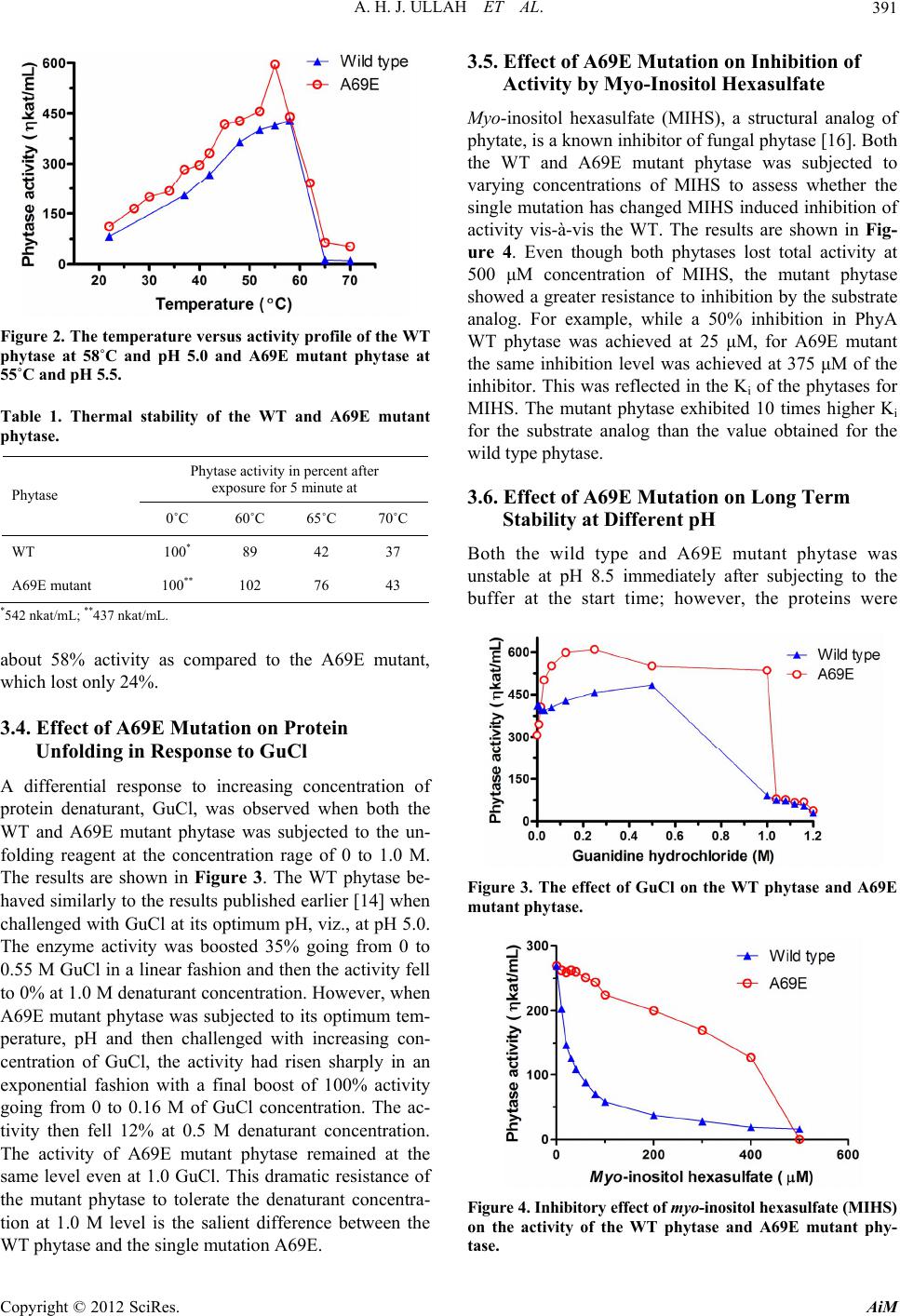 A. H. J. ULLAH ET AL. 391 Figure 2. The temperature versus activity profile of the WT phytase at 58˚C and pH 5.0 and A69E mutant phytase at 55˚C and pH 5.5. Table 1. Thermal stability of the WT and A69E mutant phytase. Phytase activity in percent after exposure for 5 minute at Phytase 0˚C 60˚C 65˚C 70˚C WT 100* 89 42 37 A69E mutant 100** 102 76 43 *542 nkat/mL; **437 nkat/mL. about 58% activity as compared to the A69E mutant, which lost only 24%. 3.4. Effect of A69E Mutation on Protein Unfolding in Response to GuCl A differential response to increasing concentration of protein denaturant, GuCl, was observed when both the WT and A69E mutant phytase was subjected to the un- folding reagent at the concentration rage of 0 to 1.0 M. The results are shown in Figure 3. The WT phytase be- haved similarly to the results published earlier [14] when challenged with GuCl at its optimum pH, viz., at pH 5.0. The enzyme activity was boosted 35% going from 0 to 0.55 M GuCl in a linear fashion and then the activity fell to 0% at 1.0 M denaturant concentration. However, when A69E mutant phytase was subjected to its optimum tem- perature, pH and then challenged with increasing con- centration of GuCl, the activity had risen sharply in an exponential fashion with a final boost of 100% activity going from 0 to 0.16 M of GuCl concentration. The ac- tivity then fell 12% at 0.5 M denaturant concentration. The activity of A69E mutant phytase remained at the same level even at 1.0 GuCl. This dramatic resistance of the mutant phytase to tolerate the denaturant concentra- tion at 1.0 M level is the salient difference between the WT phytase and the single mutation A69E. 3.5. Effect of A69E Mutation on Inhibition of Activity by Myo-Inositol Hexasulfate Myo-inositol hexasulfate (MIHS), a structural analog of phytate, is a known inhibitor of fungal phytase [16]. Both the WT and A69E mutant phytase was subjected to varying concentrations of MIHS to assess whether the single mutation has changed MIHS induced inhibition of activity vis-à-vis the WT. The results are shown in Fig- ure 4. Even though both phytases lost total activity at 500 μM concentration of MIHS, the mutant phytase showed a greater resistance to inhibition by the substrate analog. For example, while a 50% inhibition in PhyA WT phytase was achieved at 25 μM, for A69E mutant the same inhibition level was achieved at 375 μM of the inhibitor. This was reflected in the Ki of the phytases for MIHS. The mutant phytase exhibited 10 times higher Ki for the substrate analog than the value obtained for the wild type phytase. 3.6. Effect of A69E Mutation on Long Term Stability at Different pH Both the wild type and A69E mutant phytase was unstable at pH 8.5 immediately after subjecting to the buffer at the start time; however, the proteins were Figure 3. The effect of GuCl on the WT phytase and A69E mutant phytase. Figure 4. Inhibitory effect of myo-inositol hexasulfate (MIH S) on the activity of the WT phytase and A69E mutant phy- tase. Copyright © 2012 SciRes. AiM  A. H. J. ULLAH ET AL. Copyright © 2012 SciRes. AiM 392 remarkably stable at pH 7.2 and below down to pH 2.5 (Figure 5 panel A and panel B). For WT phytase the activity stayed the same up to 7 days and then lost 60% activity on day 12. The activity declined further until day 20 when the experiment was terminated. The mutant phytase retained activity slightly better going from day 12 to day 20. In summary, both WT and A69E mutant phytase showed loss of activity in the pH range 2.5 to 7.2. 4. Discussion From structural standpoint phytases belonging to HAP class of acid phosphomonoesterase have been studied exhaustively. The first reporting of any three-dimen- sional structure in phytase took place over a decade ago when PhyA phytase’s structure was elucidated [17]. This was followed by several reporting of X-ray deduced tertiary structure elucidation in other HAP phytases [18- 20]. These revelations of structures set the stage for knowledge-based protein engineering via point mutations in the active center of phytases [21-23]. In this communication, we presented evidences in support of the conclusion that a single mutation in the active site of PhyA phytase had affected a host of bio- chemical properties such as pH optima, temperature optima, kinetic parameters, reaction to GuCl and MIHS (Table 2). This is very similar to the concept of “pleio- tropic effect” in single gene mutation where the mutation of a gene leads to a host of phenotypic changes. In the case of A69E mutation in the fungal PhyA phytase, a single substitution of amino acid the substitution has caused a myriad of changes in both physicochemical and catalytic properties in A. niger phytase. The original amino acid in the hepta-peptide amino acid region, where the active site resides, an A (alanine) residue is located (10). We mutated A to E (glutamic acid) to incorporate an extra negative charge in the active site of the protein which is based on the observation that PhyB phytase, which has higher turnover number than the phyA phytase, had E residue in the identical position [10]. The newly incorporated negative charge in the active site of phytase will have an unintended conse- quence vis-à-vis the accommodation of the substrate, phytate, which has six negatively charged phosphate group. The newly acquired negative charge in the active site should repel phytate as compared to the WT phytase; therefore, the Km is expected to rise in the mutant phytase. That is what is being observed in Table 1. By the same token, the Vmax should improve somewhat because the negative charge brought on by E into the active site should facilitate the release of negatively charged phos- phate group. The turnover number increased 6.6% in the mutant phytase over the wild type phytase (Table 1). The substrate analog, MIHS, has six negatively charged sulfate groups, which should not bind as efficiently in the mutant phytases’ active site as compared to the wild Figure 5. The long-term stability of the WT phytase (panel A) and A69E mutant phytase (panel B). Table 2. Kinetic properties of A. niger WT and A69E phytase. Properties WT phytase A69E phytase Km (μM) 50 170 Specific activity at Vma x (ηkat/mg) 1950 2116 Turnover number (sec–1) 120 128 Kinetic efficiency (mol–1·sec–1) 2.3 × 10⁶ 0.74 × 10⁶ Ki for MIHS (μM) 8 83  A. H. J. ULLAH ET AL. 393 type phytase’s active site. This has been confirmed by the Ki values for MIHS in the mutant phytase. It took over 10 times concentration of MIHS in the mutant phytase to effect the same inhibition done by the wild type phytase. Therefore, incorporation of a negatively charged amino acid into the hepta-peptide active site region of the phytase has decreased the affinity for not only the substrate but also the inhibitor. In X-ray crystallographic studies with HAP type phytases re- searches have shown that sulphate groups of MIHS bind to the substrate-binding domain that is in the active center of protein [20,24]. Both the pH optima and temperature versus activity profile had changed in the mutant phytase. The me- chanism for such changes is difficult to explain due to the lack of X-ray crystallographic data; nonetheless, the charged amino acid, E, replacing the aliphatic amino acid A may allow new hydrogen bonding sites in the active site area. Also, the domain flexibility in the active center of the protein possibly brought on by the charged amino acid may explain these far-reaching effects on pH optima and temperature optima profiles. The native phytases belonging to HAP family of pho- sphatases have already been unfolded using GuCl and then refolded by the removal of denaturant by dialysis [14,25]. However, a differential response has been ob- served when the WT and A69E mutant phytases were treated with varying concentrations of GuCl. Initially, the activity was boosted 35% when GuCl concentration was raised to 0.55 M. We explained this phenomenon of rise in the Vmax in the light of domain flexibility brought on by GuCl. However, in case of the mutant phytase, the activity had risen in exponential manner in response to GuCl at the concentration of 0.16 M. This is in sharp contrast to the WT phytase’s reaction to increasing concentration of GuCl. At higher concentration of the denaturant, when WT phytase had lost over 75% of the activity, the mutant phytase was holding onto the 100% activity gain it achieved earlier due to addition of small amounts of GuCl. One possible reason for such discre- pancy on the part of the mutant phytase could be that the altered structure containing the flexible domain was resistant to chemical denaturation. The long-term stability of both the WT and A69E phytases in various buffers were observed; there were no discernible difference between the two phytases under the conditions. The fungal phytase was very susceptible to catalytic demise when subjected to pH 8.5 in contrast to acidic pH where the proteins remain stable for days. The biochemical basis of the vulnerability of fungal phytase in alkaline pH is not understood at this time. In summary, a single mutation in the hepta-peptide region that constitutes the active site resulted a myriad of both chemical and catalytic changes, which could not have been predicted. The results presented in these stud- ies have indicated that in this class of proteins a know- ledge-based approach to structural change may offer a better choice than random mutagenesis through error prone PCR technique [26,27]. 5. Conclusion The results of the present communication had clearly demonstrated the power of chemical probing of the ac- tive site by site-directed mutagenesis. By mutating the fourth residue A to E in the hepta-peptide active site of A. niger PhyA the mutant protein exhibited a myriad of new physico-chemical properties. Not only the pH optimum shifted 0.5 pH unit, the temperature optimum also shifted from 58˚C to 55˚C. Furthermore, the catalytic activity of the mutant phytase doubled as compared to the wild type phytase when subjected to 0.1 to 0.2 M GuCl. A single mutation in the active site affecting so many parameters of a biocatalyst gives insight into the complexity of pro- tein’s three-dimensional structure. REFERENCES [1] T. R. Shieh and J. H. Ware, “Survey of Microorganisms for the Production of Extracellular Phytase,” Applied Mi- crobiology, Vol. 16, No. 9, 1968, pp. 1348-1351. http://aem.asm.org/content/16/9/1348.full.pdf [2] A. H. J. Ullah and D. M. Gibson, “Extracellular Phytase (E.C. 3.1.3.8) from Aspergillus ficuum NRRL 3135: Puri- fication and Characterization,” Preparative Biochemistry, Vol. 17, No. 1, 1987, pp. 63-91. doi:10.1080/00327488708062477 [3] S. Haefner, A. Knietsch, E. Scholten, J. Braun, M. Lohs- cheidt and O. Zelder, “Biotechnological Production and Applications of Phytases,” Applied Microbiology and Bio- technology, Vol. 68, No. 5, 2005, pp. 588-997. doi:10.1007/s00253-005-0005-y [4] R. J. Wodzinski and A. H. J. Ullah, “Phytase,” Advances in Applied Microbiology, Vol. 42, 1996, pp. 263-302. doi:10.1016/S0065-2164(08)70375-7 [5] A. H. J. Ullah and B. Q. Phillippy, “Immobilization of Aspergillus ficuum Phytase: Product Characterization of the Bioreactor,” Preparative Biochemistry, Vol. 18, No. 4, 1988, pp. 483-489. doi:10.1080/00327488808062546 [6] A. H. J. Ullah, K. Sethumadhavan and E. J. Mullaney, “Salt Effect on the pH Profile and Kinetic Parameters of Microbial Phytases,” Journal of Agricultural and Food Chemistry, Vol. 56, No. 9, 2008, pp. 3398-3402. doi:10.1021/jf073137i [7] J. D. Weaver, A. H. J. Ullah, K. Sethumadhavan, E. J. Mullaney and X. G. Lei, “Impact of Assay Conditions on Activity Estimate and Kinetics Comparison of Aspergil- lus niger PhyA and Escherichia coli AppA2 Phytases,” Journal of Agricultural and Food Chemistry, Vol. 57, No. 12, 2009, pp. 5315-5320. doi:10.1021/jf900261n [8] A. H. J. Ullah, B. J. Cummins and H. C. Dischinger, Jr., Copyright © 2012 SciRes. AiM  A. H. J. ULLAH ET AL. 394 “Cyclohexanedione Modification of Arginine at the Active Site of Aspergillus ficuum Phytase,” Biochemical and Bio- physical Research Communications, Vol. 178, No. 1, 1991, pp. 45-53. doi:10.1016/0006-291X(91)91777-A [9] R. L. Van Etten, R. Davidson, P. E. Stevis, H. MacArthur and D. L. Moore, “Covalent Structure, Disulfide Bonding, and Identification of Reactive Surface and Active Site Residues of Human Prostatic Acid Phosphatase,” Journal of Biological Chemistry, Vol. 266, No. 4, 1991, pp. 2313- 2319. http://www.jbc.org/content/266/4/2313.full.pdf+html [10] A. H. J. Ullah and H. C. Dischinger, Jr., “Identification of Active Site Residues in Aspergillus ficuum Extracellular pH 2.5 Optimum Acid Phosphatase,” Biochemical and Bio- physical Research Communications, Vol. 192, No. 2, 1993, pp. 754-759. doi:10.1006/bbrc.1993.1478 [11] W. Zhang, E. J. Mullaney and X. G. Lei, “Adopting Se- lected Hydrogen Bonding and Ionic Interacttions from Aspergillum fumigatus Phytase Structure Improves the Thermostability of Aspergillus niger PhyA Phytase,” Ap- plied and Environmental Microbiology, Vol. 73, No. 9, 2007, pp. 3069-3076. doi:10.1128/AEM.02970-06 [12] T. Kim, E. J. Mullaney, J. M. Porres, R. Roneker, S. Crowe, S. Rice, T. Ko, A. H. J. Ullah, C. B. Daly, R. Welch and X. G. Lei, “Shifting the pH Profile of Asper- gillus niger PhyA Phytase to Match the Stomach pH En- hances Its Effectiveness as an Animal Feed Additive,” Applied and Environmental Microbiology, Vol. 72, No. 6, 2006, pp. 4397-4403. doi:10.1128/AEM.02612-05 [13] E. J. Mullaney, H. Locovare, K. Sethumadhavan, S. Boone, X. G. Lei and A. H. J. Ullah, “Site-Directed Mutagenesis of Disulfide Bridges in Aspergillus niger NRRL 3135 Phytase (PhyA), Their Expression in Pichia pastoris and Catalytic Characterization,” Applied Micro- biology and Biotechnology, Vol. 87, No. 4, 2010, pp. 1367-1372. doi:10.1007/s00253-010-2542-2 [14] A. H. J. Ullah, K. Sethumadhavan and E. J. Mullaney, “Monitoring of Unfolding and Refolding in Fungal Phy- tase (PhyA) by Dynamic Light Scattering,” Biochemical and Biophysical Research Communications, Vol. 327, No. 4, 2005, pp. 993-998. doi:10.1016/j.bbrc.2004.12.111 [15] J. K. Heinonen and R. J. Lahti, “A New and Convenient Calorimetric Determination of Inorganic Orthophosphate and Its Application to the Assay of Inorganic Pyrophos- phatase,” Analytical Biochemistry, Vol. 113, No. 2, 1981, pp. 313-317. doi:10.1016/0003-2697(81)90082-8 [16] A. H. J. Ullah and K. Sethumadhavan, “Myo-Inositol Hex- asulfate Is a Potent Inhibitor of Aspergillus ficuum Phy- tase,” Biochemical and Biophysical Research Communi- cations, Vol. 251, No. 1, 1998, pp. 260-263. doi:10.1006/bbrc.1998.9456 [17] D. Kostrewa, F. Grueninger-Leitch, A. D’Arcy, C. Broger, D. Mitchell and A. P. G. M. Van Loon, “Crystal Structure of Phytase from Aspergillus ficuum at 2.5 Å Resolution,” Nature Structural Biology, Vol. 4, No. 3, 1997, pp. 185- 190. doi:10.1038/nsb0397-185 [18] D. Kostrewa, M. Wyss, A. D’Arcy and A. P. G. M. Van Loon, “Crystal Structure of Aspergillus niger pH 2.5 Acid Phosphatase at 2.4 Å Resolution,” Journal of Molecular Biology, Vol. 288, No. 5, 1999, pp. 965-974. doi:10.1006/jmbi.1999.2736 [19] D. Lim, S. Golova, C. W. Forsberg and Z. Jia,” Crystal Structures of Escherichia coli Phytase and Its Complex with Phytate,” Nature Structural Biology, Vol. 7, No. 2, 2000, pp. 108-113. doi:10.1038/72371 [20] K. Böhm, T. Herter, J. Müller, R. Borriss and U. Heine- mann, “Crystal Structure of Klebsiella sp. ASR1 Phytase Suggests Substrate Binding to a Preformed Active Site that Meets the Requirements of a Plant Rhizosphere En- zyme,” Federation of European Biochemical Societies Jour- nal, Vol. 277, No. 5, 2000, pp. 1284-1296. doi:10.1111/j.1742-4658.2010.07559.x [21] E. J. Mullaney, C. B. Daly, T. Kim, J. M. Porres, X. G. Lei, K. Sethumadhavan and A. H. J. Ullah, “Site-Directed Mutagenesis of Aspergillus niger NRRL 3135 Phytase at Residue 300 to Enhance Catalysis at pH 4.0,” Biochemi- cal Biophysical Research Communications, Vol. 297, No. 4, 2002, pp. 1016-1020. doi:10.1016/S0006-291X(02)02325-2 [22] Y. S. Tian, R. H. Peng, J. Xu, W. Zhao, F. Gao, X. Fu, A. S. Xiong and Q. H. Yao, “Semi-Rational Site-Directed Mutagenesis of phyi1s from Aspergillus niger 113 at Two Residue to Improve Its Phytase Activity,” Molecular Bi- ology Reports, Vol. 38, No. 2, 2011, pp. 977-982. doi:10.1007/s11033-010-0192-1 [23] A. J. Oakley, “The Structure of Aspergillus niger Phytase PhyA in Complex with a Phytate Mimetic,” Biochemical and Biophysical Research Communications, Vol. 397, No. 4, 2010, pp. 745-749. doi:10.1016/j.bbrc.2010.06.024 [24] D. C.-C. Lim, “Bound for Catalysis. Crystal Structures of Escherichia coli Phytase and Its Complex with Phytic Acid,” M.S. Thesis, Queen’s University, Kingston, 1999. [25] A. H. J. Ullah, K. Sethumadhavan and E. J. Mullaney, “Unfolding and Refolding of Aspergillus niger PhyB Phytase: Role of Disulfide Bridges,” Journal of Agricul- tural and Food Chemistry, Vol. 56, No. 17, 2008, pp. 8179-8183. doi:10.1021/jf8013712 [26] M. S. Kim and X. G. Lei, “Enhancing Thermostability of Escherichia coli Phytase AppA2 by Error-Prone PCR,” Applied Microbiology and Biotechnology, Vol. 79, No. 1, 2008, pp. 69-75. doi:10.1007/s00253-008-1412-7 [27] W. Zhu, D. Qiao, M. Huang, G. Yang, H. Xu and Y. Cao, “Modifying Thermostability of AppA from Escherichia coli,” Current Microbiology, Vol. 61, No. 4, 2010, pp. 267- 273. doi:10.1007/s00284-010-9606-5 Copyright © 2012 SciRes. AiM |