

Paper Menu >>
Journal Menu >>
 Advances in Microbiology, 2012, 2, 345-353 http://dx.doi.org/10.4236/aim.2012.23042 Published Online September 2012 (http://www.SciRP.org/journal/aim) Antibacterial Activity of Extracts from Some Bryophytes Vizma Nikolajeva1*, Ligita Liepina1, Zaiga Petrina1, Guntra Krumina1, Mara Grube2, Indrikis Muiznieks1 1Faculty of Biology, University of Latvia, Riga, Latvia 2Institute of Microbiology and Biotechnology, University of Latvia, Riga, Latvia Email: *[email protected] Received June 25, 2012; revised July 23, 2012; accepted August 4, 2012 ABSTRACT The antimicrobial activity of aqueous and ethanolic extracts of 11 Bryophyta species and 9 Marchantiophyta species collected in Latvia was tested against Staphylococcus aureus, Escherichia coli and Bacillus cereus. The extract of Lo- phocolea heterophylla inhibited the growth of B. cereus, but none of the tested extracts inhibited the growth of E. coli. 70% of bryophyte species demonstrated certain activity in relation to S. aureus. In general, 73% of ethanolic extracts and 39% of aqueous extracts exhibited antibacterial activity against S. aureus. The highest degree of antibacterial ac- tivity against S. aureus was shown by the ethanolic extract of Dicranum scoparium and aqueous extracts of Atrichum undulatum and Rhytidiadelphus squarrosus. The bactericidal action was not ascertained. For the first time antimicrobial activity has been proved for three moss species—Eurhynchium angustirete, Rhytidiadelphus squarrosus and Rhodo- bryum roseum, and for two liverwort species Frullania dilatata and Lophocolea heterophylla. Qualitative and quantita- tive differences of plant extracts were evaluated by FT-IR spectra. Keywords: Bryophyta; Marchantiophyta; Antimicrobial Activity 1. Introduction Bryophytes, including liverworts (Marchantiophyta), ho- rnworts (Anthocerotophyta), and mosses (Bryophyta), are a diverse group of land plants that usually colonize habi- tats with moist or extremely variable conditions. Tradi- tionally, because of their antimicrobial activity, mosses were used as a natural medicine in the Indian culture [1] and as natural diapers [2]. Today, mosses and liverworts are interesting for biotechnological use in medicine, ag- riculture, and pharmacology [1,3,4]. Liverworts have been proposed as ideal models for genetic studies and biotechnological applications [3]. The search for plants with antimicrobial activity has grown in importance in recent years, due to a growing concern about increase in the rate of infection caused by antibiotic-resistant microorganisms. Asakawa [5,6] has analyzed approximately 1000 bryophyte species from the world total of 27,000. However, few studies have been carried out about the antimicrobial properties of Euro- pean bryophytes. In literature, reports have been found about antibacterial activity of 23 bryophyte species [7-15] that are common in Latvia [16] and other European countries [17-20]. In presented paper, the antimicrobial activity of 20 bryophyte species collected in Latvia was evaluated. 2. Materials and Methods 2.1. Plant Material Samples of all tested plants were collected from their native habitats in Salaspils, Kemeri, Iecava and Ropazi (Latvia) in August and September and the specimens were identified. Taxonomic references used were Abolina [16] and Smith [21-23] for liverworts and mosses, and Ignatov and Ignatova [24,25] for mosses. Once harvested, the plant material was maintained in the refrigerator (+4˚C) and processed in five days to obtain extracts. 2.2. Preparation of the Extracts At first, plants were washed with sterile water to remove attached litter, dead material and fragments of epiphytic hosts. One gram of plant material per repetition was finely ground with a pestle and mortar, and then extract was made using 10 ml of sterile water or 50% ethanol. The suspensions were kept in refrigerator for 18 hours and then centrifuged (Eppendorf, 4000 rpm, 4˚C, 30 min). Autoclaved (121˚C, 15 min) aqueous extracts were used. 2.3. Antimicrobial Activity Antimicrobial assays were performed on three species of microorganisms maintained in the Microbial Strain Col- lection of Latvia (MSCL). Following strains were used: *Corresponding author. C opyright © 2012 SciRes. AiM  V. NIKOLAJEVA ET AL. 346 Bacillus cereus MSCL 330, Escherichia coli MSCL 332 and Staphylococcus aureus MSCL 334. Gentamicin (KRKA, Slovenia) 10 mg/ml was used as a positive con- trol. For the evaluation of antimicrobial activity two methods were used: the agar-well diffusion method [26] and broth microdilution assay [27]. The tests were per- formed in triplicates for each microorganism evaluated. The final results were presented as the arithmetic aver- ages. 2.3.1. A ga r-Well Dif fusion Met h o d Agar diffusion test was performed on Müller-Hinton Agar (Oxoid). Fresh inoculum approximately 106 CFU (colony-forming units)/ml of tested microorganisms was used. Aliquots of 70 μl of each test-sample solution and control (distilled water and 50% ethanol) were applied into 6.0 mm diameter wells. After incubation at 37˚C 1˚C for 18 hours the inhibition zone corresponding to the halo formed from well edge to the beginning of the zone of microbial growth was measured. 2.3.2. Broth Microdilution Assay Müller-Hinton Broth (BD DifcoTM) was used. Test strains were suspended in broth to obtain a final density of approximately 106 CFU/ml. To confirm the initial bacterial counts, serially diluted bacterial cultures were plated on the Müller-Hinton Agar plates and enumerated. The test was performed using three concentrations of each extract (3%, 17%, and 33%, v/v) in test tubes, in- cluding growth (in water or ethanol dilutions) and steril- ity controls. Tubes were incubated at 37˚C 1˚C for 48 h. After incubation, the mixtures were subjected to succes- sive 10-fold serial dilutions, mixed with a vortex shaker to ensure dispersion and quantitatively cultured in dupli- cate onto agar plates to determine the number of viable bacteria. Viable cell counts were expressed as CFU/ml and if applicable the minimal inhibitory concentration (MIC80) according to Qaiyumi [28] was evaluated. 2.4. Identification of Chemical Constituents by FT-IR Spectroscopy FT-IR absorption spectra of bryophyte extracts were reg- istered on a microplate reader HTS-XT (Bruker, Ger- many). 50 - 330 µl of each sample were dried on a 96- place silicon plate at <50˚C. Spectra were collected over the wave-number range of 4000 - 600 cm–1, 32 scans, resolution 4 cm–1. Data were processed with OPUS 6.0 (Bruker, Germany) software. Spectra were Vector nor- malized and baseline corrected by the rubber-band me- thod. 2.5. Statistics Statistical analysis was done by analysis of variance and by Chi-square ( 2) significance test. P < 0.05 was con- sidered statistically significant. 3. Results The agar-well diffusion method did not show any anti- bacterial effect of the tested extracts against the investi- gated microorganisms, and for that reason in subsequent experiments we used the method of broth microdilution assay. Using this method, in many cases significant in- fluence of bryophytes on the growth of microorganisms was found (Table 1). None of the tested bryophyte spe- cies (Lophocolea heterophylla, Nowellia curvifolia, Po- lytrichum commune, Rhodobryum roseum) had a signifi- cant influence (P > 0.05) on the growth of E. coli. The growth of Bacillus cereus was inhibited by the aqueous extracts of L. heterophylla (MIC80 27%), and P. com- mune (MIC80 was not achieved) but was not inhibited by the aqueous extracts of N. curvifolia and R. roseum. The most comprehensive researches have been made about the influence of bryophyte extracts on the growth of Staphylococcus aureus. 3.1. Antibacterial Activity of Mosses against Staphylococcus aureus Of the 12 species, 7 species (58%) exhibited antimicro- bial activity against S. aureus (Figure 1). Antibacterial activity against S. aureus was not established for Fis- sidens taxifolius, Hypnum cupressiforme, Plagiomnium undulatum, Pleurozium schreberi and Sphagnum girgen- sohnii aqueous extracts. On the contrary, H. cupressi- forme and S. girgensohnii extracts stimulated the growth of S. aureus (F i g ure 2). 3.2. Antibacterial Activity of Liverworts against Staphylococcus aureus Of the 8 species, 3 species (38%) exhibited antimicrobial activity against S. aureus (Figure 3). Antibacterial activ- ity against S. aureus was not established for Lepidozia reptans, Marchantia polymorpha, Nowellia curvifolia, Plagiochila asplenioides and Radula complanata aque- ous extracts. The extract of N. curvifolia stimulated the growth of S. aureus (Figure 2). 3.3. Main Constituents of Bryophytes Extracts FT-IR spectra of extracts showed intensive band of C=C group at 1600 cm–1 and characteristic stretching bands of C=O in 1300 - 1720 cm–1 region indicating the carbonyl groups of phenolic esters [29], and C-O stretching bands of esters and phenols at 1240 and 1052 cm–1 [30]. The intensities of absorption bands in 1500 - 1720 cm–1 re- gion varied in different samples thus indicating qualita- ive and quantitative differences of the biochemical com- t Copyright © 2012 SciRes. AiM 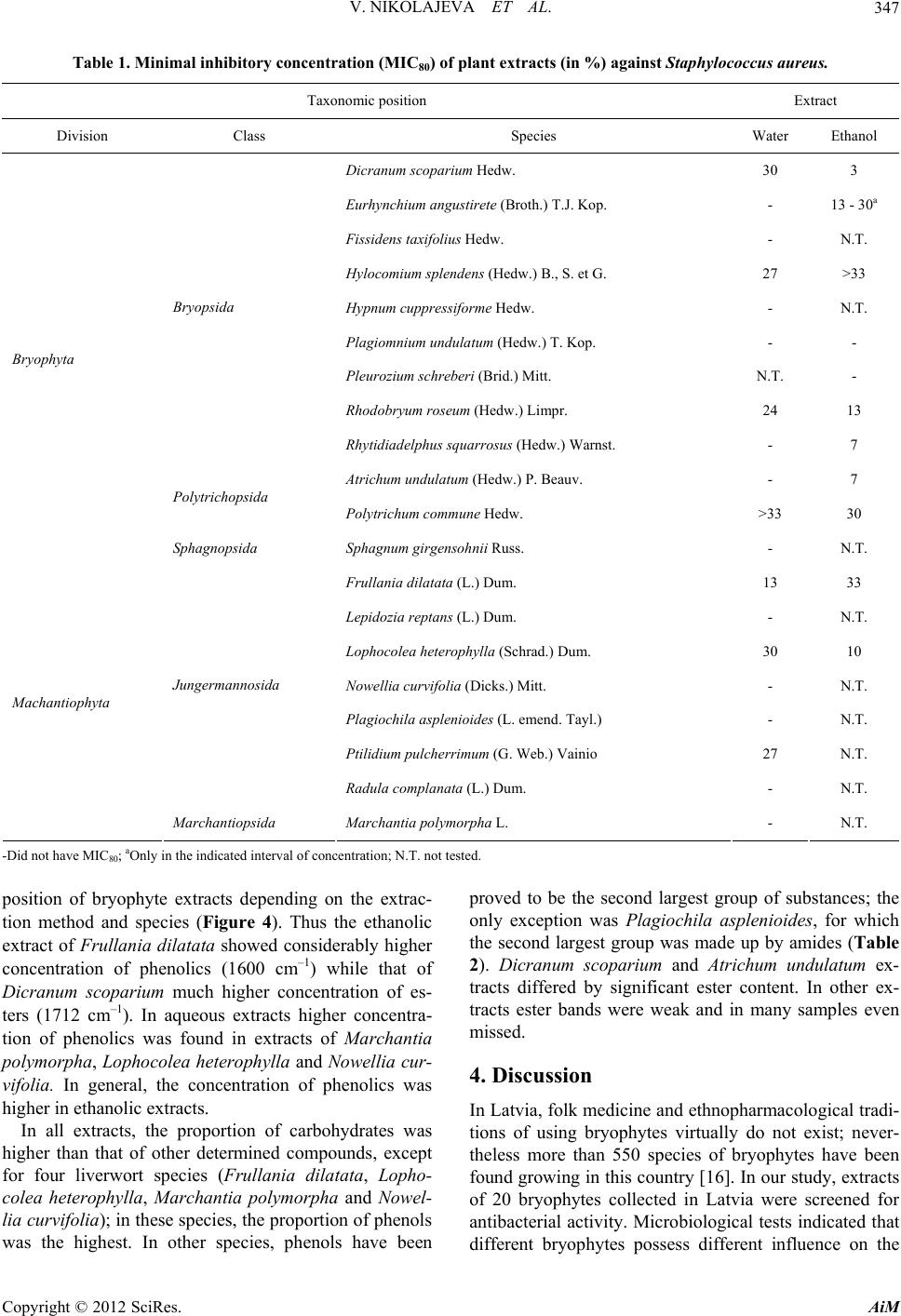 V. NIKOLAJEVA ET AL. Copyright © 2012 SciRes. AiM 347 Table 1. Minimal inhibitory concentration (MIC80) of plant extracts (in %) against Staphylococcus aureus. Taxonomic position Extract Division Class Species Water Ethanol Dicranum scoparium Hedw. 30 3 Eurhynchium angustirete (Broth.) T.J. Kop. - 13 - 30a Fissidens taxifolius Hedw. - N.T. Hylocomium splendens (Hedw.) B., S. et G. 27 >33 Hypnum cuppressiforme Hedw. - N.T. Plagiomnium undulatum (Hedw.) T. Kop. - - Pleurozium schreberi (Brid.) Mitt. N.T. - Rhodobryum roseum (Hedw.) Limpr. 24 13 Bryopsida Rhytidiadelphus squarrosus (Hedw.) Warnst. - 7 Atrichum undulatum (Hedw.) P. Beauv. - 7 Polytrichopsida Polytrichum commune Hedw. >33 30 Bryophyta Sphagnopsida Sphagnum girgensohnii Russ. - N.T. Frullania dilatata (L.) Dum. 13 33 Lepidozia reptans (L.) Dum. - N.T. Lophocolea heterophylla (Schrad.) Dum. 30 10 Nowellia curvifolia (Dicks.) Mitt. - N.T. Plagiochila asplenioides (L. emend. Tayl.) - N.T. Ptilidium pulcherrimum (G. Web.) Vainio 27 N.T. Jungermannosida Radula complanata (L.) Dum. - N.T. Machantiophyta Marchantiopsida Marchantia polymorpha L. - N.T. -Did not have MIC80; aOnly in the indicated interval of concentration; N.T. not tested. position of bryophyte extracts depending on the extrac- tion method and species (Figure 4). Thus the ethanolic extract of Frullania dilatata showed considerably higher concentration of phenolics (1600 cm–1) while that of Dicranum scoparium much higher concentration of es- ters (1712 cm–1). In aqueous extracts higher concentra- tion of phenolics was found in extracts of Marchantia polymorpha, Lophocolea heterophylla and Nowellia cur- vifolia. In general, the concentration of phenolics was higher in ethanolic extracts. In all extracts, the proportion of carbohydrates was higher than that of other determined compounds, except for four liverwort species (Frullania dilatata, Lopho- colea heterophylla, Marchantia polymorpha and Nowe l - lia curvifolia); in these species, the proportion of phenols was the highest. In other species, phenols have been proved to be the second largest group of substances; the only exception was Plagiochila asplenioides, for which the second largest group was made up by amides (Table 2). Dicranum scoparium and Atrichum undulatum ex- tracts differed by significant ester content. In other ex- tracts ester bands were weak and in many samples even missed. 4. Discussion In Latvia, folk medicine and ethnopharmacological tradi- tions of using bryophytes virtually do not exist; never- theless more than 550 species of bryophytes have been found growing in this country [16]. In our study, extracts of 20 bryophytes collected in Latvia were screened for antibacterial activity. Microbiological tests indicated that different bryophytes possess different influence on the 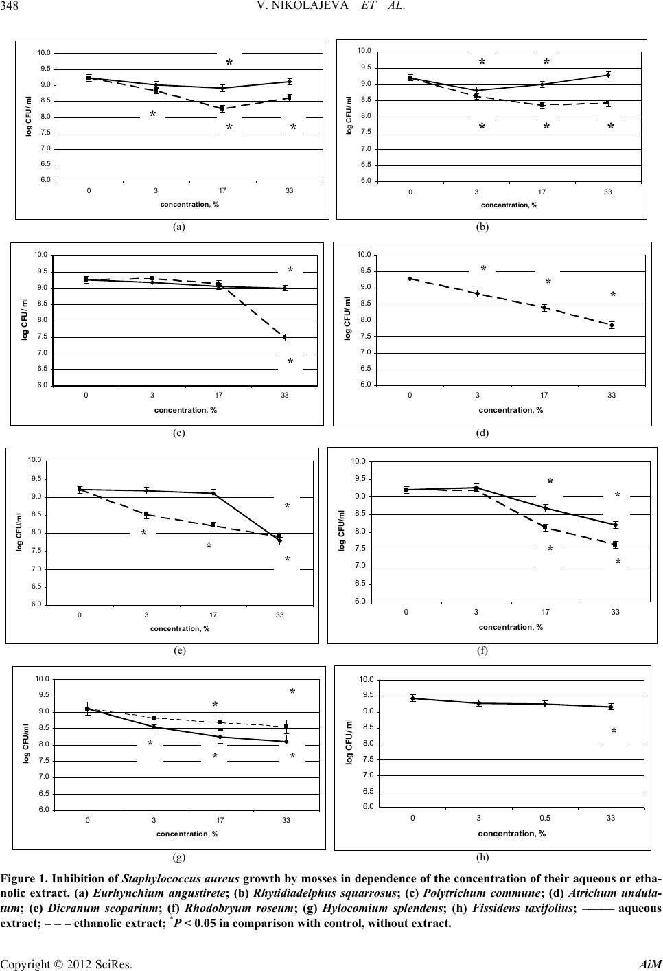 V. NIKOLAJEVA ET AL. 348 6.0 6.5 7.0 7.5 8.0 8.5 9.0 9.5 10.0 031 conc entration, % log CFU/ ml 733 * 6.0 6.5 7.0 7.5 8.0 8.5 9.0 9.5 10.0 031733 co n cent ration, % l og CFU/ m * * * l * * * * * (a) (b) 6.0 6.5 7.0 7.5 8.0 8.5 9.0 9.5 10.0 031 concentration, % l og CFU/ m 733 l * * 6.0 6.5 7.0 7.5 8.0 8.5 9.0 9.5 10.0 031733 co ncent ration, % l og CFU/ m l *** (c) (d) 6.0 6.5 7.0 7.5 8.0 8.5 9.0 9.5 10.0 031 concentration, % log CFU/ml 733 * * 6.0 6.5 7.0 7.5 8.0 8.5 9.0 9.5 10.0 0317 33 concentration, % log CFU/ml * * * * * * (e) (f) 6.0 6.5 7.0 7.5 8.0 8.5 9.0 9.5 10.0 03 17 concentration, % log CFU/ml 33 * * 6.0 6.5 7.0 7.5 8.0 8.5 9.0 9.5 10.0 030.533 concentration, % l og CFU/ m * * * l * (g) (h) Figure 1. Inhibition of Staphylococcus aureus growth by mosses in dependence of the conce ntration of their aqueous or etha- nolic extract. (a) Eurhynchium angustirete; (b) Rhytidiadelphus squarrosus; (c) Polytrichum commune; (d) Atrichum undula- tum; (e) Dicranum scoparium; (f) Rhodobryum roseum; (g) Hylocomium splendens; (h) Fissidens taxifolius; aqueous extract; ethanolic extract; *P < 0.05 in comparison with control, without extract. Copyright © 2012 SciRes. AiM 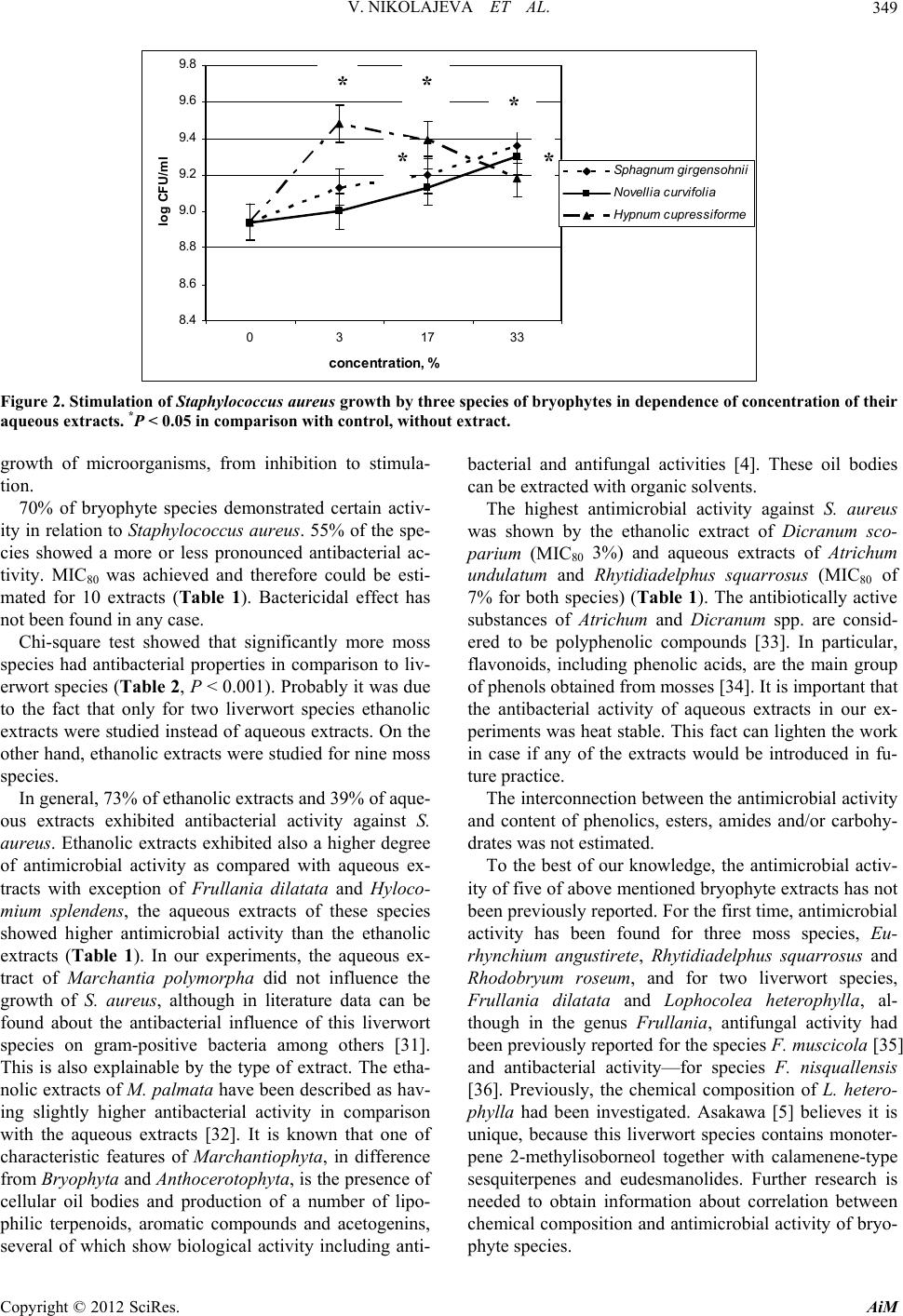 V. NIKOLAJEVA ET AL. 349 8.4 8.6 8.8 9.0 9.2 9.4 9.6 9.8 0317 33 co ncentration, % log CFU/ml Sphagnum girgensohnii Novellia curvifolia Hypn um cupressifo rme ** * * * Figure 2. Stimulation of Staphylococcus aureus growth by three species of bryophy tes in dependenc e of concentration of their aqueous extracts. *P < 0.05 in comparison with control, without extract. growth of microorganisms, from inhibition to stimula- tion. 70% of bryophyte species demonstrated certain activ- ity in relation to Staphylococcus aureus. 55% of the spe- cies showed a more or less pronounced antibacterial ac- tivity. MIC80 was achieved and therefore could be esti- mated for 10 extracts (Table 1). Bactericidal effect has not been found in any case. Chi-square test showed that significantly more moss species had antibacterial properties in comparison to liv- erwort species (Table 2, P < 0.001). Probably it was due to the fact that only for two liverwort species ethanolic extracts were studied instead of aqueous extracts. On the other hand, ethanolic extracts were studied for nine moss species. In general, 73% of ethanolic extracts and 39% of aque- ous extracts exhibited antibacterial activity against S. aureus. Ethanolic extracts exhibited also a higher degree of antimicrobial activity as compared with aqueous ex- tracts with exception of Frullania dilatata and Hyloco- mium splendens, the aqueous extracts of these species showed higher antimicrobial activity than the ethanolic extracts (Table 1). In our experiments, the aqueous ex- tract of Marchantia polymorpha did not influence the growth of S. aureus, although in literature data can be found about the antibacterial influence of this liverwort species on gram-positive bacteria among others [31]. This is also explainable by the type of extract. The etha- nolic extracts of M. palmata have been described as hav- ing slightly higher antibacterial activity in comparison with the aqueous extracts [32]. It is known that one of characteristic features of Marchantiophyta, in difference from Bryophyta and Anthocerotophyta, is the presence of cellular oil bodies and production of a number of lipo- philic terpenoids, aromatic compounds and acetogenins, several of which show biological activity including anti- bacterial and antifungal activities [4]. These oil bodies can be extracted with organic solvents. The highest antimicrobial activity against S. aureus was shown by the ethanolic extract of Dicranum sco- parium (MIC80 3%) and aqueous extracts of Atrichum undulatum and Rhytidiadelphus squarrosus (MIC80 of 7% for both species) (Table 1). The antibiotically active substances of Atrichum and Dicranum spp. are consid- ered to be polyphenolic compounds [33]. In particular, flavonoids, including phenolic acids, are the main group of phenols obtained from mosses [34]. It is important that the antibacterial activity of aqueous extracts in our ex- periments was heat stable. This fact can lighten the work in case if any of the extracts would be introduced in fu- ture practice. The interconnection between the antimicrobial activity and content of phenolics, esters, amides and/or carbohy- drates was not estimated. To the best of our knowledge, the antimicrobial activ- ity of five of above mentioned bryophyte extracts has not been previously reported. For the first time, antimicrobial activity has been found for three moss species, Eu- rhynchium angustirete, Rhytidiadelphus squarrosus and Rhodobryum roseum, and for two liverwort species, Frullania dilatata and Lophocolea heterophylla, al- though in the genus Frullania, antifungal activity had been previously reported for the species F. muscicola [35] and antibacterial activity—for species F. nisquallens is [36]. Previously, the chemical composition of L. hetero- phylla had been investigated. Asakawa [5] believes it is unique, because this liverwort species contains monoter- pene 2-methylisoborneol together with calamenene-type sesquiterpenes and eudesmanolides. Further research is needed to obtain information about correlation between chemical composition and antimicrobial activity of bryo- phyte species. Copyright © 2012 SciRes. AiM 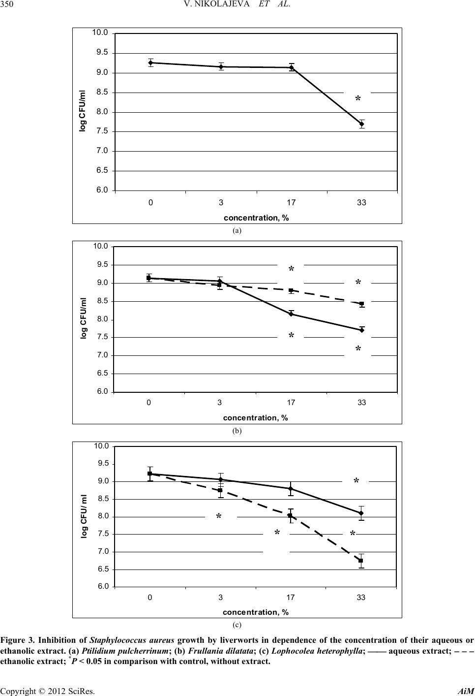 V. NIKOLAJEVA ET AL. 350 6.0 6.5 7.0 7.5 8.0 8.5 9.0 9.5 10.0 03 17 co ncen t rat ion, % l og CF U/m 33 l * (a) 6.0 6.5 7.0 7.5 8.0 8.5 9.0 9.5 10.0 0317 c oncentration, % log CFU/ml 33 ** * * (b) 6.0 6.5 7.0 7.5 8.0 8.5 9.0 9.5 10.0 03 17 c oncentration, % log CFU/ ml 33 ** * * (c) Figure 3. Inhibition of Staphylococcus aureus growth by liverworts in dependence of the concentration of their aqueous or ethanolic extract. (a) Ptilidium pulcherrinum; (b) Frullania dilatata; (c) Lophocolea heterophylla; aqueous extract; ethanolic extract; *P < 0.05 in comparison with control, without extract. Copyright © 2012 SciRes. AiM 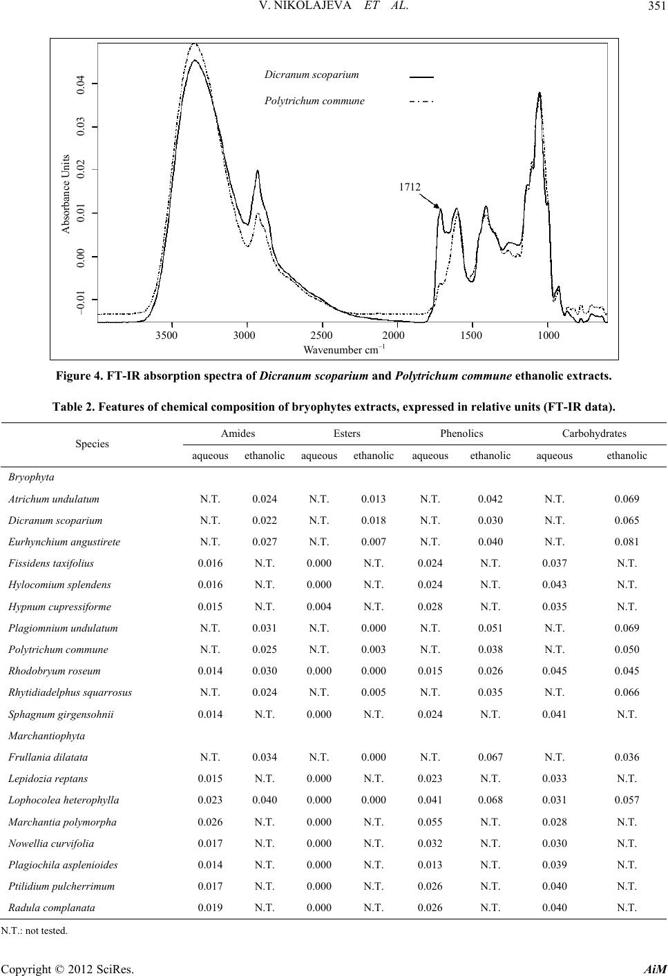 V. NIKOLAJEVA ET AL. 351 3500 3000 2500 2000 1500 1000 Absorbance Units Wavenumber cm –1 –0.01 0.00 0.01 0.02 0.03 0.04 D ic r anum scoparium P olytrichum commune 1712 Figure 4. FT-IR absorption spectra of Dicranum scoparium and Polytrichum commune ethanolic extracts. Table 2. Features of chemical composition of bryophytes extracts, expressed in relative units (FT-IR data). Amides Esters Phenolics Carbohydrates Species aqueous ethanolic aqueous ethanolicaqueousethanolic aqueous ethanolic Bryophyta Atrichum undulatum N.T. 0.024 N.T. 0.013 N.T. 0.042 N.T. 0.069 Dicranum scoparium N.T. 0.022 N.T. 0.018 N.T. 0.030 N.T. 0.065 Eurhynchium angustirete N.T. 0.027 N.T. 0.007 N.T. 0.040 N.T. 0.081 Fissidens taxifolius 0.016 N.T. 0.000 N.T. 0.024 N.T. 0.037 N.T. Hylocomium splendens 0.016 N.T. 0.000 N.T. 0.024 N.T. 0.043 N.T. Hypnum cupressiforme 0.015 N.T. 0.004 N.T. 0.028 N.T. 0.035 N.T. Plagiomnium undulatum N.T. 0.031 N.T. 0.000 N.T. 0.051 N.T. 0.069 Polytrichum commune N.T. 0.025 N.T. 0.003 N.T. 0.038 N.T. 0.050 Rhodobryum roseum 0.014 0.030 0.000 0.000 0.015 0.026 0.045 0.045 Rhytidiadelphus squarrosus N.T. 0.024 N.T. 0.005 N.T. 0.035 N.T. 0.066 Sphagnum girgensohnii 0.014 N.T. 0.000 N.T. 0.024 N.T. 0.041 N.T. Marchantiophyta Frullania dilatata N.T. 0.034 N.T. 0.000 N.T. 0.067 N.T. 0.036 Lepidozia reptans 0.015 N.T. 0.000 N.T. 0.023 N.T. 0.033 N.T. Lophocolea heterophylla 0.023 0.040 0.000 0.000 0.041 0.068 0.031 0.057 Marchantia polymorpha 0.026 N.T. 0.000 N.T. 0.055 N.T. 0.028 N.T. Nowellia curvifolia 0.017 N.T. 0.000 N.T. 0.032 N.T. 0.030 N.T. Plagiochila asplenioides 0.014 N.T. 0.000 N.T. 0.013 N.T. 0.039 N.T. Ptilidium pulcherrimum 0.017 N.T. 0.000 N.T. 0.026 N.T. 0.040 N.T. Radula complanata 0.019 N.T. 0.000 N.T. 0.026 N.T. 0.040 N.T. N.T.: not tested. Copyright © 2012 SciRes. AiM 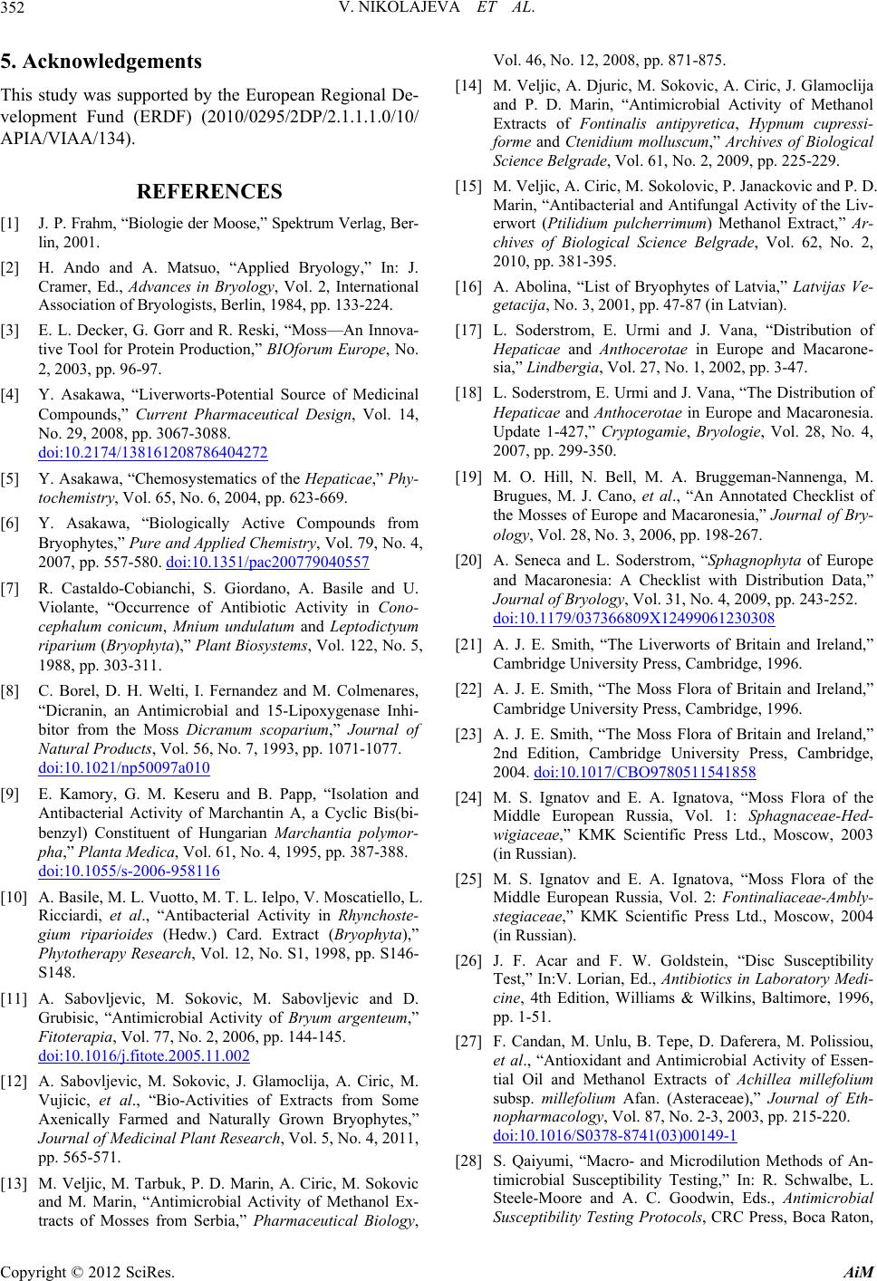 V. NIKOLAJEVA ET AL. Copyright © 2012 SciRes. AiM 352 5. Acknowledgements This study was supported by the European Regional De- velopment Fund (ERDF) (2010/0295/2DP/2.1.1.1.0/10/ APIA/VIAA/134). REFERENCES [1] J. P. Frahm, “Biologie der Moose,” Spektrum Verlag, Ber- lin, 2001. [2] H. Ando and A. Matsuo, “Applied Bryology,” In: J. Cramer, Ed., Advances in Bryology, Vol. 2, International Association of Bryologists, Berlin, 1984, pp. 133-224. [3] E. L. Decker, G. Gorr and R. Reski, “Moss—An Innova- tive Tool for Protein Production,” BIOforum Europe, No. 2, 2003, pp. 96-97. [4] Y. Asakawa, “Liverworts-Potential Source of Medicinal Compounds,” Current Pharmaceutical Design, Vol. 14, No. 29, 2008, pp. 3067-3088. doi:10.2174/138161208786404272 [5] Y. Asakawa, “Chemosystematics of the Hepaticae,” Phy- tochemistry, Vol. 65, No. 6, 2004, pp. 623-669. [6] Y. Asakawa, “Biologically Active Compounds from Bryophytes,” Pure and Applied Chemistry, Vol. 79, No. 4, 2007, pp. 557-580. doi:10.1351/pac200779040557 [7] R. Castaldo-Cobianchi, S. Giordano, A. Basile and U. Violante, “Occurrence of Antibiotic Activity in Cono- cephalum conicum, Mnium undulatum and Leptodictyum riparium (Bryophyta),” Plant Biosystems, Vol. 122, No. 5, 1988, pp. 303-311. [8] C. Borel, D. H. Welti, I. Fernandez and M. Colmenares, “Dicranin, an Antimicrobial and 15-Lipoxygenase Inhi- bitor from the Moss Dicranum scoparium,” Journal of Natural Products, Vol. 56, No. 7, 1993, pp. 1071-1077. doi:10.1021/np50097a010 [9] E. Kamory, G. M. Keseru and B. Papp, “Isolation and Antibacterial Activity of Marchantin A, a Cyclic Bis(bi- benzyl) Constituent of Hungarian Marchantia polymor- pha,” Planta Medica, Vol. 61, No. 4, 1995, pp. 387-388. doi:10.1055/s-2006-958116 [10] A. Basile, M. L. Vuotto, M. T. L. Ielpo, V. Moscatiello, L. Ricciardi, et al., “Antibacterial Activity in Rhynchoste- gium riparioides (Hedw.) Card. Extract (Bryophyta),” Phytotherapy Research, Vol. 12, No. S1, 1998, pp. S146- S148. [11] A. Sabovljevic, M. Sokovic, M. Sabovljevic and D. Grubisic, “Antimicrobial Activity of Bryum argenteum,” Fitoterapia, Vol. 77, No. 2, 2006, pp. 144-145. doi:10.1016/j.fitote.2005.11.002 [12] A. Sabovljevic, M. Sokovic, J. Glamoclija, A. Ciric, M. Vujicic, et al., “Bio-Activities of Extracts from Some Axenically Farmed and Naturally Grown Bryophytes,” Journal of Medicinal Plant Research, Vol. 5, No. 4, 2011, pp. 565-571. [13] M. Veljic, M. Tarbuk, P. D. Marin, A. Ciric, M. Sokovic and M. Marin, “Antimicrobial Activity of Methanol Ex- tracts of Mosses from Serbia,” Pharmaceutical Biology, Vol. 46, No. 12, 2008, pp. 871-875. [14] M. Veljic, A. Djuric, M. Sokovic, A. Ciric, J. Glamoclija and P. D. Marin, “Antimicrobial Activity of Methanol Extracts of Fontinalis antipyretica, Hypnum cupressi- forme and Ctenidium molluscum,” Archives of Biological Science Belgrade, Vol. 61, No. 2, 2009, pp. 225-229. [15] M. Veljic, A. Ciric, M. Sokolovic, P. Janackovic and P. D. Marin, “Antibacterial and Antifungal Activity of the Liv- erwort (Ptilidium pulcherrimum) Methanol Extract,” Ar- chives of Biological Science Belgrade, Vol. 62, No. 2, 2010, pp. 381-395. [16] A. Abolina, “List of Bryophytes of Latvia,” Latvijas Ve- getacija, No. 3, 2001, pp. 47-87 (in Latvian). [17] L. Soderstrom, E. Urmi and J. Vana, “Distribution of Hepaticae and Anthocerotae in Europe and Macarone- sia,” Lindbergia, Vol. 27, No. 1, 2002, pp. 3-47. [18] L. Soderstrom, E. Urmi and J. Vana, “The Distribution of Hepaticae and Anthocerotae in Europe and Macaronesia. Update 1-427,” Cryptogamie, Bryologie, Vol. 28, No. 4, 2007, pp. 299-350. [19] M. O. Hill, N. Bell, M. A. Bruggeman-Nannenga, M. Brugues, M. J. Cano, et al., “An Annotated Checklist of the Mosses of Europe and Macaronesia,” Journal of Bry- ology, Vol. 28, No. 3, 2006, pp. 198-267. [20] A. Seneca and L. Soderstrom, “Sphagnophyta of Europe and Macaronesia: A Checklist with Distribution Data,” Journal of Bryology, Vol. 31, No. 4, 2009, pp. 243-252. doi:10.1179/037366809X12499061230308 [21] A. J. E. Smith, “The Liverworts of Britain and Ireland,” Cambridge University Press, Cambridge, 1996. [22] A. J. E. Smith, “The Moss Flora of Britain and Ireland,” Cambridge University Press, Cambridge, 1996. [23] A. J. E. Smith, “The Moss Flora of Britain and Ireland,” 2nd Edition, Cambridge University Press, Cambridge, 2004. doi:10.1017/CBO9780511541858 [24] M. S. Ignatov and E. A. Ignatova, “Moss Flora of the Middle European Russia, Vol. 1: Sphagnaceae-Hed- wigiaceae,” KMK Scientific Press Ltd., Мoscow, 2003 (in Russian). [25] M. S. Ignatov and E. A. Ignatova, “Moss Flora of the Middle European Russia, Vol. 2: Fontinaliaceae-Ambly- stegiaceae,” KMK Scientific Press Ltd., Мosсow, 2004 (in Russian). [26] J. F. Acar and F. W. Goldstein, “Disc Susceptibility Test,” In:V. Lorian, Ed., Antibiotics in Laboratory Medi- cine, 4th Edition, Williams & Wilkins, Baltimore, 1996, pp. 1-51. [27] F. Candan, M. Unlu, B. Tepe, D. Daferera, M. Polissiou, et al., “Antioxidant and Antimicrobial Activity of Essen- tial Oil and Methanol Extracts of Achillea millefolium subsp. millefolium Afan. (Asteraceae),” Journal of Eth- nopharmacology, Vol. 87, No. 2-3, 2003, pp. 215-220. doi:10.1016/S0378-8741(03)00149-1 [28] S. Qaiyumi, “Macro- and Microdilution Methods of An- timicrobial Susceptibility Testing,” In: R. Schwalbe, L. Steele-Moore and A. C. Goodwin, Eds., Antimicrobial Susceptibility Testing Protocols, CRC Press, Boca Raton,  V. NIKOLAJEVA ET AL. 353 London, New York, 2007, pp. 75-79. doi:10.1201/9781420014495.ch4 [29] M. D. Guillen and N. Cabo, “Some of the Most Signifi- cant Changes in the Fourier Transform Infrared Spectra of Edible Oils under Oxidative Conditions,” Journal of the Science of Food and Agriculture, Vol. 80, No. 14, 2000, pp. 2028-2036. [30] N. Vlachos, Y. Skopelitis, M. Psaroudaki, V. Konstanti- nidou, A. Chatzilazarou and E. Tegou, “Applications of Fourier Transform-Infrared Spectroscopy to Edible Oils,” Analytica Chimica Acta, Vol. 573-574, 2006, pp. 459-465. doi:10.1016/j.aca.2006.05.034 [31] Y. Asakawa, “Highlights in Phytochemistry of Hepati- cae—Biologically Active Terpenoids and Aromatic Com- pounds,” P ure and Applied Che mistry, Vol. 66, No. 10-11, 1994, pp. 2193-2196. doi:10.1351/pac199466102193 [32] R. Khanam, B. L. Chaudhary, S. Khanam and P. Kumar, “Antibacterial Activity of Marchantia palmata Nees,” Asian Journal of Biochemical and Pharmaceutical Re- search, Vol. 1, No. 2, 2011, pp. 27-36. [33] J. A. McCleary and D. L. Walkington, “Mosses and Anti- biosis,” Revue Bryologique et Lichenologique, Vol. 34, 1966, pp. 309-314. [34] A. Basile, S. Giordano, J. A. Lopez-Saez and R. C. Co- bianchi, “Antibacterial Activity of Pure Flavonoids Iso- lated from Mosses,” Phytochemistry, Vol. 52, No. 8, 1999, pp. 1479-1482. [35] H. X. Lou, G. Y. Li and F. Q. Wang, “A Cytotoxic Diterpenoid and Antifungal Phenolic Compounds from Frullania muscicola Steph.,” Journal of Asian Natural Products Research, Vol. 4, No. 2, 2002, pp. 87-94. doi:10.1080/10286020290027353 [36] M. D. Russel, “Antibiotic Activity of Extracts from some Bryophytes in South Western British Columbia,” Medical Student Journal of Australia, Vol. 2, No. 1, 2010, pp. 9- 14. Copyright © 2012 SciRes. AiM |