

Paper Menu >>
Journal Menu >>
 Neuroscience & Medicine, 2012, 3, 281-286 http://dx.doi.org/10.4236/nm.2012.33032 Published Online September 2012 (http://www.SciRP.org/journal/nm) 281 Effect of Nicotine on Dopamine and Glutathione Levels in Presence of Oligoelements in Brain Regions of Young Rats* ——Effect of Nicotine on Brain Regions of Rat David Calderón Guzmán1, Ernestina Hernández García1, Francisca Trujillo Jiménez2, Gerardo Barragán Mejía1, Hugo Juárez Olguín2,3#, José A. Saldivar González3, Daniel Santamaria del Angel1, Norma Osnaya Brizuela4 1Laboratorio de Neuroquímica, Instituto Nacional de Pediatría (INP), México City, México; 2Laboratorio de Farmacología, INP, México City, México; 3Departamento de Farmacología, Facultad de Medicina, Universidad Nacional Autónoma de México, México City, México; 4Laboratorio de Patología Experimental, México City, México. Email: #[email protected] Received May 25th, 2012; revised June 29th, 2012; accepted July 5th, 2012 ABSTRACT Aim: The purpose of this study was to understand the mechanism of nicotine mediated addiction and the role of oli- goelements in reducing its effect. Methods: Male Wistar rats (weight 80 g) were treated with single and repeated doses of nicotine and/or oligoelements as follows: group 1 (control) NaCl 0.9%; group 2, nicotine (1 mg/kg); group 3, oli- goelements (50 µl/rat); and group 4, nicotine (1 mg/kg) + oligoelements (50 µl/rat). All drugs were intraperitoneally administered for 4 days. Blood for the measurement of glucose was obtained from all the animals. Samples of the brain regions (cortex, hemispheres and cerebellum + medulla oblongata) of each rat were obtained and used to measure the concentrations of dopamine, GSH levels, and lipid peroxidation (TBARS) using fluorescence and spectrophotometric methods. Results: Glucose level increased in rats treated with nicotine and oligoelements (p < 0.05), while GSH level decreased in cerebellum/medulla oblongata and hemispheres (p < 0.05) of the same animals. TBARS levels increased in cerebellum/medulla oblongata and hemispheres of animals treated with nicotine and oligoelements, but decreased in the same regions (p < 0.05) in rats treated only with oligoelements. The levels of dopamine decreased in cortex and hemi- spheres, but increased in cerebellum/medulla and oblongata regions of rats treated with both compounds (p < 0.05). Conclusions: Nicotine and oligoelements are associated with increase in the level of glucose, an effect that was more pronounced in the group treated with both drugs. Reduction of oxidative stress and dopamine metabolism may be in- volved in this effect. Keywords: Glutathione; Lipid Peroxidation; Oxidative Stress 1. Introduction In Mexico, drug addictions in adolescents (53.5% in men and 63.2% in women) begin with tobacco. In our country, smoking usually begins in mid-adolescence (15 years old) and smokers between 18 and 65 years old represent 20.6% of the population [1], a figure which is presently with an increasing tendency. Nicotine is an alkaloid found in tobacco plant (Nicotiana tabacum). In low con- centrations, nicotine has stimulating effect and consti- tutes one of the principal causes of tobacco addiction. Majority of cigarettes in the world contain between 1 and 2 mg or more of nicotine. On smoking, a smoker usually inhales 0.8 mg of nicotine per cigarette [2]. It is known that dopamine is an appetite suppressant, and the administration of nicotine, regardless of the way, acutely increases the levels of neurotransmitters in the brain, thereby reducing the need for energy intake and consequently suppressing the appetite [3], although the mechanisms are unclear. Nicotine has been documented as a possible contrib- uting factor to the current nutrition-related chronic dis- eases epidemic [1]. However, recent studies have shown that micronutrient deficiencies are still significant public health problems, affecting the most vulnerable age groups and gender [4]. Likewise, there are some food rich in oligoelements like milk for daily intake [5], to offset this deficiency. *Declaration of interest: We declare that there is no conflict of interest. #Corresponding author. The vital biological processes need oligoelements as Zinc (Zn), Manganese (Mn), Copper (Cu), and others, all Copyright © 2012 SciRes. NM  Effect of Nicotine on Dopamine and Glutathione Levels in Presence of Oligoelements in Brain Regions of Young Rats 282 of which are integral part of many important enzymes. Cu is essential for life and their excessive accumulation in the brain decreases GSH levels [6], while exposure to Mn differentially affects GSH in brain regions [7]. On one part, smoking is associated with increased plasma homocysteine levels [8], a precursor of GSH and a tripeptide known as reduced glutathione, which is also a ubiquitous reducing agent whose absence induces se- vere oxidative stress (OS). It has been suggested that GSH interacts with nitric oxide (NO) by giving S-nitro- soglutathione (GNSO), in the presence of endogenously produced oxygen [9]. GSH is the main redox equilibrium regulator. It plays an important role in the protection of tissues suffering from damage by oxidative agents. Recent studies indicated that the use of Zn induces de- fensive mechanisms in the brain by diminishing free radical-induced lipid peroxidation [10]. Free radicals are reactive oxygen or nitrogen species with impaired elec- trons, which may induce oxidative damage to biologi- cally important molecules. Although membrane lipids are the main target [11], the central nervous system (CNS) is particularly susceptible to this type of damage, because membrane lipids are known to strongly interact with the lipid bilayer structural proteins [12]. It is necessary to determine the effects of nicotine and oligoelements, be- cause the coexistence of nicotine and micronutrients’ deficiency complicates the health of young people. For this, we aim at determining the effect of these substances on dopamine levels and some biomarkers of oxidative stress in juvenile rat brain regions. 2. Methods Fifty six male Wistar rats each with a weight of 80g were used in the study. They were divided in single dose and repeated doses, into four experimental groups for each dose and treated as follows: Group 1 (n = 7), control (NaCl 0.9%); group 2 (n = 7), nicotine (1mg/rat); group 3 (n = 7), oligoelements 50 µl/mg/kg b.w; and group 4 (n = 7), nicotine (1 mg/rat) + oligoelements 50 µl/mg/kg b.w. All treatments were administered intraperitoneally every 24 hours for 4 days. Every 100 mL of oligoelemental solution contains Zinc 55 mg, copper 16.90 mg, manga- nese 38.10 mg, sodium 163.9 mg, fluoride 14 mg, iodide 1.3 mg, and chloride 25.6 mg. The animals were sacri- ficed by decapitation 30 min after receiving the last dose of nicotine and oligoelements, and fresh blood was with- drawn to measure their glucose levels. The brains were extracted and stored in NaCl 0.9% at 4˚C. Brain sections obtained from cortex, hemispheres, and cerebellum/me- dulla oblongata were each homogenized in 5 volumes of TRIS-HCl 0.05 M, pH 7.4 for assessment of lipid peroxi- dation (TBARS). An aliquot of this tissue was mixed (50:50) with perchloric acid (HClO4) 0.1 M, to evaluate the levels of Glutathione (GSH) and dopamine. All ex- perimental procedures were done with authorization of the Laboratory Animals Use and Care Committee of our institution. The treatments given to animals were as follow: Groups Single dose Repeated doses Control (NaCl 0.9%) (7) (7) Nicotine (1 mg/rat) (7) (7) Oligoelements (50 µl/mg/kg) (7) (7) Nicotine (1 mg/rat) + oligoelements (50 µl/mg/kg) (7) (7) ( ) number of animals. 2.1. Measurement of Blood Glucose The procedure to measure glucose in blood was followed in all the animals at the moment of their sacrifice. 20 µl of non-anticoagulant fresh blood was placed in a reactive paper of Accu-Chek active equipment (Roche Mannheim Germany). The concentration in mg/dl was obtained. 2.2. Measurement of Glutathione (GSH) in Brain The levels of GSH were measured from a sample of the homogenized tissue in HClO4 which was obtained after being centrifuged at 9000 rpm for 5 min. (in a microcen- trifuge Mikro 12 - 42, Germany), using the technique re- ported by Hissin and Hilf [13]. 1.8 ml of Phosphate Buffer at pH 8.0 with EDTA at 0.2%, an aliquot of 20 µl of the homogenized tissue in HClO4, and 100 µl of or- tho-phtaldialdehyde (OPT) in concentration of 1mg/ml in methanol, were put in an assay tube and incubated for 15 min. at ambient temperature in total darkness. At the end of incubation, the samples were read in a PERLIN ELMER LS 55 spectrofluorometer with excitation wave- length of 350mm and emission of 420 nm. FL Win Lab version 4.00.02 software was used. The values were in- ferred in a previously standardized standard curve and were reported in nM/g of wet tissue. 2.3. Measurement of Lipid Peroxidation (TBARS) in Brain TBARS determination was carried out based on the tech- nique reported by Gutteridge and Halliwell [14], as de- scribed below: from the homogenized brain in tris-HCl 0.05 M pH 7.4, 1 ml was taken and to it was added 2 ml of thiobarbaturic acid (Tba) containing 1.25 g of Tba, 40 g of trichloroacetic acid (TCA), and 6.25 ml of concen- trated hydrochloric acid (HCL) diluted in 250 ml of de- ionized H2O. They were heated to boiling point for 30 min. (Thermomix 1420). The samples were then sub- jected to an ice bath for 5 min and were centrifuged at 700 g for 15 min. (Sorvall RC-5B Dupont). The absorb- Copyright © 2012 SciRes. NM 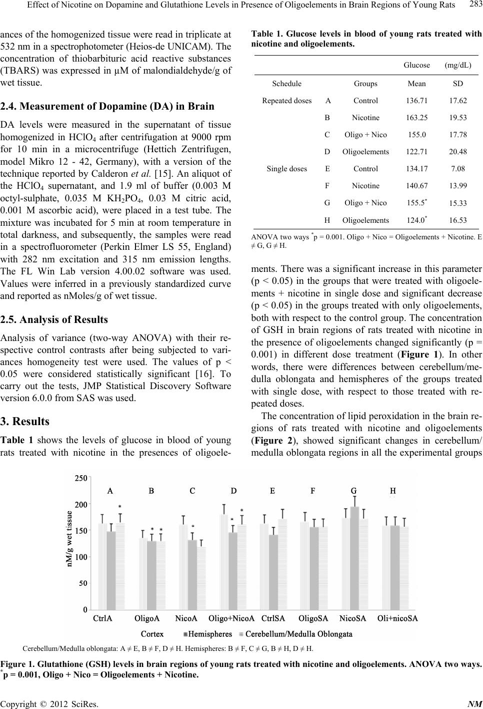 Effect of Nicotine on Dopamine and Glutathione Levels in Presence of Oligoelements in Brain Regions of Young Rats Copyright © 2012 SciRes. NM 283 Table 1. Glucose levels in blood of young rats treated with nicotine and oligoelements. ances of the homogenized tissue were read in triplicate at 532 nm in a spectrophotometer (Heios-de UNICAM). The concentration of thiobarbituric acid reactive substances (TBARS) was expressed in µM of malondialdehyde/g of wet tissue. Glucose (mg/dL) Schedule Groups Mean SD Repeated dosesAControl 136.71 17.62 BNicotine 163.25 19.53 COligo + Nico 155.0 17.78 DOligoelements 122.71 20.48 Single doses EControl 134.17 7.08 FNicotine 140.67 13.99 GOligo + Nico 155.5* 15.33 HOligoelements 124.0* 16.53 2.4. Measurement of Dopamine (DA) in Brain DA levels were measured in the supernatant of tissue homogenized in HClO4 after centrifugation at 9000 rpm for 10 min in a microcentrifuge (Hettich Zentrifugen, model Mikro 12 - 42, Germany), with a version of the technique reported by Calderon et al. [15]. An aliquot of the HClO4 supernatant, and 1.9 ml of buffer (0.003 M octyl-sulphate, 0.035 M KH2PO4, 0.03 M citric acid, 0.001 M ascorbic acid), were placed in a test tube. The mixture was incubated for 5 min at room temperature in total darkness, and subsequently, the samples were read in a spectrofluorometer (Perkin Elmer LS 55, England) with 282 nm excitation and 315 nm emission lengths. The FL Win Lab version 4.00.02 software was used. Values were inferred in a previously standardized curve and reported as nMoles/g of wet tissue. ANOVA two ways *p = 0.001. Oligo + Nico = Oligoelements + Nicotine. E ≠ G, G ≠ H. ments. There was a significant increase in this parameter (p < 0.05) in the groups that were treated with oligoele- ments + nicotine in single dose and significant decrease (p < 0.05) in the groups treated with only oligoelements, both with respect to the control group. The concentration of GSH in brain regions of rats treated with nicotine in the presence of oligoelements changed significantly (p = 0.001) in different dose treatment (Figure 1). In other words, there were differences between cerebellum/me- dulla oblongata and hemispheres of the groups treated with single dose, with respect to those treated with re- peated doses. 2.5. Analysis of Results Analysis of variance (two-way ANOVA) with their re- spective control contrasts after being subjected to vari- ances homogeneity test were used. The values of p < 0.05 were considered statistically significant [16]. To carry out the tests, JMP Statistical Discovery Software version 6.0.0 from SAS was used. The concentration of lipid peroxidation in the brain re- gions of rats treated with nicotine and oligoelements (Figure 2), showed significant changes in cerebellum/ medulla oblongata regions in all the experimental groups 3. Results Table 1 shows the levels of glucose in blood of young rats treated with nicotine in the presences of oligoele- Cerebellum/Medulla oblongata: A ≠ E, B ≠ F, D ≠ H. Hemispheres: B ≠ F, C ≠ G, B ≠ H, D ≠ H. Figure 1. Glutathione (GSH) levels in brain regions of young rats treated with nicotine and oligoelements. ANOVA tw o way s. *p = 0.001, Oligo + Nico = Oligoelements + Nicotine. 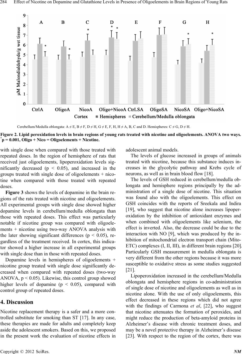 Effect of Nicotine on Dopamine and Glutathione Levels in Presence of Oligoelements in Brain Regions of Young Rats 284 Cerebellum/Medulla oblongata: A ≠ E, B ≠ F, D ≠ H, G ≠ E, F, H, H ≠ A, B, C and D. Hemispheres: C ≠ G, D ≠ H. Figure 2. Lipid peroxidation levels in brain regions of young rats treated w ith nicotine and oligoelements. ANOVA tw o ways. *p = 0.001, Oligo + Nico = Oligoelements + Nicotine. with single dose when compared with those treated with repeated doses. In the region of hemisphere of rats that received just oligoelements, lipoperoxidation levels sig- nificantly decreased (p < 0.05), and increased in the groups treated with single dose of oligoelements + nico- tine when compared with those treated with repeated doses. Figure 3 shows the levels of dopamine in the brain re- gions of the rats treated with nicotine and oligoelements. All experimental groups with single dose showed higher dopamine levels in cerebellum/medulla oblongata than those with repeated doses. This effect was particularly notable if nicotine group was compared with oligoele- ments + nicotine using two-way ANOVA analysis with the later showing significant differences (p < 0.05), re- gardless of the treatment received. In cortex, this indica- tor showed a higher increase in all experimental groups with single dose than in those with repeated doses. Dopamine levels in hemispheres of oligoelements + nicotine group treated with single dose significantly de- creased when compared with repeated doses (two-way ANOVA, p < 0.05). Likewise, this control group showed higher levels of dopamine (p < 0.05), compared with control group of repeated doses. 4. Discussion Nicotine replacement therapy is a safer and a more con- trolled substitute for smoking than ST [17]. In any case, these therapies are made for adults and completely keep aside the adolescent smokers. Based on this, we proposed in the present work the evaluation of nicotine effects in adolescent animal models. The levels of glucose increased in groups of animals treated with nicotine, because this substance induces in- creases in the glycolytic pathway and Krebs cycle of neurons, as well as in brain blood flow [18]. The levels of GSH reduced in cerebellum/medulla ob- longata and hemisphere regions principally by the ad- ministration of a single dose of nicotine. This situation was found also with the oligoelements. This effect on GSH coincides with the reports of Sreekala and Indira [19], who suggest that nicotine alone increases lipoper- oxidation by the inhibition of antioxidant enzymes and when combined with oligoelements like selenium, the effect is inverted. Also, the decrease could be due to the interaction with NO [9], which was produced by the in- hibition of mitochondrial electron transport chain (Mito- ETC) complexes (I, II, III), in different brain regions [20]. Particularly GSH measurement in medulla oblongata is very different from the other regions because it was more susceptible to oxidative stress as some studies suggested [21]. Lipoperoxidation increased in the cerebellum/Medulla oblongata and hemisphere regions in co-administration of single dose of nicotine and oligoelements as well as in nicotine alone. With the use of only oligoelements, this effect decreased in these regions which did not agree with the findings of Carmona et al, [22], who suggest that nicotine attenuates the formation of peroxides, and might reduce the production of beta-amyloid proteins in Alzheimer’s disease with chronic treatment doses, and may be a novel protective therapy in Alzheimer’s disease [23]. With respect to the region of the cortex, there was Copyright © 2012 SciRes. NM  Effect of Nicotine on Dopamine and Glutathione Levels in Presence of Oligoelements in Brain Regions of Young Rats 285 Cerebellum/Medulla oblongata: E ≠ G, F ≠ G. Cortex: A ≠ E, B ≠ F, C ≠ G, D ≠ H. Hemispheres: A ≠ E and G. Figure 3. Dopamine levels in brain regions of young rats treated with nicotine and oligoelements. ANOVA two ways. *p = 0.0001, Oligo + Nico = Oligoelements + Nicotine. no finding of any effect to the treatment in all the animals, although recent studies suggest that the presence of nico- tine increases the formation of peroxides in this region [24]. In general, dopamine levels decreased in cortex and hemisphere regions with co-administration of nicotine and oligoelements in unique doses. On the other hand, this effect was not found in cerebellum/medulla oblon- gata where there was an increase of dopamine by the combined administration of the two substances in the same doses. This effect was in accordance with the find- ings of Tariq et al. [25], who suggested that inhibition of nicotine on decreasing DA, which is induced by 3-nitro- propionic acid, is due thanks to nicotinic acetylcholine receptors (nAChRs). These receptors could be responsi- ble for the absence of significant statistical difference in the indicators evaluated in this study with chronic dosis of nicotine, probably due to the modulation of these re- ceptors by oligoelements like zinc and the concentration, and time of exposure to nicotine, suggesting that zinc potency depends on the degree of desensitization [26]. In the present study developed with adolescent animal model and based on the results of some studies, the mechanism proposed for the inhibition of tobacco (nico- tine) consumption in adolescent subjects is to reduce sugar-rich food consumption and substitute it with oli- goelement-rich food [5], because if this source of sugar is reduced, dopamine (DA) systems are activated and le- sions of dopaminergic neurons or pharmacological blockade of DA receptors seem to reduce the reward value of both sweet tastes and drugs of abuse [27]. Con- sidering the above results, we conclude that nicotine and oligoelements are associated with increase in glucose levels, an effect which was more pronounced in the group of animals treated with single dose of both drugs, probably by no inhibition of fatty acid oxidation on brain regions [28], because glucose determine fatty acid oxida- tion by controlling the rate of long-chain fatty acid en- trance into the mitochondria [29]. Reduction of oxidative stress and dopamine metabolism may be involved in these effects. 5. Conclusion Nicotine and oligoelements are associated with increase in glucose levels on blood. It induces reduction of oxida- tive stress and dopamine metabolism on brain regions. 6. Acknowledgements We thank Dr. Cyril Ndidi Nwoye, who helps us to trans- late the manuscript. REFERENCES [1] ENA, “National Survey of Addictions,” SSA, Conadic, INP, DGE, INSP, 2008. [2] C. Kalpana, A. R. Sudheer, K. N. Rajasekharan and V. P. Menon, “Comparative Effects of Curcumin and Its Syn- thetic Analogue on Tissue Lipid Peroxidation and Anti- oxidant Status during Nicotine-Induced Toxicity,” Singa- pore Medical Journal, Vol. 48, No. 2, 2007, pp. 124-130. [3] R. Chatkin and J. M. Chatkin, “Smoking and Changes in Body Weight: Can Physiopathology and Genetics Explain This Association?” Journal of Brasileiro de Pneumologia, Vol. 33, No. 6, 2007, pp. 712-719. [4] M. Flores, N. Macías, M. Rivera, S. Barquera, L. Her- nández, G. A. García and J. Rivera, “Energy and Nutrient Intake among Mexican School-Aged Children, Mexican National Health and Nutrition Survey 2006,” Salud Pub- lica de Mexico, Vol. 51, No. 4, 2009, pp. S540-S550. [5] G. D. Calderón, G. E. Hernández, M. G. Barragán and A. Copyright © 2012 SciRes. NM 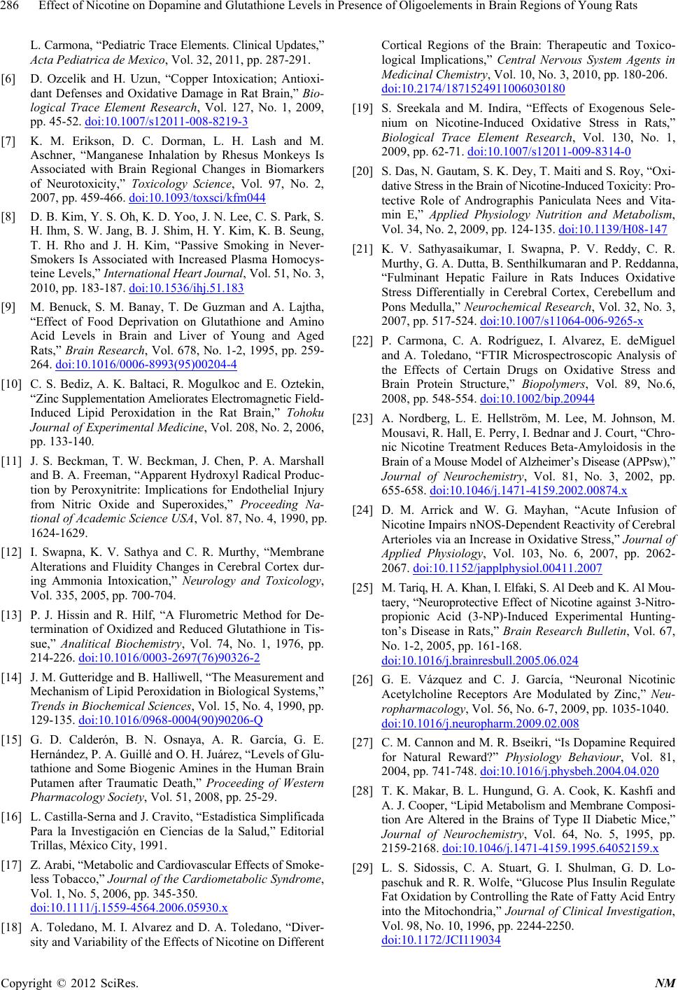 Effect of Nicotine on Dopamine and Glutathione Levels in Presence of Oligoelements in Brain Regions of Young Rats 286 L. Carmona, “Pediatric Trace Elements. Clinical Updates,” Acta Pediatrica de Mexico, Vol. 32, 2011, pp. 287-291. [6] D. Ozcelik and H. Uzun, “Copper Intoxication; Antioxi- dant Defenses and Oxidative Damage in Rat Brain,” Bio- logical Trace Element Research, Vol. 127, No. 1, 2009, pp. 45-52. doi:10.1007/s12011-008-8219-3 [7] K. M. Erikson, D. C. Dorman, L. H. Lash and M. Aschner, “Manganese Inhalation by Rhesus Monkeys Is Associated with Brain Regional Changes in Biomarkers of Neurotoxicity,” Toxicology Science, Vol. 97, No. 2, 2007, pp. 459-466. doi:10.1093/toxsci/kfm044 [8] D. B. Kim, Y. S. Oh, K. D. Yoo, J. N. Lee, C. S. Park, S. H. Ihm, S. W. Jang, B. J. Shim, H. Y. Kim, K. B. Seung, T. H. Rho and J. H. Kim, “Passive Smoking in Never- Smokers Is Associated with Increased Plasma Homocys- teine Levels,” Intern ational Heart Journal, Vol. 51, No. 3, 2010, pp. 183-187. doi:10.1536/ihj.51.183 [9] M. Benuck, S. M. Banay, T. De Guzman and A. Lajtha, “Effect of Food Deprivation on Glutathione and Amino Acid Levels in Brain and Liver of Young and Aged Rats,” Brain Research, Vol. 678, No. 1-2, 1995, pp. 259- 264. doi:10.1016/0006-8993(95)00204-4 [10] C. S. Bediz, A. K. Baltaci, R. Mogulkoc and E. Oztekin, “Zinc Supplementation Ameliorates Electromagnetic Field- Induced Lipid Peroxidation in the Rat Brain,” Tohoku Journal of Experimental Medicine, Vol. 208, No. 2, 2006, pp. 133-140. [11] J. S. Beckman, T. W. Beckman, J. Chen, P. A. Marshall and B. A. Freeman, “Apparent Hydroxyl Radical Produc- tion by Peroxynitrite: Implications for Endothelial Injury from Nitric Oxide and Superoxides,” Proceeding Na- tional of Academic Science USA, Vol. 87, No. 4, 1990, pp. 1624-1629. [12] I. Swapna, K. V. Sathya and C. R. Murthy, “Membrane Alterations and Fluidity Changes in Cerebral Cortex dur- ing Ammonia Intoxication,” Neurology and Toxicology, Vol. 335, 2005, pp. 700-704. [13] P. J. Hissin and R. Hilf, “A Flurometric Method for De- termination of Oxidized and Reduced Glutathione in Tis- sue,” Analitical Biochemistry, Vol. 74, No. 1, 1976, pp. 214-226. doi:10.1016/0003-2697(76)90326-2 [14] J. M. Gutteridge and B. Halliwell, “The Measurement and Mechanism of Lipid Peroxidation in Biological Systems,” Trends in Biochemical Sciences, Vol. 15, No. 4, 1990, pp. 129-135. doi:10.1016/0968-0004(90)90206-Q [15] G. D. Calderón, B. N. Osnaya, A. R. García, G. E. Hernández, P. A. Guillé and O. H. Juárez, “Levels of Glu- tathione and Some Biogenic Amines in the Human Brain Putamen after Traumatic Death,” Proceeding of Western Pharmacology Society, Vol. 51, 2008, pp. 25-29. [16] L. Castilla-Serna and J. Cravito, “Estadística Simplificada Para la Investigación en Ciencias de la Salud,” Editorial Trillas, México City, 1991. [17] Z. Arabi, “Metabolic and Cardiovascular Effects of Smoke- less Tobacco,” Journal of the Cardiometabolic Syndrome, Vol. 1, No. 5, 2006, pp. 345-350. doi:10.1111/j.1559-4564.2006.05930.x [18] A. Toledano, M. I. Alvarez and D. A. Toledano, “Diver- sity and Variability of the Effects of Nicotine on Different Cortical Regions of the Brain: Therapeutic and Toxico- logical Implications,” Central Nervous System Agents in Medicinal Chemistry, Vol. 10, No. 3, 2010, pp. 180-206. doi:10.2174/1871524911006030180 [19] S. Sreekala and M. Indira, “Effects of Exogenous Sele- nium on Nicotine-Induced Oxidative Stress in Rats,” Biological Trace Element Research, Vol. 130, No. 1, 2009, pp. 62-71. doi:10.1007/s12011-009-8314-0 [20] S. Das, N. Gautam, S. K. Dey, T. Maiti and S. Roy, “Oxi- dative Stress in the Brain of Nicotine-Induced Toxicity: Pro- tective Role of Andrographis Paniculata Nees and Vita- min E,” Applied Physiology Nutrition and Metabolism, Vol. 34, No. 2, 2009, pp. 124-135. doi:10.1139/H08-147 [21] K. V. Sathyasaikumar, I. Swapna, P. V. Reddy, C. R. Murthy, G. A. Dutta, B. Senthilkumaran and P. Reddanna, “Fulminant Hepatic Failure in Rats Induces Oxidative Stress Differentially in Cerebral Cortex, Cerebellum and Pons Medulla,” Neurochemical Research, Vol. 32, No. 3, 2007, pp. 517-524. doi:10.1007/s11064-006-9265-x [22] P. Carmona, C. A. Rodríguez, I. Alvarez, E. deMiguel and A. Toledano, “FTIR Microspectroscopic Analysis of the Effects of Certain Drugs on Oxidative Stress and Brain Protein Structure,” Biopolymers, Vol. 89, No.6, 2008, pp. 548-554. doi:10.1002/bip.20944 [23] A. Nordberg, L. E. Hellström, M. Lee, M. Johnson, M. Mousavi, R. Hall, E. Perry, I. Bednar and J. Court, “Chro- nic Nicotine Treatment Reduces Beta-Amyloidosis in the Brain of a Mouse Model of Alzheimer’s Disease (APPsw),” Journal of Neurochemistry, Vol. 81, No. 3, 2002, pp. 655-658. doi:10.1046/j.1471-4159.2002.00874.x [24] D. M. Arrick and W. G. Mayhan, “Acute Infusion of Nicotine Impairs nNOS-Dependent Reactivity of Cerebral Arterioles via an Increase in Oxidative Stress,” Journal of Applied Physiology, Vol. 103, No. 6, 2007, pp. 2062- 2067. doi:10.1152/japplphysiol.00411.2007 [25] M. Tariq, H. A. Khan, I. Elfaki, S. Al Deeb and K. Al Mou- taery, “Neuroprotective Effect of Nicotine against 3-Nitro- propionic Acid (3-NP)-Induced Experimental Hunting- ton’s Disease in Rats,” Brain Research Bulletin, Vol. 67, No. 1-2, 2005, pp. 161-168. doi:10.1016/j.brainresbull.2005.06.024 [26] G. E. Vázquez and C. J. García, “Neuronal Nicotinic Acetylcholine Receptors Are Modulated by Zinc,” Neu- ropharmacology, Vol. 56, No. 6-7, 2009, pp. 1035-1040. doi:10.1016/j.neuropharm.2009.02.008 [27] C. M. Cannon and M. R. Bseikri, “Is Dopamine Required for Natural Reward?” Physiology Behaviour, Vol. 81, 2004, pp. 741-748. doi:10.1016/j.physbeh.2004.04.020 [28] T. K. Makar, B. L. Hungund, G. A. Cook, K. Kashfi and A. J. Cooper, “Lipid Metabolism and Membrane Composi- tion Are Altered in the Brains of Type II Diabetic Mice,” Journal of Neurochemistry, Vol. 64, No. 5, 1995, pp. 2159-2168. doi:10.1046/j.1471-4159.1995.64052159.x [29] L. S. Sidossis, C. A. Stuart, G. I. Shulman, G. D. Lo- paschuk and R. R. Wolfe, “Glucose Plus Insulin Regulate Fat Oxidation by Controlling the Rate of Fatty Acid Entry into the Mitochondria,” Journal of Clinical Investigation, Vol. 98, No. 10, 1996, pp. 2244-2250. doi:10.1172/JCI119034 Copyright © 2012 SciRes. NM |