

Paper Menu >>
Journal Menu >>
 International Journal of Clinical Medicine, 2012, 3, 387-393 http://dx.doi.org/10.4236/ijcm.2012.35073 Published Online September 2012 (http://www.SciRP.org/journal/ijcm) 1 Oral Administration Recombinant Bifidobacterium-LTB (B Subunit of Heat-Labile Enterotoxin) Enhances the OVA (Ovalbumin)-Specific sIgA in Jejunal Mucosa of Sprague-Dawley (SD) Rat Yong-Ping Ma*, Ya-Ning Hao, Wei Tang, Rong-Rong Wang, Fa-Ping Yi, You-Quan Bu, Lu-Yu Zhang, Fang-Zhou Song Key Laboratory of Biochemistry and Molecular Biology, The Molecular Medicine and Cancer Research Center, Chongqing Medical University, Chongqing, China. Email: *[email protected] Received June 19th, 2012; revised July 20th, 2012; accepted July 30th, 2012 ABSTRACT The LTB of enterotoxigenic Escherichia coli (ETEC) expressed in Bifidobacterium infantis (BI) has been testified as mucosal adjuvant with co-vaccination BI-CfaB (the major fimbrial subunit) together in vivo in our previous study. In order to investigate the mucosal adjuvant effect of BI-LTB to purified antigens, we oral vaccinated SD rats with recom- binant BI-LTB plus OVA (rBI-LTB + OVA), and wild type BI plus OVA (wBI + OVA), OVA and PBS (Phosphate buffered saline) were vaccinated as controls, respectively. The OVA-specific sIgA in jejunal mucosa and specific IgG in serium were measured with ELISA (Enzyme-linked immunosorbent assay) and the sIgA producing cells were de- tected with immunohistochemistry technology (IHC) and Qwin image manipulation tools subsequently. The results shown rBI-LTB could stimulate SD rats produce high titer OVA-specific sIgA in rBI-LTB + OVA group and the OVA-specific sIgA titer in rBI-LTB + OVA group was found significant greater than that of the wBI + OVA group or OVA single group (p < 0.05). However no such significant difference was detected between the group wBI + OVA and OVA. IHC results suggested that intestinal mucosa and submucosa was the main field of sIgA secretion. These results suggested that recombinant LTB expression in BI could be used as a wide range mucosal adjuvant with different form antigens. Keywords: Bifidobacterium infantis; LTB; OVA; Mucosal Adjuvant 1. Introduction The LTB of Escherichia coli is one of bacterial products with the greatest potential to function as mucosal adju- vant. LTB belongs to ADP-ribosylating enterotoxins (cholera toxin and the heat-labile enterotoxin of E. coli). Both LTB and CTB (cholera toxin B subunit) recognize and bind to mucosal cell surface via their receptor GM1-ganglioside existed ubiquitously on the surface of mammalian cells [1-3]. The adjuvanticity of LTB has been directly related to GM1-binding activity and the interaction between LTB and the receptor activates B and CD4+ T– cells; and enhances antigen presentation by ac- tivating DCs (Dendritic cells) and other APCs (antigen presenting cells) through receptor-mediated endocytosis mechanisms [4]. Scientists explain that these molecules exert their adjuvant function by interacting with a variety of cell types, including epithelial cells and DCs, etc. [5]. LTB activates selective differentiation of lymphocyte populations and increases presentation on MHC (Major histocompatibility complex) class II, which may be the basis for its adjuvant effect [6-8]. LTB and cholera toxin B (CTB) proteins have been successfully expressed in different expression systems, such as intracellular production in Bifidobacterium infan- tis (BI), Mycobacterium bovis and Lactobacillus or Ba- cillus brevis surface-displaced on Staphylococcus xylosus and S. carnosus, or as a secreted protein in a yeast ex- pression system [9-15]. Lactic acid bacteria (LAB) have proved to be effective mucosal delivery vehicles that overcome the problem of delivering functional proteins to the mucosal tissues. BI, a special LAB, is a Gram-positive, probiotic and ana- erobic bacterium belonging to Bifidobacterium in human intestinal [16]. We testified BI is suitable to human oral *Corresponding author. Copyright © 2012 SciRes. IJCM  Oral Administration Recombinant Bifidobacterium-LTB (B Subunit of Heat-Labile Enterotoxin) Enhances the OVA (Ovalbumin)-Specific sIgA in Jejunal Mucosa of Sprague-Dawley (SD) Rat 388 vaccine development and the recombinant BI-LTB (rBI-LTB) co-vaccination with recombinant BI-CfaB of ETEC in vivo has mucosal adjuvant function in previous study [9]. However, we do not know whether rBI-LTB confers adjuvantity to purified antigens. The aim of the present study is to investigate the mucosal adjuvant ef- fect of oral administration rBI-LTB on purified antigen OVA. 2. Materials and Methods Bacterial strains and growth conditions. E. coli DH5α strain was propagated in LB medium (Luria-Bertani) and used as host cell for molecular cloning; pBEX-LTB was constructed as described previously [9]. BI strain was cultured in MRS broth (Difco) con- taining 0.25% l-cysteine-HCl (w/v, pH 7), at 37˚C under anaerobic conditions. 50 μg·mL–1 of ampicillin was added to both recombinant BI and E. coli strains when required. Animals and Immunization. Sprague-Dawley (SD) rats of 3 - 4 weeks of age (male) were obtained from the experimental animal center (Chongqing Medical Univer- sity, China) and divided into four groups as A, B, C and D. After treating with saturated sodium bicarbonate (0.3 ml each rat), the rats were intragastrically vaccinated three times on day 0, 7 and 14 with rBI-LTB + OVA, wBI + OVA, OVA and PBS, The BI dose was 1.0 1010 CFU mL–1 and that of OVA was 10 μg per rat on 3 occa- sions (Table 1). Immunological assay (IMA). Blood samples were individually collected from immunized rats by a tail bleed on days 0, 7, 14 and 21 for the analysis of systemic OVA-specific antibodies (10e10 bateria, on 3 occasions). Fresh fecal pellets were individually collected lyophi- lized from the same rat groups on days 0, 7, 14 and 21. The samples were treated as previous described [9] (Ma et al., 2011). The supernatants were analyzed for OVA-specific sIgA levels to study the secretory mucosal immune response. Both the serum was analyzed with goat anti-rat IgG and the fecal specific antibody titers were analyzed with goat anti-rat IgA by ELISA, respec- tively. OVA (1.0 μg·mL–1) was diluted in a coating buffer (50 mM sodium carbonate, 30 mM sodium bicar- bonate, pH 9.6) and allowed to adsorb to 96-well plates Table 1. The SD rats and the vaccination groups. Groups (rats) Vaccinations agents Dose ((C.F.U. mL–1) + μg OVA) A (n = 12) rBI-LTB + OVA 1.0 1010 + 10 μg B (n= 12) wBI + OVA 1.0 1010 + 10 μg C (n = 12) OVA 10 μg D (n = 10) PBS / (1.0 μg each well) overnight at 4˚C. The plates were blocked with 5% skim milk for 1h at room temperature and a twofold serial dilution of serum was added. After incubation, the plates were washed three times with PBS with 0.05% Tween 20 (PBS-Tween), and then 1.0 mg· mL –1 of goat anti-rat IgG mAb or anti-rat sIgA mAb conjugated to HRP (1.0 μg·mL–1, BOSTER, China) was added and incubated again. The OD450 value was measured on a Molecular De- vices SpectroMax Plus spectrophotometer. Endpoint titers were determined as the dilution of each sample showing a 2.1-fold higher level of absorbance at 450 nm than that of the negative control samples. Average OD450 values for the animals were calculated. Tissue samples preparation. Following collection of blood and fecal samples, two of vaccinated rats were randomly anaesthetised and sacrificed from each group and the jejunum samples were isolated from each rat on days 7, 14 and 21, respectively. Following wash with PBS, the jejunum samples were fixed with 4% polyoxy- methylene for 24 h. Then the fixed jejunums were rinsed with 95% ethyl and successively dehydrated 10 m with ethyl from concentration 90%, 95% and 100 %, respec- tively. After treating with dimethylbenzene, the samples were embedded 4 h with paraffins at 56˚C and cut into slices (<5 μm). Then the slices were treated with acetone containing 1% APES (3-aminopropyl triethoxysilane) and dried at 37˚C. Immunohistochemical staining sections (IHCS). 1) The prepared slice samples were twice treated 40 m with dimethylbenzene to dewax and then successively dehy- drated with ethyl from concentration 100%, 95%, 90%, 80% to 70 % for 30 s, respectively; 2) Following rinse 5 m with PBS three times, each slice was incubated 30 m with 50 μL peroxidase blocking solution (198 mL metha- nol + 2 mL 30 % hydrogen peroxide) at 25˚C; 3) After rinsing 5 m with distilled water, the slices were put into 0.01 M citric acid solution (pH 6.0) and heat 15 m at 95˚C. Then, the slices were naturally cooled to 25˚C and rinsed with PBS three times; 4) Incubated with OVA (1.0 μg·mL–1) at 37˚C for 3 h and then rinsed 5 m with PBS three times; 5) Following blocked with 5% skim milk, each slice was incubated at 37˚C for 30 m and then sucked up the serum with filter paper; 6) Incubated with OVA antibody (goat anti-OVA/HRP) at 37˚C for 3 h and then rinsed 5 m with PBS three times; 7) Incubated 3 m with 100 μL DBA solution and rinsed with tap water; 8) Following counterstain 3 m with hematine, then rinsed with tap water and overnight at 50˚C; 9) Coverslip with neutral balsam. ICH analysis (ICHA). All slices were surveyed and took pictures with Leica DM2000 microscope. Each slice randomly selected ten visual fields to statistical analysis Copyright © 2012 SciRes. IJCM  Oral Administration Recombinant Bifidobacterium-LTB (B Subunit of Heat-Labile Enterotoxin) Enhances the OVA (Ovalbumin)-Specific sIgA in Jejunal Mucosa of Sprague-Dawley (SD) Rat 389 by Qwin image manipulation tools. The yellowish-brown value stands for the positive cell area. Statistical analysis. The data was statistically evalu- ated by the SPSS 19.0 statistical software package (SPSS Inc., Chicago, IL) and a value of p < 0.05 was considered significant. 3. Results IMA. Serum and mucosal OVA-specific antibody levels were measured by ELISA. High levels of serum OVA- specific IgG were observed on day 14 after the secondary booster vaccination, with an endpoint titer of 206 and reached a novel increased level in following booster dose on day 21 with the titer of 566 in groups A (n = 12). However, in group B (n = 12), the OVA-specific IgG titer just reached to 95 on 14 d and to 183 on 21 d. In the other way, we did not find the OVA-specific IgG in group C (n = 12) and D (n = 10). Statistically, there was a significant difference in the IgG titer between the groups A and B for the adjuvant function of LTB (p < 0.05) (Figure 1). The level of variation in the responses was 20.48 in group A and 20.18 in group B between in- dividual animals on day 21. Similar to the serum IgG titer variation, the fecal sIgA titer in group A was greater than that in group B. High levels of OVA-specific sIgA was observed on day 14 after the secondary booster vaccination, with an endpoint titer of 7.57 and reached a novel increased level in fol- lowing booster dose on day 21 with the titer of 54.67 in groups A (n = 12). However, in group B (n = 12), the OVA-specific sIgA titer just reached to 3.02 on 14 d and to 9.70 on 21 d. However, we did not find the OVA- specific sIgA in group C (n = 12) and D (n = 10). The Group A Group B Group C Group D 7 14 21 day (d) 700 600 500 400 300 200 100 0 Sorum l g G tite r Figure 1. The serum IgG titer in different groups post-im- munization. The group A rats were immunized orally with rBI-LTB + OVA, group B with wBI + OVA, group C with OVA, and group D were treated with PBS (10e10 bateria, on 3 occasions). The group A shown significant difference, compared with group B (p < 0.05). However, there was no statistically significant difference IgG titer between the group C and group D for no detectable spec i fic IgG. results suggest that rBI-LTB expressed in BI had muco- sal adjuvant activity in group A. Statistically, there was a significant difference in the sIgA titer between the groups A and B for the adjuvant function of LTB (p < 0.05) (Figure 2). The level of variation in the responses was 10.88 in group A and 0.61 in group B between indi- vidual animals on day 21. ICHA. Examined under a microscope, the OVA-spe- cific sIgA producing cells were stained in yellow- ish-brown and distributed in jejunal mucosa, submucosa, external and internal of intestinal crypt, intestinal lamina propria and serous membrane (Figures 3 (a)-(h)). How- ever, the colored positive cells in serous membrane were obviously fewer than that in other type cells. So the re- sults suggest that intestinal mucosa and submucosa was the main field of sIgA secretion. Analysis the specific sIgA producing cells on 7 d post-vaccination statistically, we found that the positive cells were increased appreciably group A than that in group B, but there was no significant difference each other. However, the positive cells were increased sig- nificantly from secondary and third booster in group A than that in group B (p < 0.05). As was expected, the positive cells did not find in group C and D (Figure 4). 4. Discussion Appropriate mucosal adjuvant is essential for oral immu- nization to elicit immune response. Besides of the ADP-ribosylating enterotoxins (CT and LTB), the other two bacterial products, synthetic oligodeoxy-nucleotides containing unmethylated CpG dinucleotides (CpG ODN), and mono-phosphoryl lipid A (MPL), were used As mucosal adjuvants. Both MPL and CpG act through Group A Group B Group C Group D 7 14 21 day (d) 60 50 40 30 20 10 0 Fecal sIgA tite r Figure 2. The fecal sIgA titer in different immunization groups in SD rat. The group A rats were immunized orally with rBI-LTB + OVA, group B with wBI + OVA, group C with OVA, and group D were treated with PBS (10e10 bateria, on 3 occasions). The group A shown significant difference, compared with group B (p < 0.05). However, there was no statistically significant difference sIgA titer between the group C and group D for no detectable specific sIgA. Copyright © 2012 SciRes. IJCM  Oral Administration Recombinant Bifidobacterium-LTB (B Subunit of Heat-Labile Enterotoxin) Enhances the OVA (Ovalbumin)-Specific sIgA in Jejunal Mucosa of Sprague-Dawley (SD) Rat Copyright © 2012 SciRes. IJCM 390 (a) (b) (c) (d) (e) (f) (g) (h) Figure 3. Immunohistochemic al evaluation of OVA-specific producing cells in jenunal mucosa. (a), (b) Jenunal mucosal sec- tions on 14 d and 21 d in group A (magnification 200 and 400, respectively). (c), (d) Sections on 14 d and 21 d in group B (magnification 200 and 400, respectively). (e), (f) Sections on 14 d and 21 d in group C (magnification 200 and 400, respec- tively). (g), (h) sections on 14 d and 21 d in group D (magnification 400 and 200, respectively). The OVA-specific sIgA pro- ducing cells were stained with goat anti-OVA/HRP in yellowish-brown and distributed in jejunal mucosa, submucosa (arrows indicating mucosal locations of sIgA producing cells). 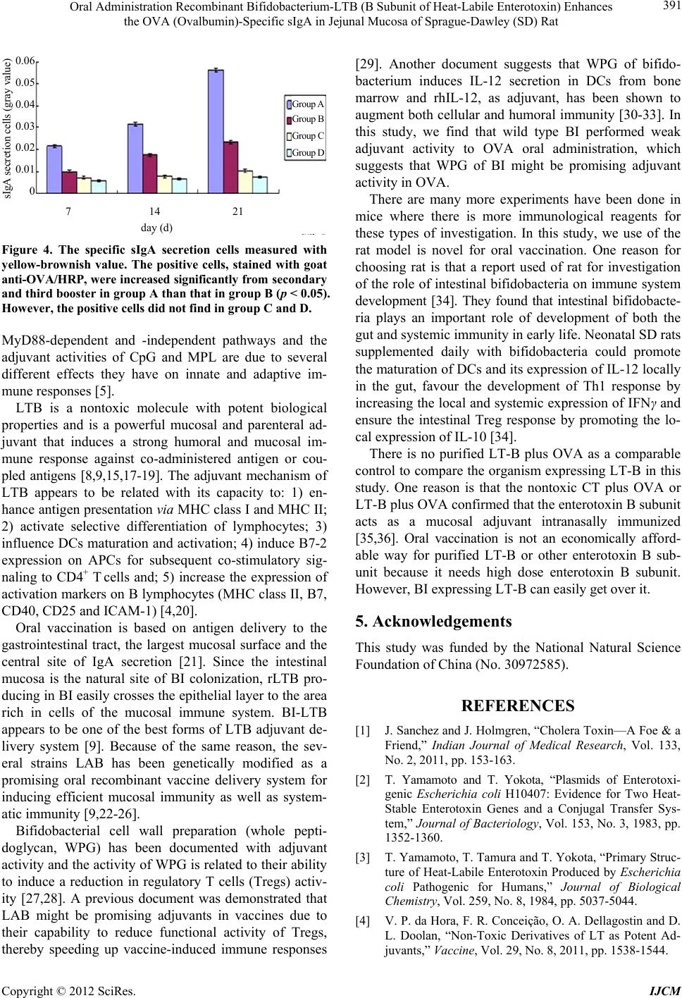 Oral Administration Recombinant Bifidobacterium-LTB (B Subunit of Heat-Labile Enterotoxin) Enhances the OVA (Ovalbumin)-Specific sIgA in Jejunal Mucosa of Sprague-Dawley (SD) Rat 391 7 14 21 day (d) 0.06 0.05 0.04 0.03 0.02 0.01 0 sIgA secretion cells (gray value) Group A Group B Group C Group D Figure 4. The specific sIgA secretion cells measured with yellow-brownish value. The positive cells, stained with goat anti-OVA/HRP, were increased significantly from sec ondar y and third booster in group A than that in group B ( p < 0.05). However, the positive cells did not find in group C and D. MyD88-dependent and -independent pathways and the adjuvant activities of CpG and MPL are due to several different effects they have on innate and adaptive im- mune responses [5]. LTB is a nontoxic molecule with potent biological properties and is a powerful mucosal and parenteral ad- juvant that induces a strong humoral and mucosal im- mune response against co-administered antigen or cou- pled antigens [8,9,15,17-19]. The adjuvant mechanism of LTB appears to be related with its capacity to: 1) en- hance antigen presentation via MHC class I and MHC II; 2) activate selective differentiation of lymphocytes; 3) influence DCs maturation and activation; 4) induce B7-2 expression on APCs for subsequent co-stimulatory sig- naling to CD4+ T cells and; 5) increase the expression of activation markers on B lymphocytes (MHC class II, B7, CD40, CD25 and ICAM-1) [4,20]. Oral vaccination is based on antigen delivery to the gastrointestinal tract, the largest mucosal surface and the central site of IgA secretion [21]. Since the intestinal mucosa is the natural site of BI colonization, rLTB pro- ducing in BI easily crosses the epithelial layer to the area rich in cells of the mucosal immune system. BI-LTB appears to be one of the best forms of LTB adjuvant de- livery system [9]. Because of the same reason, the sev- eral strains LAB has been genetically modified as a promising oral recombinant vaccine delivery system for inducing efficient mucosal immunity as well as system- atic immunity [9,22-26]. Bifidobacterial cell wall preparation (whole pepti- doglycan, WPG) has been documented with adjuvant activity and the activity of WPG is related to their ability to induce a reduction in regulatory T cells (Tregs) activ- ity [27,28]. A previous document was demonstrated that LAB might be promising adjuvants in vaccines due to their capability to reduce functional activity of Tregs, thereby speeding up vaccine-induced immune responses [29]. Another document suggests that WPG of bifido- bacterium induces IL-12 secretion in DCs from bone marrow and rhIL-12, as adjuvant, has been shown to augment both cellular and humoral immunity [30-33]. In this study, we find that wild type BI performed weak adjuvant activity to OVA oral administration, which suggests that WPG of BI might be promising adjuvant activity in OVA. There are many more experiments have been done in mice where there is more immunological reagents for these types of investigation. In this study, we use of the rat model is novel for oral vaccination. One reason for choosing rat is that a report used of rat for investigation of the role of intestinal bifidobacteria on immune system development [34]. They found that intestinal bifidobacte- ria plays an important role of development of both the gut and systemic immunity in early life. Neonatal SD rats supplemented daily with bifidobacteria could promote the maturation of DCs and its expression of IL-12 locally in the gut, favour the development of Th1 response by increasing the local and systemic expression of IFNγ and ensure the intestinal Treg response by promoting the lo- cal expression of IL-10 [34]. There is no purified LT-B plus OVA as a comparable control to compare the organism expressing LT-B in this study. One reason is that the nontoxic CT plus OVA or LT-B plus OVA confirmed that the enterotoxin B subunit acts as a mucosal adjuvant intranasally immunized [35,36]. Oral vaccination is not an economically afford- able way for purified LT-B or other enterotoxin B sub- unit because it needs high dose enterotoxin B subunit. However, BI expressing LT-B can easily get over it. 5. Acknowledgements This study was funded by the National Natural Science Foundation of China (No. 30972585). REFERENCES [1] J. Sanchez and J. Holmgren, “Cholera Toxin—A Foe & a Friend,” Indian Journal of Medical Research, Vol. 133, No. 2, 2011, pp. 153-163. [2] T. Yamamoto and T. Yokota, “Plasmids of Enterotoxi- genic Escherichia coli H10407: Evidence for Two Heat- Stable Enterotoxin Genes and a Conjugal Transfer Sys- tem,” Journal of Bacteriology, Vol. 153, No. 3, 1983, pp. 1352-1360. [3] T. Yamamoto, T. Tamura and T. Yokota, “Primary Struc- ture of Heat-Labile Enterotoxin Produced by Escherichia coli Pathogenic for Humans,” Journal of Biological Chemistry, Vol. 259, No. 8, 1984, pp. 5037-5044. [4] V. P. da Hora, F. R. Conceição, O. A. Dellagostin and D. L. Doolan, “Non-Toxic Derivatives of LT as Potent Ad- juvants,” Vaccine, Vol. 29, No. 8, 2011, pp. 1538-1544. Copyright © 2012 SciRes. IJCM 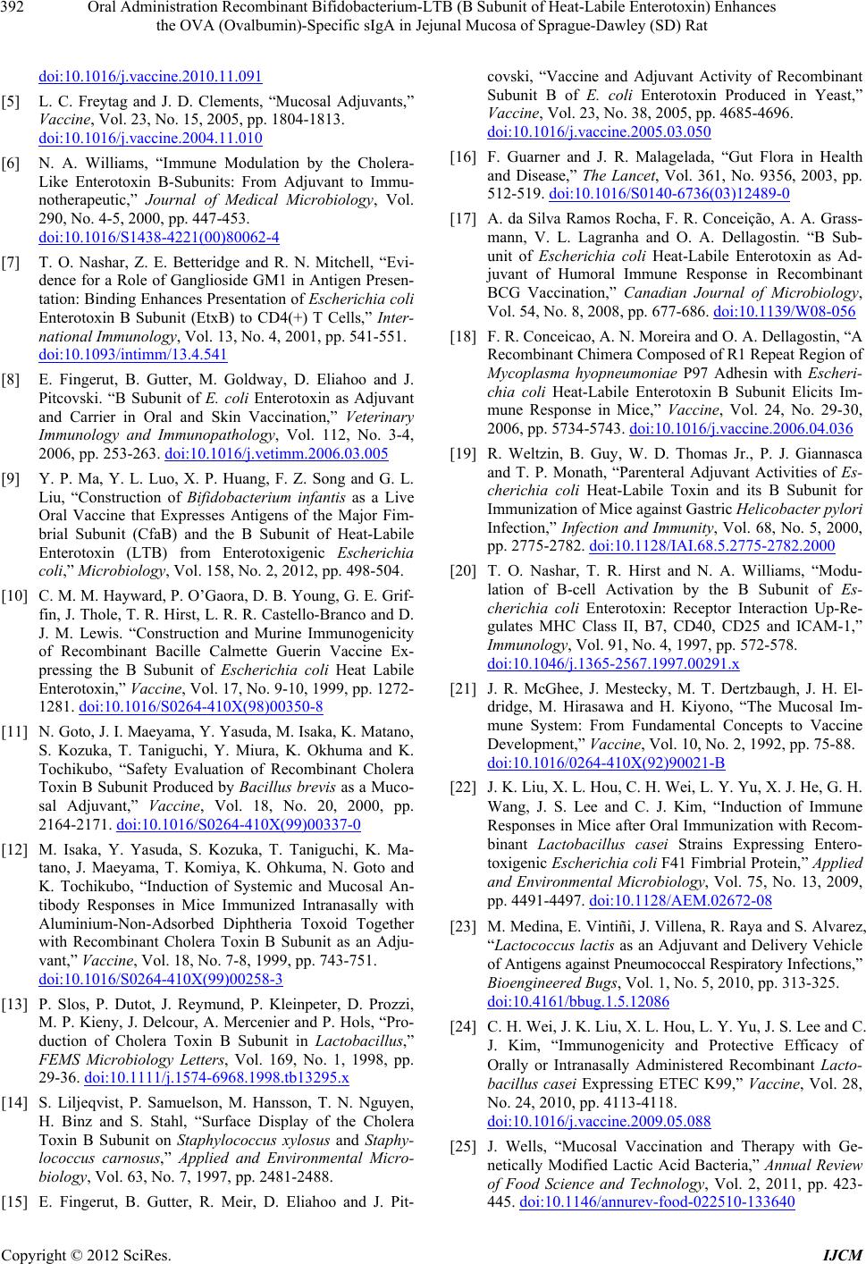 Oral Administration Recombinant Bifidobacterium-LTB (B Subunit of Heat-Labile Enterotoxin) Enhances the OVA (Ovalbumin)-Specific sIgA in Jejunal Mucosa of Sprague-Dawley (SD) Rat 392 doi:10.1016/j.vaccine.2010.11.091 [5] L. C. Freytag and J. D. Clements, “Mucosal Adjuvants,” Vaccine, Vol. 23, No. 15, 2005, pp. 1804-1813. doi:10.1016/j.vaccine.2004.11.010 [6] N. A. Williams, “Immune Modulation by the Cholera- Like Enterotoxin B-Subunits: From Adjuvant to Immu- notherapeutic,” Journal of Medical Microbiology, Vol. 290, No. 4-5, 2000, pp. 447-453. doi:10.1016/S1438-4221(00)80062-4 [7] T. O. Nashar, Z. E. Betteridge and R. N. Mitchell, “Evi- dence for a Role of Ganglioside GM1 in Antigen Presen- tation: Binding Enhances Presentation of Escherichia coli Enterotoxin B Subunit (EtxB) to CD4(+) T Cells,” Inter- national Immunology, Vol. 13, No. 4, 2001, pp. 541-551. doi:10.1093/intimm/13.4.541 [8] E. Fingerut, B. Gutter, M. Goldway, D. Eliahoo and J. Pitcovski. “B Subunit of E. coli Enterotoxin as Adjuvant and Carrier in Oral and Skin Vaccination,” Veterinary Immunology and Immunopathology, Vol. 112, No. 3-4, 2006, pp. 253-263. doi:10.1016/j.vetimm.2006.03.005 [9] Y. P. Ma, Y. L. Luo, X. P. Huang, F. Z. Song and G. L. Liu, “Construction of Bifidobacterium infantis as a Live Oral Vaccine that Expresses Antigens of the Major Fim- brial Subunit (CfaB) and the B Subunit of Heat-Labile Enterotoxin (LTB) from Enterotoxigenic Escherichia coli,” Microbiology, Vol. 158, No. 2, 2012, pp. 498-504. [10] C. M. M. Hayward, P. O’Gaora, D. B. Young, G. E. Grif- fin, J. Thole, T. R. Hirst, L. R. R. Castello-Branco and D. J. M. Lewis. “Construction and Murine Immunogenicity of Recombinant Bacille Calmette Guerin Vaccine Ex- pressing the B Subunit of Escherichia coli Heat Labile Enterotoxin,” Vaccine, Vol. 17, No. 9-10, 1999, pp. 1272- 1281. doi:10.1016/S0264-410X(98)00350-8 [11] N. Goto, J. I. Maeyama, Y. Yasuda, M. Isaka, K. Matano, S. Kozuka, T. Taniguchi, Y. Miura, K. Okhuma and K. Tochikubo, “Safety Evaluation of Recombinant Cholera Toxin B Subunit Produced by Bacillus brevis as a Muco- sal Adjuvant,” Vaccine, Vol. 18, No. 20, 2000, pp. 2164-2171. doi:10.1016/S0264-410X(99)00337-0 [12] M. Isaka, Y. Yasuda, S. Kozuka, T. Taniguchi, K. Ma- tano, J. Maeyama, T. Komiya, K. Ohkuma, N. Goto and K. Tochikubo, “Induction of Systemic and Mucosal An- tibody Responses in Mice Immunized Intranasally with Aluminium-Non-Adsorbed Diphtheria Toxoid Together with Recombinant Cholera Toxin B Subunit as an Adju- vant,” Vaccine, Vol. 18, No. 7-8, 1999, pp. 743-751. doi:10.1016/S0264-410X(99)00258-3 [13] P. Slos, P. Dutot, J. Reymund, P. Kleinpeter, D. Prozzi, M. P. Kieny, J. Delcour, A. Mercenier and P. Hols, “Pro- duction of Cholera Toxin B Subunit in Lactobacillus,” FEMS Microbiology Letters, Vol. 169, No. 1, 1998, pp. 29-36. doi:10.1111/j.1574-6968.1998.tb13295.x [14] S. Liljeqvist, P. Samuelson, M. Hansson, T. N. Nguyen, H. Binz and S. Stahl, “Surface Display of the Cholera Toxin B Subunit on Staphylococcus xylosus and Staphy- lococcus carnosus,” Applied and Environmental Micro- biology, Vol. 63, No. 7, 1997, pp. 2481-2488. [15] E. Fingerut, B. Gutter, R. Meir, D. Eliahoo and J. Pit- covski, “Vaccine and Adjuvant Activity of Recombinant Subunit B of E. coli Enterotoxin Produced in Yeast,” Vaccine, Vol. 23, No. 38, 2005, pp. 4685-4696. doi:10.1016/j.vaccine.2005.03.050 [16] F. Guarner and J. R. Malagelada, “Gut Flora in Health and Disease,” The Lancet, Vol. 361, No. 9356, 2003, pp. 512-519. doi:10.1016/S0140-6736(03)12489-0 [17] A. da Silva Ramos Rocha, F. R. Conceição, A. A. Grass- mann, V. L. Lagranha and O. A. Dellagostin. “B Sub- unit of Escherichia coli Heat-Labile Enterotoxin as Ad- juvant of Humoral Immune Response in Recombinant BCG Vaccination,” Canadian Journal of Microbiology, Vol. 54, No. 8, 2008, pp. 677-686. doi:10.1139/W08-056 [18] F. R. Conceicao, A. N. Moreira and O. A. Dellagostin, “A Recombinant Chimera Composed of R1 Repeat Region of Mycoplasma hyopneumoniae P97 Adhesin with Escheri- chia coli Heat-Labile Enterotoxin B Subunit Elicits Im- mune Response in Mice,” Vaccine, Vol. 24, No. 29-30, 2006, pp. 5734-5743. doi:10.1016/j.vaccine.2006.04.036 [19] R. Weltzin, B. Guy, W. D. Thomas Jr., P. J. Giannasca and T. P. Monath, “Parenteral Adjuvant Activities of Es- cherichia coli Heat-Labile Toxin and its B Subunit for Immunization of Mice against Gastric Helicobacter pylori Infection,” Infection and Immunity, Vol. 68, No. 5, 2000, pp. 2775-2782. doi:10.1128/IAI.68.5.2775-2782.2000 [20] T. O. Nashar, T. R. Hirst and N. A. Williams, “Modu- lation of B-cell Activation by the B Subunit of Es- cherichia coli Enterotoxin: Receptor Interaction Up-Re- gulates MHC Class II, B7, CD40, CD25 and ICAM-1,” Immunology, Vol. 91, No. 4, 1997, pp. 572-578. doi:10.1046/j.1365-2567.1997.00291.x [21] J. R. McGhee, J. Mestecky, M. T. Dertzbaugh, J. H. El- dridge, M. Hirasawa and H. Kiyono, “The Mucosal Im- mune System: From Fundamental Concepts to Vaccine Development,” Vaccine, Vol. 10, No. 2, 1992, pp. 75-88. doi:10.1016/0264-410X(92)90021-B [22] J. K. Liu, X. L. Hou, C. H. Wei, L. Y. Yu, X. J. He, G. H. Wang, J. S. Lee and C. J. Kim, “Induction of Immune Responses in Mice after Oral Immunization with Recom- binant Lactobacillus casei Strains Expressing Entero- toxigenic Escherichia coli F41 Fimbrial Protein,” Applied and Environmental Microbiology, Vol. 75, No. 13, 2009, pp. 4491-4497. doi:10.1128/AEM.02672-08 [23] M. Medina, E. Vintiñi, J. Villena, R. Raya and S. Alvarez, “Lactococcus lactis as an Adjuvant and Delivery Vehicle of Antigens against Pneumococcal Respiratory Infections,” Bioengineered Bugs, Vol. 1, No. 5, 2010, pp. 313-325. doi:10.4161/bbug.1.5.12086 [24] C. H. Wei, J. K. Liu, X. L. Hou, L. Y. Yu, J. S. Lee and C. J. Kim, “Immunogenicity and Protective Efficacy of Orally or Intranasally Administered Recombinant Lacto- bacillus casei Expressing ETEC K99,” Vaccine, Vol. 28, No. 24, 2010, pp. 4113-4118. doi:10.1016/j.vaccine.2009.05.088 [25] J. Wells, “Mucosal Vaccination and Therapy with Ge- netically Modified Lactic Acid Bacteria,” Annual Review of Food Science and Technology, Vol. 2, 2011, pp. 423- 445. doi:10.1146/annurev-food-022510-133640 Copyright © 2012 SciRes. IJCM  Oral Administration Recombinant Bifidobacterium-LTB (B Subunit of Heat-Labile Enterotoxin) Enhances the OVA (Ovalbumin)-Specific sIgA in Jejunal Mucosa of Sprague-Dawley (SD) Rat Copyright © 2012 SciRes. IJCM 393 [26] S. Yamamoto, J. Wada, T. Katayama, T. Jikimoto, M. Nakamura, S. Kinoshita, K. M. Lee, M. Kawabata and T. Shirakawa, “Genetically Modified Bifidobacterium Dis- playing Salmonella-Antigen Protects Mice from Lethal Challenge of Salmonella Typhimurium in a Murine Ty- phoid Fever Model,” Vaccine, Vol. 28, No. 41, 2010, pp. 6684-6691. doi:10.1016/j.vaccine.2010.08.007 [27] K. Sekine, E. Watanabe-Sekine, T. Toida, T. Kasashima, T. Kataoka and Y. Hashimoto, “Adjuvant Activity of the Cell Wall of Bifidobacterium infantis for in Vivo Immune Responses in Mice,” Immunopharmacol Immunotoxicol, Vol. 16, No. 4, 1994, pp. 589-609. doi:10.3109/08923979409019741 [28] E. G. Schmidt, M. H. Claesson, S. S. Jensen, P. Ravn and N. N. Kristensen, “Antigen-Presenting Cells Exposed to Lactobacillus acidophilus NCFM, Bifidobacterium bifidum BI-98, and BI-504 Reduce Regulatory T Cell Activity,” Inflammatory Bowel Diseases, Vol. 16, No. 3, 2010, pp. 390-400. [29] D. Paineau, D. Carcano, G. Leyer, S. Darquy, M. A. Al- yanakian, G. Simoneau, J. F. Bergmann, D. Brassart, F. Bornet and A. C. Ouwehand, “Effects of Seven Potential Probiotic Strains on Specific Immune Responses in Healthy Adults: A Double-Blind, Randomized, Con- trolled Trial,” FEMS Immunology and Medical Microbi- ology, Vol. 53, No. 1, 2008, pp.107-113. doi:10.1111/j.1574-695X.2008.00413.x [30] L. L. Zhao and Q. Cheng, “The Influence of Whole Pep- tidoglycan of Bifidobacterium on Interleukin-12 Produced by Dendritic Cells Derived from Bone Marrow of Mice in Vitro,” Chinese Journal of Microecology, Vol. 21, No. 10, 2009, pp. 896-901. [31] O. Hamid, J. C. Solomon, R. Scotland, M. Garcia, S. Sian, W. Ye, S. L. Groshen and J. S. Weber, “Alum with Inter- leukin-12 Augments Immunity to a Melanoma Peptide Vaccine: Correlation with Time to Relapse in Patients with Resected High-Risk Disease,” Clinical Cancer Re- search, Vol. 13, No. 1, 2007, pp. 215-222. doi:10.1158/1078-0432.CCR-06-1450 [32] M. A. Jacobson, E. Sinclair, B. Bredt, L. Agrillo, D. Black, C. L. Epling, A. Carvidi, T. Ho, R. Bains, V. Girling and S. P. Adler, “Safety and Immunogenicity of Towne Cytomegalovirus Vaccine with or without Adju- vant Recombinant Interleukin-12,” Vaccine, Vol. 24, No. 25, 2006, pp. 5311-5319. doi:10.1016/j.vaccine.2006.04.017 [33] A. K. Wright, I. Christopoulou, S. El Batrawy, J. Limer and S. B. Gordon, “rhIL-12 as Adjuvant Augments Lung Cell Cytokine Responses to Pneumococcal Whole Cell Antigen,” Immunobiology, Vol. 216, No. 10, 2011, pp. 1143-1147. [34] P. Dong, Y. Yang and W. P. Wang, “The Role of Intesti- nal Bifidobacteria on Immune System Development in Young Rats,” Early Human Development, Vol. 86, No. 1, 2010, pp. 51-58. doi:10.1016/j.earlhumdev.2010.01.002 [35] P. N. Boyaka, M. Ohmura, K. Fujihashi, T. Koga, M. Yamamoto, M. N. Kweon, Y. Takeda, R. J. Jackson, H. Kiyono, Y. Yuki and J. R. McGhee, “Chimeras of Labile Toxin One and Cholera Toxin Retain Mucosal Adjuvan- ticity and Direct Th Cell Subsets via Their B Subunit,” Journal of Immunology, Vol. 170, No. 1, 2003, pp. 454- 462. [36] S. Yamamoto, H. Kiyono, M. Yamamoto, K. Imaoka, K. Fujihashi, F. W. Van Ginkel, M. Noda, Y. Takeda and J. R. McGhee, “A Nontoxic Mutant of Cholera Toxin Elic- its Th2-Type Responses for Enhanced Mucosal Immu- nity,” Proceedings of the National Academy of Sciences USA, Vol. 94, No. 10, 1997, pp. 5267-5272. doi:10.1073/pnas.94.10.5267 |