

Paper Menu >>
Journal Menu >>
 International Journal of Clinical Medicine, 2012, 3, 431-432 http://dx.doi.org/10.4236/ijcm.2012.35080 Published Online September 2012 (http://www.SciRP.org/journal/ijcm) 1 Antenatal Son ographic Diagnosis of a Case of Alobar Holoprosencephaly: A Case Report Rizwan Ahmad Khan1, Manjari Thapa2, Shagufta Wahab2* 1Division of Pediatric Sugery, Department of Surgery, JN Medical College, Aligarh Muslim University, Aligarh, India; 2Department of Radiodiagnosis, JN Medical College, Aligarh Muslim University, Aligarh, India. Email: *[email protected] Received May 2nd, 2012; revised June 6th, 2012; accepted July 19th, 2012 ABSTRACT Holoprosencephaly is a rare congenital brain malformation resulting from failure of diverticulation and cleavage of primitive prosencephalon which occurs at 4th - 8th week of gestation and is usually associated with multiple midline facial anomalies. Herein we report an antenatal case of such patient. Patient was evaluated and because of the magni- tude of the problem induction was done which led to a still born baby. Keywords: Holoprosencephaly; Alob ar; Antenatal Detection; Developing Countries; Prognosis 1. Introduction Holoprosencephaly is a rare congenital brain malforma- tion resulting fro m failure of diverticulation and cleavage of primitive prosencephalon which occurs at 4th - 8th week of gestation and is usually associated with multiple midline facial anomalies. This disorder is either incom- patible with life or infants suffer from varying grade of mental retardation. Sonography is an excellent non-invasive tool for pre- natal diagnosis of holoprosencephaly. This case report describes a case of alobar holoprosencephaly diagnosed prenatally with ultrasound and highlights the importance of high-resolution real time imaging. We want to stress that the conditio n is rare and therefore lack of familiarity may lead to diagnostic confusion. 2. Case Report A 24-year-old, gravida 2 and para 1 underwent antenatal ultrasonography at 24 menstrual weeks as determined by dates, which disclosed increased intracranial fluid in the fetus. Past medical history and family history were un- remarkable, with the previous child being born at term with no congenital anomaly. A repeat ultrasound exami- nation demonstrated a monoventricle fluid-filled cavity. The cerebral cortex was represented by thinned out man- tle. Two orbits were seen; no clear evidence of proboscis was seen. The midbrain and posterior fossa appeared normal (Figures 1). An increased amount of amniotic fluid (polyhydramnios) was present. Based on the ante- natal sonographic findings a diagnosis of alobar holo- prosencephaly was made. Preterm induction of vaginal delivery resulted in a stillborn fetus. Examination of the fetus demonstrated only proptosis but no gross facial anomaly. Brain morphology could not be assessed due to autolysis. Chromosomal analysis disclosed a trisomy 13 karyotype. Patient was investigated as per the guidelines set by the Ethics committee of our hospital. 3. Discussion The incidence of holoprosencephaly is approximately 1 - 1.4 per 10,000 live births [1] but as the incidence of Figure 1. Antenatal sonogram of fetal head (coronal view) with alobar holoprosencephaly showing monoventricular cavity with surrounding thinned cerebral tissue, fused tha- lami and absence of midline structures such as falx cerebri. Axial view of the same patient showing fused thalami and thinned out cortical mantle of the fetal head. *Corresponding a uthor. Copyright © 2012 SciRes. IJCM 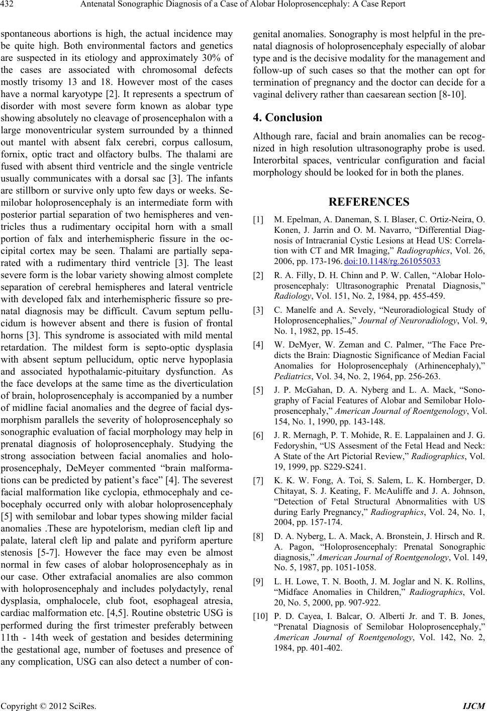 Antenatal Sonographic Diagnosis of a Case of Alobar Holoprosencephaly: A Case Report 432 spontaneous abortions is high, the actual incidence may be quite high. Both environmental factors and genetics are suspected in its etiology and approximately 30% of the cases are associated with chromosomal defects mostly trisomy 13 and 18. However most of the cases have a normal karyotype [2]. It represents a spectrum of disorder with most severe form known as alobar type showing absolutely no cleavage of prosencephalon with a large monoventricular system surrounded by a thinned out mantel with absent falx cerebri, corpus callosum, fornix, optic tract and olfactory bulbs. The thalami are fused with absent third ventricle and the single ventricle usually communicates with a dorsal sac [3]. The infants are stillborn or survive only upto few d ays or weeks. Se- milobar holoprosencephaly is an intermediate form with posterior partial separation of two hemispheres and ven- tricles thus a rudimentary occipital horn with a small portion of falx and interhemispheric fissure in the oc- cipital cortex may be seen. Thalami are partially sepa- rated with a rudimentary third ventricle [3]. The least severe form is the lobar variety showing almost complete separation of cerebral hemispheres and lateral ventricle with developed falx and interhemispheric fissure so pre- natal diagnosis may be difficult. Cavum septum pellu- cidum is however absent and there is fusion of frontal horns [3]. This syndrome is associated with mild mental retardation. The mildest form is septo-optic dysplasia with absent septum pellucidum, optic nerve hypoplasia and associated hypothalamic-pituitary dysfunction. As the face develops at the same time as the diverticulation of brain, holoprosencephaly is accompanied by a number of midline facial anomalies and the degree of facial dys- morphism parallels the severity of holoprosencephaly so sonographic evaluatio n of facial morphology may he lp in prenatal diagnosis of holoprosencephaly. Studying the strong association between facial anomalies and holo- prosencephaly, DeMeyer commented “brain malforma- tions can be predicted by patient’s face” [4]. The severest facial malformation like cyclopia, ethmocephaly and ce- bocephaly occurred only with alobar holoprosencephaly [5] with semilobar and lobar types showing milder facial anomalies .These are hypotelorism, median cleft lip and palate, lateral cleft lip and palate and pyriform aperture stenosis [5-7]. However the face may even be almost normal in few cases of alobar holoprosencephaly as in our case. Other extrafacial anomalies are also common with holoprosencephaly and includes polydactyly, renal dysplasia, omphalocele, club foot, esophageal atresia, cardiac malformation etc. [4,5]. Routine obstetric USG is performed during the first trimester preferably between 11th - 14th week of gestation and besides determining the gestational age, number of foetuses and presence of any complication, USG can also detect a number of con- genital ano malies. Sonography is most helpful in th e pre- natal diagnosis of holoprosencephaly especially of alobar type and is the decisive modality for the management and follow-up of such cases so that the mother can opt for termination of pregnancy and the doctor can decide for a vaginal delivery rather than caesarean section [8-10]. 4. Conclusion Although rare, facial and brain anomalies can be recog- nized in high resolution ultrasonography probe is used. Interorbital spaces, ventricular configuration and facial morphology should be looked for in both the planes. REFERENCES [1] M. Epelman, A. Daneman, S. I. Blaser, C. Ortiz-Neira, O. Konen, J. Jarrin and O. M. Navarro, “Differential Diag- nosis of Intracranial Cystic Lesions at Head US: Correla- tion with CT and MR Imaging,” Radiographics, Vol. 26, 2006, pp. 173-196. doi:10.1148/rg.261055033 [2] R. A. Filly, D. H. Chinn and P. W. Callen, “Alobar Holo- prosencephaly: Ultrasonographic Prenatal Diagnosis,” Radiology, Vol. 151, No. 2, 1984, pp. 455-459. [3] C. Manelfe and A. Sevely, “Neuroradiological Study of Holoprosencephalies,” Journal of Neuroradiology, Vol. 9, No. 1, 1982, pp. 15-45. [4] W. DeMyer, W. Zeman and C. Palmer, “The Face Pre- dicts the Brain: Diagnostic Significance of Median Facial Anomalies for Holoprosencephaly (Arhinencephaly),” Pediatrics, Vol. 34, No. 2, 1964, pp. 256-263. [5] J. P. McGahan, D. A. Nyberg and L. A. Mack, “Sono- graphy of Facial Features of Alobar and Semilobar Holo- prosencephaly,” American Journal of Roentgenology, Vol. 154, No. 1, 1990, pp. 143-148. [6] J. R. Mernagh, P. T. Mohide, R. E. Lappalainen and J. G. Fedoryshin, “US Assesment of the Fetal Head and Neck: A State of the Art Pictorial Review,” Radiographics, Vol. 19, 1999, pp. S229-S241. [7] K. K. W. Fong, A. Toi, S. Salem, L. K. Hornberger, D. Chitayat, S. J. Keating, F. McAuliffe and J. A. Johnson, “Detection of Fetal Structural Abnormalities with US during Early Pregnancy,” Radiographics, Vol. 24, No. 1, 2004, pp. 157-174. [8] D. A. Nyberg, L. A. Mack, A. Bronstein, J. Hirsch and R. A. Pagon, “Holoprosencephaly: Prenatal Sonographic diagnosis,” American Journal of Roentgenology, Vol. 149, No. 5, 1987, pp. 1051-1058. [9] L. H. Lowe, T. N. Booth, J. M. Joglar and N. K. Rollins, “Midface Anomalies in Children,” Radiographics, Vol. 20, No. 5, 2000, pp. 907-922. [10] P. D. Cayea, I. Balcar, O. Alberti Jr. and T. B. Jones, “Prenatal Diagnosis of Semilobar Holoprosencephaly,” American Journal of Roentgenology, Vol. 142, No. 2, 1984, pp. 401-402. Copyright © 2012 SciRes. IJCM |