

Paper Menu >>
Journal Menu >>
 Neuroscience & Medicine, 2012, 3, 321-326 http://dx.doi.org/10.4236/nm.2012.33037 Published Online September 2012 (http://www.SciRP.org/journal/nm) 321 Human Embryo Neuronal Culture in Vitro: A Model to Study Cellular Physiology, Receptors, Power and Toxicity of Cytostatic Drugs for Human Use Stabile Mariano1, Monaco Roberto2, Iuorio Tina1, Buoninfante Luca3, Marino Lucia1, Altieri Vincenzo4, Della Ragione Carlo2, Masillo Francesco3 1ZigoteGenetic and Prenatal Diagnosis Centre, Salerno, Italy; 2Anatomy Pathology Department, Hospital “A. Cardarelli”, Naples, Italy; 3Department of Gynecology and Obstetrics, Hospital “Maria SS Addolorata”, Eboli, Italy; 4Genetic Department, Hospital “Elenad’ Aosta”, Naples, Italy. Email: [email protected] Received May 14th, 2012; revised June 14th, 2012; accepted June 22nd, 2012 ABSTRACT Neural cells cultures from human embryo brain of 9˚ - 11˚W gestational age have been used to study ERα (Estrogens Receptor α) and to perform toxicity test for Mitomycin C and Methotrexate. Histochemical confirmation of cellular neuronal phenotype was based on histochemical evidence of NSE (Neuron Specific Enolase).The detection of ERα in neuronal cells was perfo rmed with a rabbit Monoclonal Antibody. ERα was absen t both on neurons grown in vitro an d on tissue brain specimens. This finding is apparently in contrast with the positive immunoreactivity of ERα and ERβ reported by other Authors on foetal and adult CNS (Central Nervous Sys tem). The absence of nuclear ERα on neurons in culture and in brain tissue specimens in our expe riment is not in contrast with the relevant physiologic role of estro- gens on nervous central system, but it could be correlated to the embryonic period of life and could represent a protec- tion of male brain from an undue estrogens imprinting. The mitomycin C, alkylation agent, has shown in our experi- ment a major neurotoxic and cytostatic power in comparison with methotrexate. Our conclusion is that human embryo neuronal culture in vitro is a powerful instrument for physiology and human therapy for cancer and neurodegenerative diseases. Keywords: Human Embryo Neuronal Culture; ERα on Embryonic Brain; Mitomycin C Toxicity Test in Vitro; Methotrexat e To xi ci t y Test in Vitro 1. Introduction Neural cells culture has been successfully accomplished from neural precursors from marine embryonic stem cell [1] and from human ones [2]. The aim o f the above me ntioned r esearch es was to ob- tain neural progenitors fit to integrate in vivo into a host tissue in order to replace damaged or absent neurons [3-6]. Our in vitro mod el, that is the culture of well differen- tiated neurons and not stem cells, from human embryo brain, is important for the research on hormones, drugs, neuromediators receptors with the aim to understand their role in morphogenesis and a potential therapeutic use or neuroto xi c i t y . In addition, an original con tribution of our paper is the study of estrogens receptor ERα on neurons in culture and toxicity test for Mitomycin C and Methotrexate on neurons in vitro. 2. Materials and Methods 2.1. Embryos Collection We have obtained the positive judgement by the ethic committee (Pro t. n . 47 519 /I3D7 A sl Sa 2 the 25 .0 8.2005) for use of embryonic material from voluntary abortion. The gestational age of the pregnant women were be- tween 9˚ - 11˚W; an expert pathologist was in operating room in order to collect the embryo just delivered, enu- cleate brain and spinal medulla and, after washing, place the material in medium culture. Fragmented embryos were not selected for experiments. 2.2. Neurons in Vitro Culture Brain and spinal medulla were gently cutted in a Petri dish, put in a sterile solution of Collagen ase Type I 176.0 units/mg (Gibco) at a concentration of 1 mg/100 ml. Af- ter 2 hours of incubation at 37˚C in atmosphere of CO2 Copyright © 2012 SciRes. NM 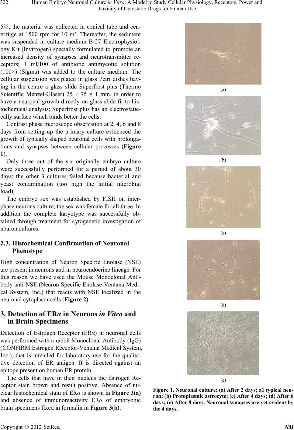 Human Embryo Neuronal Culture in Vitro: A Model to Study Cellular Physiology, Receptors, Power and Toxicity of Cytostatic Drugs for Human Use 322 5%, the material was collected in conical tube and cen- trifuge at 1500 rpm for 10 m’. Thereafter, the sediment was suspended in culture medium B-27 Electrophysiol- ogy Kit (Invitrogen) specially formulated to promote an increased density of synapses and neurotransmitter re- ceptors; 1 ml/100 of antibiotic antimycotic solution (100×) (Sigma) was added to the culture medium. The cellular suspension was plated in glass Petri dishes hav- ing in the centre a glass slide Superfrost plus (Thermo Scientific Menzel-Glaser) 25 × 75 × 1 mm, in order to have a neuronal growth directly on glass slide fit to his- tochemical analysis; Superfrost plus has an electrostatic- cally surface which binds better the cells. Contrast phase microscope observation at 2, 4, 6 and 8 days from setting up the primary culture evidenced the growth of typically shaped neuronal cells with prolonga- tions and synapses between cellular processes (Figure 1). Only three out of the six originally embryo culture were successfully performed for a period of about 30 days; the other 3 cultures failed because bacterial and yeast contamination (too high the initial microbial load). The embryo sex was established by FISH on inter- phase neurons culture; the sex was female for all three. In addition the complete karyotype was successfully ob- tained through treatment for cytogenetic investigation of neuron cultures. 2.3. Histochemical Confirmation of Neuronal Phenotype High concentration of Neuron Specific Enolase (NSE) are present in neurons and in neuroendocrine lineage. For this reason we have used the Mouse Monoclonal Anti- body anti-NSE (Neuron Specific Enolase-Ventana Medi- cal System, Inc.) that reacts with NSE localized in the neuronal cytoplasm cells (Figure 2). 3. Detection of ERα in Neurons in Vitro and in Brain Specimens Detection of Estrogen Receptor (ERα) in neuronal cells was performed with a rabbit Mo noclonal Antibod y (IgG) (CONFIRM Estrogen Receptor-Ventana Medical System, Inc.), that is intended for laboratory use for the qualita- tive detection of ER antigen. It is directed against an epitope present on human ER protein. The cells that have in their nucleus the Estrogen Re- ceptor stain brown and result positive. Absence of nu- clear histochemical stain of ERα is shown in Figure 3(a) and absence of immunoreactivity ERα of embryonic brain specimens fixed in formalin in Figure 3 (b). (a) (b) (c) (d) (e) Figure 1. Neuronal culture: (a) After 2 days; a1 typical neu- ron; (b) Protoplasmic astrocyte; (c) After 4 days; (d) After 6 days; (e) After 8 days. Neur onal synapses are yet evident by the 4 days. Copyright © 2012 SciRes. NM  Human Embryo Neuronal Culture in Vitro: A Model to Study Cellular Physiology, Receptors, Power and Toxicity of Cytostatic Drugs for Human Use 323 Figure 2. Immunohistochemical positivity for NSE (Neu- ronal Specific Enolase) of the neuron grown on glass slide: staining with anti-NSE antibody is uniformly intense on the cytosol of the cells confirming the neur onal phenotype. 4. Toxicity Test in Vitro for Two Antineoplastic Drugs: Mitomycin C and Methotrexate The toxicity of the two drugs has been evaluated on two distinct subcultures in flasks from a primary culture of embryonic neurons. Both has been added to the culture medium with a fi- nal concentration of 50 µg/ml, comparable to blood con- centrations used in human cancer therapy [7]. After 48 hours from the administration of Mitomycin C (Mitomycin C 10 mg, Kyowa Pharmaceuticals), the culture showed clear signs of cellular suffering with most of the cells in suspension (Figure 4). Instead, the culture with Methotrexate (Metotrexate 5 mg, Lederle Pharmaceuticals) did not show, after 48 hours, any sign of cytopathy or detachment from the sur- face monolayer and proliferation was non apparently inhibited (Figure 5). (a) (b) Figure 3. Histochemical stain of ERα with a rabbit Mono- clonal Antibody (IgG) (CONFIRM Estrogen Receptor- Ventana Medical System, Inc.): (a) Absence of brown col- oration indicating absence of ERα protein, in neurons in vitro; (b) In brain tissue. 5. Discussion Human embryonic neurons from spinal cord in culture have been obtained by Kato A. C. et al. 1985 [8]: bio- chemical studies demonstrate a prevalence of cholinergic and GABAergic neurons. Sah D. W. 1995 [9] has studied voltage and ligand- gated currents in human foetal central neurons in culture. Human embryonic neuronal cultures have been estab- lished in our laboratory without particular difficulty. The more critical step of the procedure is the collection of material: an expert operator must be present in the su rgi- cal room and has to select embryonic material immedi- ately after the expulsion. A further careful micro-dissect- tion of the material has been performed in laboratory; it is better to initiate the cultu re with a redu ced quantity (5 - 10 mg) of embryonic nervous tissue material instead of a contamination with other cellular type. More than 90 % of Copyright © 2012 SciRes. NM  Human Embryo Neuronal Culture in Vitro: A Model to Study Cellular Physiology, Receptors, Power and Toxicity of Cytostatic Drugs for Human Use 324 Figure 4. Mitomycin C added neuronal culture: reduction of cellular prolongations and many cells are in suspension was yet evident at 2 days from subc ultur es. Figure 5. Me totrexate added neuronal culture: no cytotoxic effect of metotrexate was apparent at 2, 4, 6 days from subculture. cells in vitro in our experiment are neurons, astrocytes, oligodendrocytes and Neural Stem Cell on the basis of morphological aspect and histochemical study. A protective rule of estrogens in neurodegenerative diseases has been postulated [10,11]. The two types of Estrogens Receptors (ERs), ERα and ERβ, belong to the nuclear receptor superfamily, a family of ligand-regulated transcription factor. Both receptors are coexpressed in a number of tissue and form homo- dimers (α/α, β/β) and heterodimers (α/β) [12]. When co- expressed, ERβ inhibits the ERα mediated gene expres- sion. In mice ERα is expressed primarily in the uterus, liver, kidney, and heart, whereas ERβ is expressed pri- marily in the ovary, prostate, lung, gastrointestinal tract, bladder, hematopoietic and central nervous systems [13-15]. Cells containing immunoreactive estrogen re- ceptor-alpha have been found in the human basal fore- brain [16,17]. Differential Expression of Estrogen Re- ceptor α and β immunoreactivity has been found in the Human Supraoptic Nucleus (dl-SON) in Relation to Sex and Aging [18]. Significant correlations between the percentage of ERβ- and ERα-positive and -negative AVP (Plasma Arginine Vasopressin) neurons and age were found in women, but not in men: a strong decrease of ER β and an in crease of ERα immunoreactivity in AVP neu- rons of the dl-SON of postmenopausal women. Both re- ceptor changes could participate in the activation of the AVP neurons in postmenopausal wome n. Estrogen receptors localization in the human spinal trigeminal nucleus has been evidenced by Fenzi e Rizzuto [19]. Although ER subtypes may be expressed in the same tissue, they may not be expressed in the same cell type. Nonetheless, ERα and ERβ proteins have been simulta- neously detected in many cell types including neurons. The absence of nuclear ERα on neurons in culture and in brain tissue specimens in our experiment is not in con- trast with the relevant physiologic role of estrogens on nervous central system, but it could be correlated to the embryonic period of life and could represent a protection of male brain from an undue estrogens imprinting. A role of progesterone in inducing human embryonic stem cell proliferation and differentiation into neuroectodermal rosettes has been established [20].Considering that the two hormones, estrogens and progesterone, often have an antagonistic effect, the absence of ERα receptor in em- bryonic brain could have its significance in non-antago- nize the morphogenetic effect of progesterone. Mitomycin C and metotrexate are antineoplastic drugs present in many polichemiotherapic schedules, including brain cancer. The mitomycin C, alkylating agent, has shown in our experiment a major neurotoxic and cytostatic power in comparison with methotrexate. Metotrexate, competitive antagonist of the folic acid on catalytic site of the dihydrofolate reductase (DHFR), has an action which can be weakened by three factors: the reduction of polyglutamate inside the cell, increase of DHFR by genetic amplification, reduction of intracellular transport [7]. Therefore the apparent absence of neuro- toxic effect in our model need a more complex experi- mental design to be explained. Nevertheless, the thera- peutic index of Metotrexate is better than Mitomycin C on the basis of our experi mental findi n g. Figure 6. Metaphase with GTL banding from human neu- rons in culture. Copyright © 2012 SciRes. NM  Human Embryo Neuronal Culture in Vitro: A Model to Study Cellular Physiology, Receptors, Power and Toxicity of Cytostatic Drugs for Human Use 325 The cytogenetic investigation has easily been per- formed on our human embryonic neurons (Figure 6); the analysis of chromosomal fragility and SCE (Sister Chromatide Exchange) is another powerful method to test the clastogenic activity of many substances for hu- man therapy [21,22]. REFERENCES [1] H. W. Li, H. Liu, C. E. Corrales, J. R. Risner, J. Forrester, J. R. Holt, S. Heller and A. S. B. Edge, “Differentiation of neurons from Neural Precursors Generated in Floating Spheres from Embryonic Stem Cells,” BMC Neurosci- ence, Vol. 10, 2009, p. 122. doi:10.1186/1471-2202-10-122 [2] M. Schuldiner, O. Yanuka, J. Itskovitz-Eldor, D. A. Mel- ton and N. Benvenisty, “Effects of Eight Growth Factors on the Differentiation of Cells Derived from Human Em- bryonic Stem Cells,” Proceedings of the National Acad- emy of Sciences of USA, Vol. 97, No. 21, 2000, pp. 11307- 11312. doi:10.1073/pnas.97.21.11307 [3] U. S. Sohur, J. G. Emsley, B. D. Mitchell and J. D. Mack- lis, “Adult Neurogenesis and Cellular Brain Repair with Neural Progenitors, Precursors and Stem Cells,” Phi- losophical Transactions of the Royal Society B: Biologi- cal Sciences, Vol. 361, No. 1473, 2006, pp. 1477-1497. doi:10.1098/rstb.2006.1887 [4] C. E. Corrales, L. Pan, H. W. Li, M. C. Liberman, S. Heller and A. S. B. Edge, “Engraftment and Differentia- tion of Embryonic Stem Cell-Derived Neural Progenitor Cells in the Cochlear Nerve Trunk: Growth of Processes into the Organ of Corti,” Journal of Neurobiology, Vol. 66, No. 13, 2006, pp. 1489-1500. doi:10.1002/neu.20310 [5] M. Emgard, L. Holmberg, E. B. Samuelsson, B. A. Bahr, S. Falci, A. Seiger and E. Sundström, “Human Neural Precursor Cells Continue to Proliferate and Exhibit Low Cell Death after Transplantation to the Injured Rat Spinal Cord,” Brain Research, Vol. 1278, 2009, pp. 15-26. doi:10.1016/j.brainres.2009.04.012 [6] Y.-K. Chang, M.-H. Chen, Y.-H. Chiang, Y.-F. Chen, W.-H. Ma, C.-Y. Tseng, B.-W. Soong, J. H. Ho and O. K. Lee, “Mesenchymal Stem Cell Transplantation Amelio- ratesmotor Function Deterioration of Spinocerebellara- taxia by Rescuing Cerebellar Purkinje Cells,” Journal of Biomedical Science, Vol. 18, 2011, p. 54. doi:10.1186/1423-0127-18-54 [7] E. Chu and A. Sartorelli, “Antineoplastic Chemotherapy,” In: Katzung Basic & Clinical Pharmacology, 9th Edition, The McGraw-Hill Companies, Inc., 2004, pp. 898-932. [8] A. C. Kato, G. Touzeau, D. Bertrand and C. R. Bader, “Human Spinal Cord Neurons in Dissociated Monolayer Cultures: Morphological, Biochemical, and Electro- phy- siological Properties,” The Journal of Neuroscience Co- pyright Society for Neuroscience, Vol. 5, No. 10, 1985, pp. 2750-2761. [9] D. W. Sah, “Human Fetal Central Neurons in Culture: Voltage- and Ligand-Gated Currents,” Journal of Neuro- physiology, Vol. 74, No. 5, 1995, pp. 1889-1899. [10] L. Q. Zhao and R. D. Brinton, “Select Estrogens within the Complex Formulation of Conjugated Equine Estro- gens (Premarin®) Are Protective against Neurodegenera- tive Insults: Implications for a Composition of Estrogen Therapy to Promote Neuronal Function and Prevent Alz- heimer’s Disease,” BMC Neuroscience, Vol. 7, 2006, p. 24. doi:10.1186/1471-2202-7-24 [11] G. E. Gillies and S. McArthur, “Estrogen Actions in the Brain and the Basis for Differential Action in Men and Women: A Case for Sex-Specific Medicines,” Pharma- cological Reviews, Vol. 62, No. 2, 2010, pp. 155-198. doi:10.1124/pr.109.002071 [12] J. Matthews and J. A. Gustafsson, “Estrogen Signaling: A Subtle Balance between ER Alpha and ER Beta,” Mo- lecular Interventions, Vol. 3, No. 5, 2003, pp. 281-292. [13] Z. Sheng, J. Kawano, A. Yanai, R. Fujinaga, M. Tanaka, Y. Watanabe and K. Shinoda, “Expression of Estrogen Receptors (Alpha, Beta) and Androgen Receptor in Sero- tonin Neurons of the Rat and Mouse Dorsal Raphe Nuclei; Sex and Species Differences,” Neuroscience Research, Vol. 49, No. 2, 2004, pp. 185-196. [14] R. J. Handa, S. Ogawa, J. M. Wang and A. E. Herbison, “Roles for Estrogen Receptor Beta in Adult Brain Func- tion,” Journal of Neurobiology, Vol. 24, 2011, pp. 160- 173. [15] B. Greco, E. A. Allegretto, M. J. Tetel and J. D. Blaustein, “Coexpression of ER_ with ER and Progestin Receptor Proteins in the Female Rat Forebrain: Effects of Estradiol Treatment,” Endocrinology, Vol. 142, 2001, pp. 5172- 5181. doi:10.1210/en.142.12.5172 [16] J. E. Donahue, E. G. Stopa, R. L. Chorsky, J. C. King, H. M. Schipper, S. A. Tobet, J. D. Blaustein and S. Reichlin, “Cells Containing Immunoreactive Estrogen Receptor- Alpha in the Human Basal Forebrain,” Brain Researvh, Vol. 856, No. 1-2, 2000, pp. 142-151. doi:10.1016/S0006-8993(99)02413-0 [17] D. Montague, C. S. Weickert, E. Tomaskovic-Crook, D. A. Rothmond, J. E. Kleinman and D. R. Rubinow, “Oes- trogen Receptor Alpha Localisation in the Prefrontal Cortex of Three Mammalian Species,”Journal of Neuro- endocrinology, Vol. 20, No. 7, 2008, pp. 893-903. doi:10.1111/j.1365-2826.2008.01743.x [18] T. A. Ishunina, F. P. Kruijver, R. Balesar and D. F. Swaab, “Differential Expression of Estrogen Receptor Alpha and Beta Immunoreactivity in the Human Supra- optic Nucleus in Relation to Sex and Aging,” Journal of Clinical Endocrinology and Metabolism, Vol. 85, No. 9, 2000, pp. 3283-3291. doi:10.1210/jc.85.9.3283 [19] F. F. N. Rizzzuto, “Estrogen Receptors Localization in the Spinal Trigeminal Nucleus: An Immunohistochemical Study in Humans,” European Journal of Pain, Vol. 15, No. 10, 2011, pp. 1002-1007. [20] M. J. Gallego, P. Porayette, M. M. Kaltcheva, R. L. Bo- wen, S. V. Meethal and C. S. Atwood, “The Pregnancy Hormones Human Chorionic Gonadotropin and Proges- terone Induce Human Embryonic Stem Cell Proliferation and Differentiation into Neuroectodermal Rosettes,” Stem Copyright © 2012 SciRes. NM  Human Embryo Neuronal Culture in Vitro: A Model to Study Cellular Physiology, Receptors, Power and Toxicity of Cytostatic Drugs for Human Use Copyright © 2012 SciRes. NM 326 Cell Research & Therapy, Vol. 1, 2010, p. 28. [21] D. Jacobson-Kram, R. J. Albertini, R. F. Branda, M. T. Falta, P. T. Iype, K. Kolodner, S.-H. Liou, M. A. Mc- Diarmid, M. Morris, J. A. Nicklas, J. P. O’Neill, M. C. Poirier, D. Putman, P. T. Strickland, J. R. Williams and S. Xiao, “Measurement of Chromosomal Aberrations, Sister Chromatid Exchange, Hprt Mutations, and DNA Adducts in Peripheral Lymphocytes of Human Populations at In- creased Risk for Cancer,” Environmental Health Perspec- tives, Vol. 101, No. 3, 1993, pp. 121-125. [22] K. Siviková, J. Dianovsky and B. Holecková, “Induction of SCEs and DNA Fragmentation in Bovine Peripheral Lymphocytes by in Vitro Exposure to Tolylfluanid-Based Fungicide,” Genetics and Molecular Biology, Vol. 34, No. 1, 2011, pp. 110-115. doi:10.1590/S1415-47572010005000097 |