

Paper Menu >>
Journal Menu >>
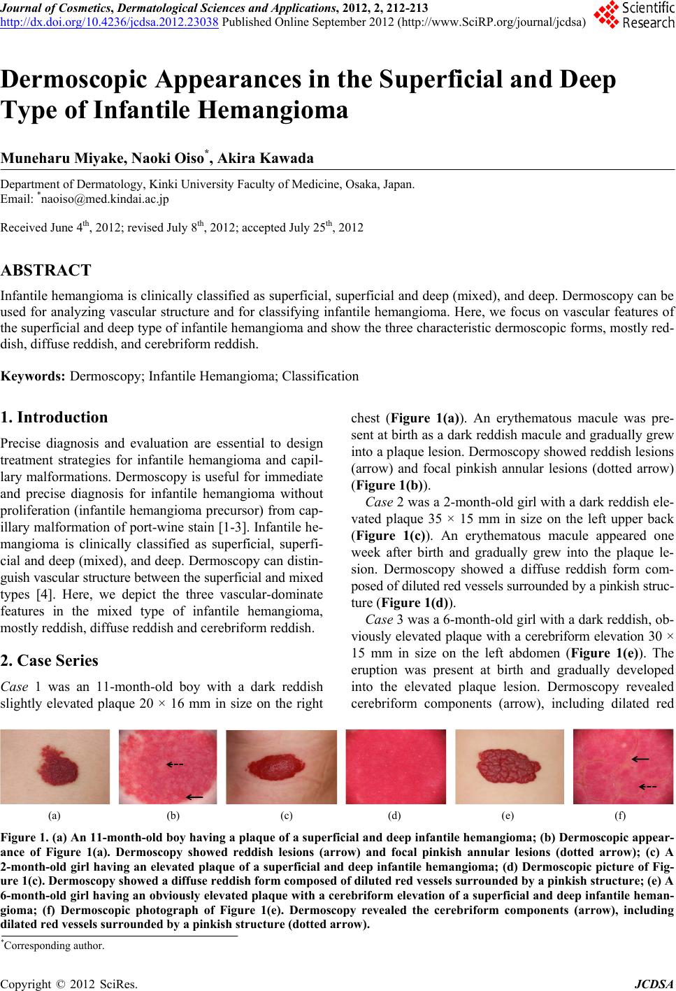 Journal of Cosmetics, Dermatological Sciences and Applications, 2012, 2, 212-213 http://dx.doi.org/10.4236/jcdsa.2012.23038 Published Online September 2012 (http://www.SciRP.org/journal/jcdsa) Dermoscopic Appearances in the Superficial and Deep Type of Infantile Hemangioma Muneharu Miyake, Naoki Oiso*, Akira Kawada Department of Dermatology, Kinki University Faculty of Medicine, Osaka, Japan. Email: *[email protected] Received June 4th, 2012; revised July 8th, 2012; accepted July 25th, 2012 ABSTRACT Infantile hemangioma is clinically classified as superficial, superficial and deep (mixed), and deep. Dermoscopy can be used for analyzing vascular structure and for classifying infantile hemangioma. Here, we focus on vascular features of the superficial and deep type of infantile hemangioma and show the three characteristic dermoscopic forms, mostly red- dish, diffuse reddish, and cerebriform reddish. Keywords: Dermoscopy; Infantile Hemangioma; Classification 1. Introduction Precise diagnosis and evaluation are essential to design treatment strategies for infantile hemangioma and capil- lary malformations. Dermoscopy is useful for immediate and precise diagnosis for infantile hemangioma without proliferation (infantile hemangioma precursor) from cap- illary malformation of port-wine stain [1-3]. Infantile he- mangioma is clinically classified as superficial, superfi- cial and deep (mixed), and deep. Dermoscopy can distin- guish vascular structure between the superficial and mixed types [4]. Here, we depict the three vascular-dominate features in the mixed type of infantile hemangioma, mostly reddish, diffuse reddish and cerebriform reddish. 2. Case Series Case 1 was an 11-month-old boy with a dark reddish slightly elevated plaque 20 × 16 mm in size on the right chest (Figure 1(a)). An erythematous macule was pre- sent at birth as a dark reddish macule and gradually grew into a plaque lesion. Dermoscopy showed reddish lesions (arrow) and focal pinkish annular lesions (dotted arrow) (Figure 1(b)). Case 2 was a 2-month-old girl with a dark reddish ele- vated plaque 35 × 15 mm in size on the left upper back (Figure 1(c)). An erythematous macule appeared one week after birth and gradually grew into the plaque le- sion. Dermoscopy showed a diffuse reddish form com- posed of diluted red vessels surrounded by a pinkish struc- ture (Figure 1(d)). Case 3 was a 6-month-old girl with a dark reddish, ob- viously elevated plaque with a cerebriform elevation 30 × 15 mm in size on the left abdomen (Figure 1(e)). The eruption was present at birth and gradually developed into the elevated plaque lesion. Dermoscopy revealed cerebriform components (arrow), including dilated red (a) (b) (c) (d) (e) (f) Figure 1. (a) An 11-month-old boy having a plaque of a superficial and deep infantile hemangioma; (b) Dermoscopic appear- ance of Figure 1(a). Dermoscopy showed reddish lesions (arrow) and focal pinkish annular lesions (dotted arrow); (c) A 2-month-old girl having an elevated plaque of a superficial and deep infantile hemangioma; (d) Dermoscopic picture of Fig- ure 1(c). Dermoscopy showed a diffuse reddish form composed of diluted red vessels surrounded by a pinkish structure; (e) A 6-month-old girl having an obviously elevated plaque with a cerebriform elevation of a superficial and deep infantile heman- gioma; (f) Dermoscopic photograph of Figure 1(e). Dermoscopy revealed the cerebriform components (arrow), including ilated red vessels surrounded by a pinkish structure (dotted arrow). d *Corresponding author. Copyright © 2012 SciRes. JCDSA  Dermoscopic Appearances in the Superficial and Deep Type of Infantile Hemangioma 213 vessels surrounded by pinkish structure (dotted arrow) (Figure 1(f)). 3. Discussion We previously summarized the dermoscopic features of polymorphous vascular structures in infantile heman- gioma, a superficial type with a polymorphous vascular structure with no obvious red linear vessels or red dilated vessels, and a mixed type with polymorphous vascular structure with red linear and red dilated vessels [4]. Here, we focus on the dermoscopic appearances of vascular- dominant features of the mixed type infantile heman- gioma. Dermoscopy can subdivide a mixed type into polymorphous vascular forms [3], mostly reddish, diffuse reddish, and cerebriform reddish. As shown previously as well as here, dermoscopy can be used to classify infantile hemangioma as either super- ficial or mixed, and to subdivide the mixed type as either mild to severe. REFERENCES [1] N. Oiso, M. Kimura, S. Kawara and A. Kawada, “Clinical, Dermoscopic and Histopathologic Features in a Case of Infantile Hemangioma without Proliferation,” Pediatric Dermatology, Vol. 28, No. 1, 2011, pp. 66-68. doi:10.1111/j.1525-1470.2010.01363.x [2] F. Toledo-Alberola, F. I. Betlloch-Mas, L. Cuesta-Mon- tero, I. Ballester-Nortes, J. Bañuls-Roca, E. Calonje and M. T. Martínez, “Abortive Hemangiomas. Description of Clinical and Pathological Findings with Special Emphasis on Dermoscopy,” European Journal of Dermatology, Vol. 20, No. 4, 2010, pp. 497-500. [3] F. Vázquez-López, P. Coto-Segura, A. Fueyo-Casado and N. Pérez-Oliva, “Dermoscopy of Port-Wine Stains,” Ar- chive of Dermatology, Vol. 143, No. 7, 2007, p. 962. doi:10.1001/archderm.143.7.962 [4] N. Oiso and A. Kawada, “The Dermoscopic Features in Infantile Hemangioma,” Pediatric Dermatology, Vol. 28, No. 1, 2011, pp. 591-593. doi:10.1111/j.1525-1470.2011.01385.x Copyright © 2012 SciRes. JCDSA |