

Paper Menu >>
Journal Menu >>
 Journal of Cosmetics, Dermatological Sciences and Applications, 2012, 2, 164-173 http://dx.doi.org/10.4236/jcdsa.2012.23032 Published Online September 2012 (http://www.SciRP.org/journal/jcdsa) Semi-Quantitative Histological Analysis of the Effect of Intense Pulsed Light (IPL) and Carbon Dioxide (CO2) Intradermic Injection on Fibroblast and Collagen Proliferation in the Skin of Wistar Rats Tamara Lemos Maia-Figueiró1, Alexandre Nakao Odashiro2, Giovanna Padoa de Menezes3, Lilian Rezende Coelho4, Ili Breda4, Bruno Areco de Souza4, Ernesto Antonio Figueiró-Filho1 1Post-Graduation Program, Faculty of Medicine, Federal University of Mato Grosso do Sul, Campo Grande, Brazil; 2Hospital Enfant-Jesus of University Laval, Department of Pathology, Quebec, Canada; 3Medical Residents, University Hospital, Federal University of Mato Grosso do Sul, Campo Grande, Brazil; 4Faculty of Medicine, Federal University of Mato Grosso do Sul, Campo Grande, Brazil. Email: tamarafigu[email protected] Received July 28th, 2012; revised August 26th, 2012; accepted September 9th, 2012 ABSTRACT Background: In recent years, so-called “non-ablative rejuvenation” has been carried out with the use of lasers or in- tense pulsed light (IPL) to stimulate collagen production by dermal fibroblasts. Intradermal infusion of CO2 stimulates fibroblasts and the synthesis of collagen and elastin, contributing to the retraction of the skin and tissue rejuvenation. Objectives: To evaluate the effects of IPL and the intradermal infusion of CO2 on fibroblast proliferation and collagen in the skin of female rats. Methods: Sixteen adult female Wistar rats were divided into two groups of eight animals. Group 1 underwent IPL and group 2 underwent intradermal CO2 infusion. There was a total of 8 weeks of treatment. We conducted a punch in each animal before any procedure (T0), another punch in the middle of treatment at 4 weeks post-procedure (T1) and a punch at the end of treatment at 8 weeks post-procedure (T2). The cells involved in inflam- mation, fibrosis and vascularization of the injured tissue by histopathology were analyzed. Results: There was statisti- cally significant fibroblast proliferation and collagen proliferation noted when analyzing all 16 animals together and also when considering the two study groups separately. In both groups, the greatest proliferation of fibroblasts coin- cided with periods of increased collagen production. Conclusion: Both IPL and intradermal CO2 infusion stimulated fibroblast and collagen proliferation in the skin of the rats studied. Keywords: Collagen; Cosmetic Dermatology; Rejuvenation; Pulsed Light; Intense Pulsed Light; Intradermal Injection 1. Introduction Collagen is the most abundant protein in the human body and is synthesized by several cell types, including fibro- blasts. As people age, there is a reduction of the carrying capacity of the skin and subsequent atrophy and loss of elasticity secondary to the reduction in collagen produc- tion [1,2]. Various proposed techniques for the rejuvena- tion of skin involve increasing the proliferation of colla- gen via laser-based methods, pulsed light and intradermal injection of CO2 [3,4]. Intense pulsed light (IPL) produces a non-coherent light beam that has a radiation spectrum that covers many wavelengths simultaneously. A polychromatic beam is captured by different chromophores associated with dif- ferent wavelengths or colors. This, together with the other aspects mentioned, increases the therapeutic spec- trum covered by IPL [5-7]. The non-ablative photoreju- venation with intense pulsed light works causing reversi- ble thermal damage of collagen by light penetration into the dermis and direct heating of these structures, sparing the epidermis [5-7]. Thus, the contraction of collagen fi- bers and proper remodeling of the fibers after the in- flammatory period is obtained. A polychromatic beam is captured by different chromophores associated with dif- ferent wavelengths or colors. This, together with the other aspects mentioned, increases the therapeutic spec- trum covered by IPL [8]. In recent years, so-called “non-ablative rejuvenation” involving the use of lasers or (IPL) to promote stimula- tion of collagen production by dermal fibroblasts has been increasingly conducted. The treatments typically are repeated monthly, and at least four sessions are necessary Copyright © 2012 SciRes. JCDSA  Semi-Quantitative Histological Analysis of the Effect of Intense Pulsed Light (IPL) and Carbon Dioxide (CO2) Intradermic Injection on Fibroblast and Collagen Proliferation in the Skin of Wistar Rats 165 to obtain efficient results [4,5,8]. Carbon dioxide (CO2) treatment involves the subcuta- neous or transcutaneous infusion of CO2 to induce thera- peutic effects that benefit microcirculation and tissue oxygenation [9,10]. Studies have demonstrated the bene- ficial effects of subcutaneous carbon dioxide therapy for various medical conditions, including functional disor- ders of blood flow, peripheral artery disease, microcircu- latory disorders, delayed healing, multiple symmetric lip- omatosis, cellulite and adiposity [2,9-12]. There are no published reports of adverse effects or complications associated with either local or systemic intradermal and subcutaneous CO2 infusion [11]. The possible side effects are limited to low-intensity pain dur- ing application, small bruises of the punch and a local crackling sensation [9]. Studies using carbon dioxide for contrast angiography attest to the safety of this gas and have shown that it is not likely to promote clot. CO2 can be used with intravascular bolus injections of up to 100 ml and continuous flows between 20 and 30 ml/second without adverse reactions [13-15]. When applied to the skin surface layer, carbon dioxide stimulates fibroblast synthesis of collagen and elastin, contributing to the retraction of the skin and resulting in the rejuvenation and the reduction of tissue laxity [9,10, 16,17]. A review of the literature concerning IPL and intra- dermal CO2 injection as applied to fibroblast and colla- gen proliferation indicates existing gaps in knowledge about this subject [2]. After consideration of the relevance of the study of such techniques for the rejuvenation, pre- vention and healing of skin aging, it is obvious that addi- tional research in this area is imperative. It is possible to experimentally evaluate the histologi- cal effects of intense pulsed light and the intradermal injection of CO2 into skin [2,4,5,8]. Therefore, in the pre- sent study, our aim was to evaluate the effect of IPL and intradermic CO2 injection on fibroblast proliferation and collagen in the skin of female Wistar rats. 2. Methods This study was an experimental, comparative, non-con- trolled trial that provides a skin semi-quantitative histo- logical analysis of 16 adult female Wistar rats with a body mass ranging between 160 and 200 grams. The animals received food and water ad libitum before and during the experimental period and underwent alternating cycles of 12 h light and 12 h darkness. The animals were kept in collective cages with rec- tangular dimensions of 49 × 34 × 16 cm (length × width × height) with eight animals in each. The environment was quiet, and the temperature was maintained between 21˚C and 25˚C, according with guidelines for the use of labo- ratory animals [18,19]. The rats were divided into two groups, each consisting of 8 animals, as described below: • Group 1 (G1): underwent to intense pulsed light (IPL); • Group 2 (G2): underwent intradermal injection of CO2. Before any procedures, the animals were anesthetized with 10% ketamine injectable solution at a dose of 10 mg/kg associated with a 1 mg/kg intramuscular xylazine dose [18-21]. All animals were anesthetized and shaved on the dorsal region to create a “punch” of approximately 6 mm in diameter to remove a circular piece of skin in the left inferior-lateral portion of the back for histological study. The skin piece was standardized as a control at time zero (T0) that corresponded to intact skin from pre- treatment and without intervention. These (T0) biopsies were also called as controls non-treated skins. After a week of completing the initial (T0) punch, the animals were subjected to treatment according to the group divisions: • G1 (IPL): Eight animals in this group were treated with intense pulsed light at a wavelength of 550 to 900 nanometers in the average pulse. Each animal was submitted to six passages. The light was applied to the right supero-lateral region of the animal’s back just after shaving. The treatment was applied once every two weeks for eight weeks; • G2 (CO2): The eight animals in this group were treated with an intradermal CO2 injection at a flow of 80 ml/min. The total volume infused by the applica- tion of CO2, in milliliters, was limited to the formula weight of the animal (in kg) × 5. The treatment was applied in the right supero-lateral region of the ani- mal’s back just after shaving once a week for eight weeks. After the fourth week of treatment, the animals under- went a new punch of approximately 6 mm in diameter to remove a circular piece of skin from the intervention site (right superolateral portion of the back of the animal). The piece was used for the histological analysis of the middle portion of the treatment (T1). At the end of eight weeks of treatment, the animals underwent a new punch of approximately 6 mm in dia- meter to remove circular piece of skin from the interven- tion site (right superolateral portion of the back of the animal) for the end treatment (T2) histological analysis. After eight weeks of treatment and the acquisition of all of the punches for histological analysis, the animals were euthanized by chemical methods (overdose of pen- tobarbital) [18-21]. The specimens were fixed in 10% buffered formalin Copyright © 2012 SciRes. JCDSA 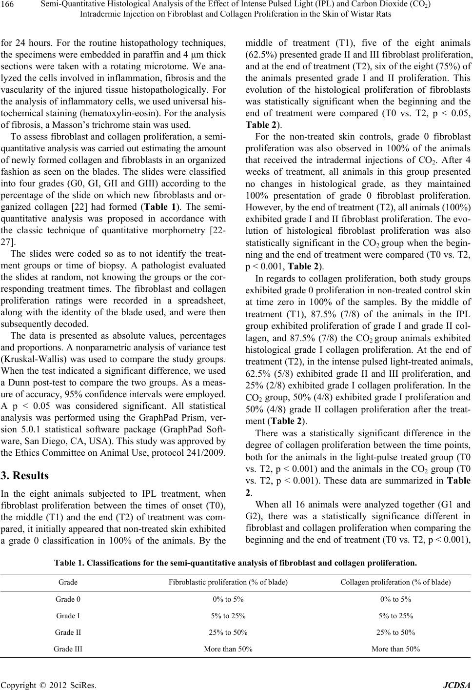 Semi-Quantitative Histological Analysis of the Effect of Intense Pulsed Light (IPL) and Carbon Dioxide (CO2) Intradermic Injection on Fibroblast and Collagen Proliferation in the Skin of Wistar Rats Copyright © 2012 SciRes. JCDSA 166 for 24 hours. For the routine histopathology techniques, the specimens were embedded in paraffin and 4 μm thick sections were taken with a rotating microtome. We ana- lyzed the cells involved in inflammation, fibrosis and the vascularity of the injured tissue histopathologically. For the analysis of inflammatory cells, we used universal his- tochemical staining (hematoxylin-eosin). For the analysis of fibrosis, a Masson’s trichrome stain was used. To assess fibroblast and collagen proliferation, a semi- quantitative analysis was carried out estimating the amount of newly formed collagen and fibroblasts in an organized fashion as seen on the blades. The slides were classified into four grades (G0, GI, GII and GIII) according to the percentage of the slide on which new fibroblasts and or- ganized collagen [22] had formed (Table 1). The semi- quantitative analysis was proposed in accordance with the classic technique of quantitative morphometry [22- 27]. The slides were coded so as to not identify the treat- ment groups or time of biopsy. A pathologist evaluated the slides at random, not knowing the groups or the cor- responding treatment times. The fibroblast and collagen proliferation ratings were recorded in a spreadsheet, along with the identity of the blade used, and were then subsequently decoded. The data is presented as absolute values, percentages and proportions. A nonparametric analysis of variance test (Kruskal-Wallis) was used to compare the study groups. When the test indicated a significant difference, we used a Dunn post-test to compare the two groups. As a meas- ure of accuracy, 95% confidence intervals were employed. A p < 0.05 was considered significant. All statistical analysis was performed using the GraphPad Prism, ver- sion 5.0.1 statistical software package (GraphPad Soft- ware, San Diego, CA, USA). This study was approved by the Ethics Committee on Animal Use, protocol 241/2009. 3. Results In the eight animals subjected to IPL treatment, when fibroblast proliferation between the times of onset (T0), the middle (T1) and the end (T2) of treatment was com- pared, it initially appeared that non-treated skin exhibited a grade 0 classification in 100% of the animals. By the middle of treatment (T1), five of the eight animals (62.5%) presented grade II and III fibroblast proliferation, and at the end of treatment (T2), six of the eight (75%) of the animals presented grade I and II proliferation. This evolution of the histological proliferation of fibroblasts was statistically significant when the beginning and the end of treatment were compared (T0 vs. T2, p < 0.05, Table 2). For the non-treated skin controls, grade 0 fibroblast proliferation was also observed in 100% of the animals that received the intradermal injections of CO2. After 4 weeks of treatment, all animals in this group presented no changes in histological grade, as they maintained 100% presentation of grade 0 fibroblast proliferation. However, by the end of treatment (T2), all animals (100%) exhibited grade I and II fibroblast proliferation. The evo- lution of histological fibroblast proliferation was also statistically significant in the CO2 group when the begin- ning and the end of treatment were compared (T0 vs. T2, p < 0.001, Table 2). In regards to collagen proliferation, both study groups exhibited grade 0 proliferation in non-treated control skin at time zero in 100% of the samples. By the middle of treatment (T1), 87.5% (7/8) of the animals in the IPL group exhibited proliferation of grade I and grade II col- lagen, and 87.5% (7/8) the CO2 group animals exhibited histological grade I collagen proliferation. At the end of treatment (T2), in the intense pulsed light-treated animals, 62.5% (5/8) exhibited grade II and III proliferation, and 25% (2/8) exhibited grade I collagen proliferation. In the CO2 group, 50% (4/8) exhibited grade I proliferation and 50% (4/8) grade II collagen proliferation after the treat- ment (Table 2). There was a statistically significant difference in the degree of collagen proliferation between the time points, both for the animals in the light-pulse treated group (T0 vs. T2, p < 0.001) and the animals in the CO2 group (T0 vs. T2, p < 0.001). These data are summarized in Table 2. When all 16 animals were analyzed together (G1 and G2), there was a statistically significance different in fibroblast and collagen proliferation when comparing the beginning and the end of treatment (T0 vs. T2, p < 0.001), Table 1. Classifications for the semi-quantitative analysis of fibroblast and collagen proliferation. Grade Fibroblastic proliferation (% of blade) Collagen proliferation (% of blade) Grade 0 0% to 5% 0% to 5% Grade I 5% to 25% 5% to 25% Grade II 25% to 50% 25% to 50% Grade III More than 50% More than 50% 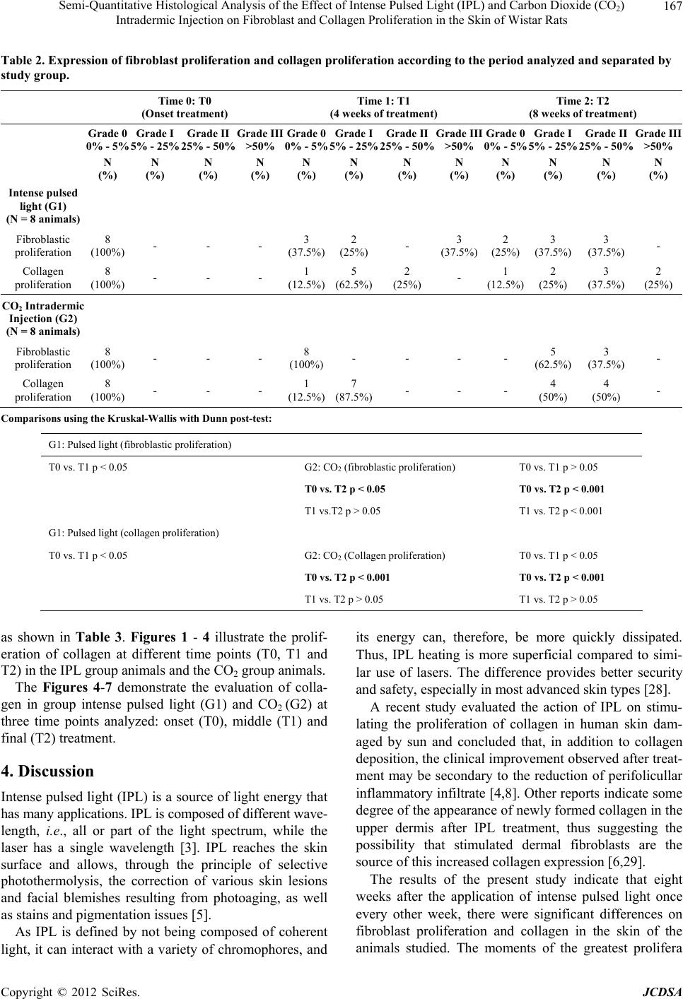 Semi-Quantitative Histological Analysis of the Effect of Intense Pulsed Light (IPL) and Carbon Dioxide (CO2) Intradermic Injection on Fibroblast and Collagen Proliferation in the Skin of Wistar Rats 167 Table 2. Expression of fibroblast proliferation and collagen proliferation according to the period analyzed and separated by study group. Time 0: T0 (Onset treatment) Time 1: T1 (4 weeks of treatment) Time 2: T2 (8 weeks of treatment) Grade 0 0% - 5% 5% - 25% Grade IGrade II 25% - 50% Grade III >50% Grade 0 0% - 5% Grade I 5% - 25% Grade II 25% - 50% Grade III >50% Grade 0 0% - 5% 5% - 25% Grade I 25% - 50% Grade IIGrade III >50% N (%) N (%) N (%) N (%) N (%) N (%) N (%) N (%) N (%) N (%) N (%) N (%) Intense pulsed light (G1) (N = 8 animals) Fibroblastic proliferation 8 (100%) - - - 3 (37.5%) 2 (25%) - 3 (37.5%) 2 (25%) 3 (37.5%) 3 (37.5%) - Collagen proliferation 8 (100%) - - - 1 (12.5%) 5 (62.5%) 2 (25%) - 1 (12.5%) 2 (25%) 3 (37.5%) 2 (25%) CO2 Intradermic Injection (G2) (N = 8 animals) Fibroblastic proliferation 8 (100%) - - - 8 (100%) - - - - 5 (62.5%) 3 (37.5%) - Collagen proliferation 8 (100%) - - - 1 (12.5%) 7 (87.5%) - - - 4 (50%) 4 (50%) - Comparisons using the Kruskal-Wallis with Dunn post-test: G1: Pulsed light (fibroblastic proliferation) T0 vs. T1 p < 0.05 G2: CO2 (fibroblastic proliferation) T0 vs. T1 p > 0.05 T0 vs. T2 p < 0.05 T0 vs. T2 p < 0.001 T1 vs.T2 p > 0.05 T1 vs. T2 p < 0.001 G1: Pulsed light (collagen proliferation) T0 vs. T1 p < 0.05 G2: CO2 (Collagen proliferation) T0 vs. T1 p < 0.05 T0 vs. T2 p < 0.001 T0 vs. T2 p < 0.001 T1 vs. T2 p > 0.05 T1 vs. T2 p > 0.05 as shown in Table 3. Figures 1 - 4 illustrate the prolif- eration of collagen at different time points (T0, T1 and T2) in the IPL group animals and the CO2 group animals. The Figures 4-7 demonstrate the evaluation of colla- gen in group intense pulsed light (G1) and CO2 (G2) at three time points analyzed: onset (T0), middle (T1) and final (T2) treatment. 4. Discussion Intense pulsed light (IPL) is a source of light energy that has many applications. IPL is composed of different wave- length, i.e., all or part of the light spectrum, while the laser has a single wavelength [3]. IPL reaches the skin surface and allows, through the principle of selective photothermolysis, the correction of various skin lesions and facial blemishes resulting from photoaging, as well as stains and pigmentation issues [5]. As IPL is defined by not being composed of coherent light, it can interact with a variety of chromophores, and its energy can, therefore, be more quickly dissipated. Thus, IPL heating is more superficial compared to simi- lar use of lasers. The difference provides better security and safety, especially in most advanced skin types [28]. A recent study evaluated the action of IPL on stimu- lating the proliferation of collagen in human skin dam- aged by sun and concluded that, in addition to collagen deposition, the clinical improvement observed after treat- ment may be secondary to the reduction of perifolicullar inflammatory infiltrate [4,8]. Other reports indicate some degree of the appearance of newly formed collagen in the upper dermis after IPL treatment, thus suggesting the possibility that stimulated dermal fibroblasts are the source of this increased collagen expression [6,29]. The results of the present study indicate that eight weeks after the application of intense pulsed light once every other week, there were significant differences on fibroblast proliferation and collagen in the skin of the animals studied. The moments of the greatest prolifera Copyright © 2012 SciRes. JCDSA 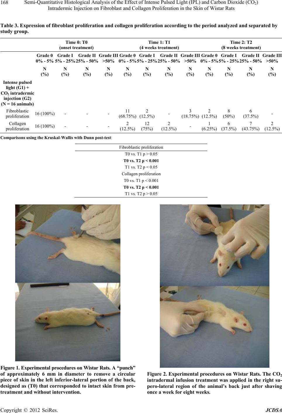 Semi-Quantitative Histological Analysis of the Effect of Intense Pulsed Light (IPL) and Carbon Dioxide (CO2) Intradermic Injection on Fibroblast and Collagen Proliferation in the Skin of Wistar Rats 168 Table 3. Expression of fibroblast proliferation and collagen proliferation according to the period analyzed and separated by study group. Time 0: T0 (onset treatment) Time 1: T1 (4 weeks treatment) Time 2: T2 (8 weeks treatment) Grade 0 0% - 5% Grade I 5% - 25% 25% - 50% Grade II Grade III >50% Grade 0 0% - 5% Grade I 5% - 25% Grade II 25% - 50% Grade III >50% Grade 0 0% - 5% 5% - 25% Grade I 25% - 50% Grade IIGrade III >50% N (%) N (%) N (%) N (%) N (%) N (%) N (%) N (%) N (%) N (%) N (%) N (%) Intense pulsed light (G1) + CO2 intradermic injection (G2) (N = 16 animals) Fibroblastic proliferation 16 (100%) - - - 11 (68.75%) 2 (12.5%)- 3 (18.75%) 2 (12.5%) 8 (50%) 6 (37.5%)- Collagen proliferation 16 (100%) - - - 2 (12.5%) 12 (75%) 2 (12.5%)- 1 (6.25%) 6 (37.5%) 7 (43.75%) 2 (12.5%) Comparisons using the Kruskal-Wallis with Dunn post-test: Fibroblastic proliferation T0 vs. T1 p > 0.05 T0 vs. T2 p < 0.001 T1 vs. T2 p < 0.05 Collagen proliferation T0 vs. T1 p < 0.001 T0 vs. T2 p < 0.001 T1 vs. T2 p > 0.05 Figure 1. Experimental procedures on Wistar Rats. A “punch” of approximately 6 mm in diameter to remove a circular piece of skin in the left inferior-lateral portion of the back, designed as (T0) that corresponded to intact skin from pre- treatment and without intervention. Figure 2. Experimental procedures on Wistar Rats. The CO2 intradermal infusion treatment was applied in the right su- pero-lateral region of the animal’s back just after shaving once a week for eight weeks. Copyright © 2012 SciRes. JCDSA 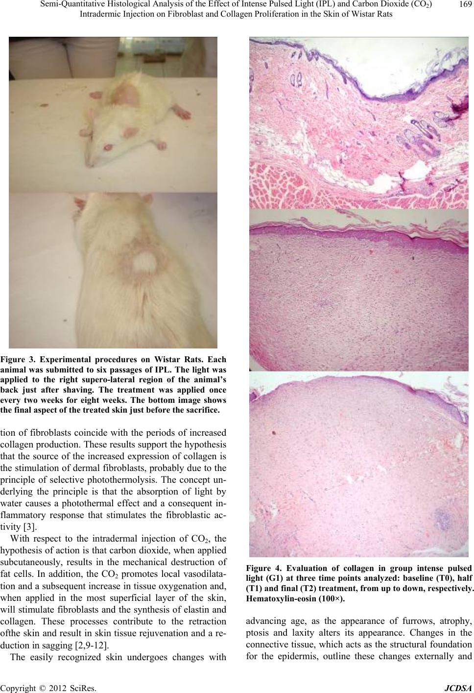 Semi-Quantitative Histological Analysis of the Effect of Intense Pulsed Light (IPL) and Carbon Dioxide (CO2) Intradermic Injection on Fibroblast and Collagen Proliferation in the Skin of Wistar Rats 169 Figure 3. Experimental procedures on Wistar Rats. Each animal was submitted to six passages of IPL. The light was applied to the right supero-lateral region of the animal’s back just after shaving. The treatment was applied once every two weeks for eight weeks. The bottom image shows the final aspect of the treated skin just before the sacrifice. tion of fibroblasts coincide with the periods of increased collagen production. These results support the hypothesis that the source of the increased expression of collagen is the stimulation of dermal fibroblasts, probably due to the principle of selective photothermolysis. The concept un- derlying the principle is that the absorption of light by water causes a photothermal effect and a consequent in- flammatory response that stimulates the fibroblastic ac- tivity [3]. With respect to the intradermal injection of CO2, the hypothesis of action is that carbon dioxide, when applied subcutaneously, results in the mechanical destruction of fat cells. In addition, the CO2 promotes local vasodilata- tion and a subsequent increase in tissue oxygenation and, when applied in the most superficial layer of the skin, will stimulate fibroblasts and the synthesis of elastin and collagen. These processes contribute to the retraction ofthe skin and result in skin tissue rejuvenation and a re- duction in sagging [2,9-12]. The easily recognized skin undergoes changes with Figure 4. Evaluation of collagen in group intense pulsed light (G1) at three time points analyzed: baseline (T0), half (T1) and final (T2) treatment, from up to down, respectively. Hematoxylin-eosin (100×). advancing age, as the appearance of furrows, atrophy, ptosis and laxity alters its appearance. Changes in the connective tissue, which acts as the structural foundation for the epidermis, outline these changes externally and Copyright © 2012 SciRes. JCDSA 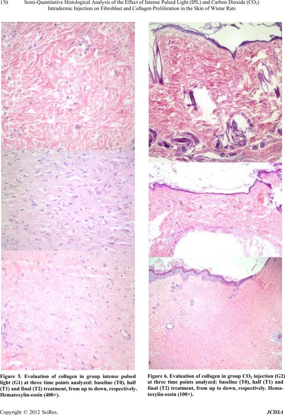 Semi-Quantitative Histological Analysis of the Effect of Intense Pulsed Light (IPL) and Carbon Dioxide (CO2) Intradermic Injection on Fibroblast and Collagen Proliferation in the Skin of Wistar Rats 170 Figure 5. Evaluation of collagen in group intense pulsed light (G1) at three time points analyzed: baseline (T0), half (T1) and final (T2) treatment, from up to down, respectively. Hematoxylin-eosin (400×). Figure 6. Evaluation of collagen in group CO2 injection (G2) at three time points analyzed: baseline (T0), half (T1) and final (T2) treatment, from up to down, respectively. Hema- toxylin-eosin (100×). Copyright © 2012 SciRes. JCDSA 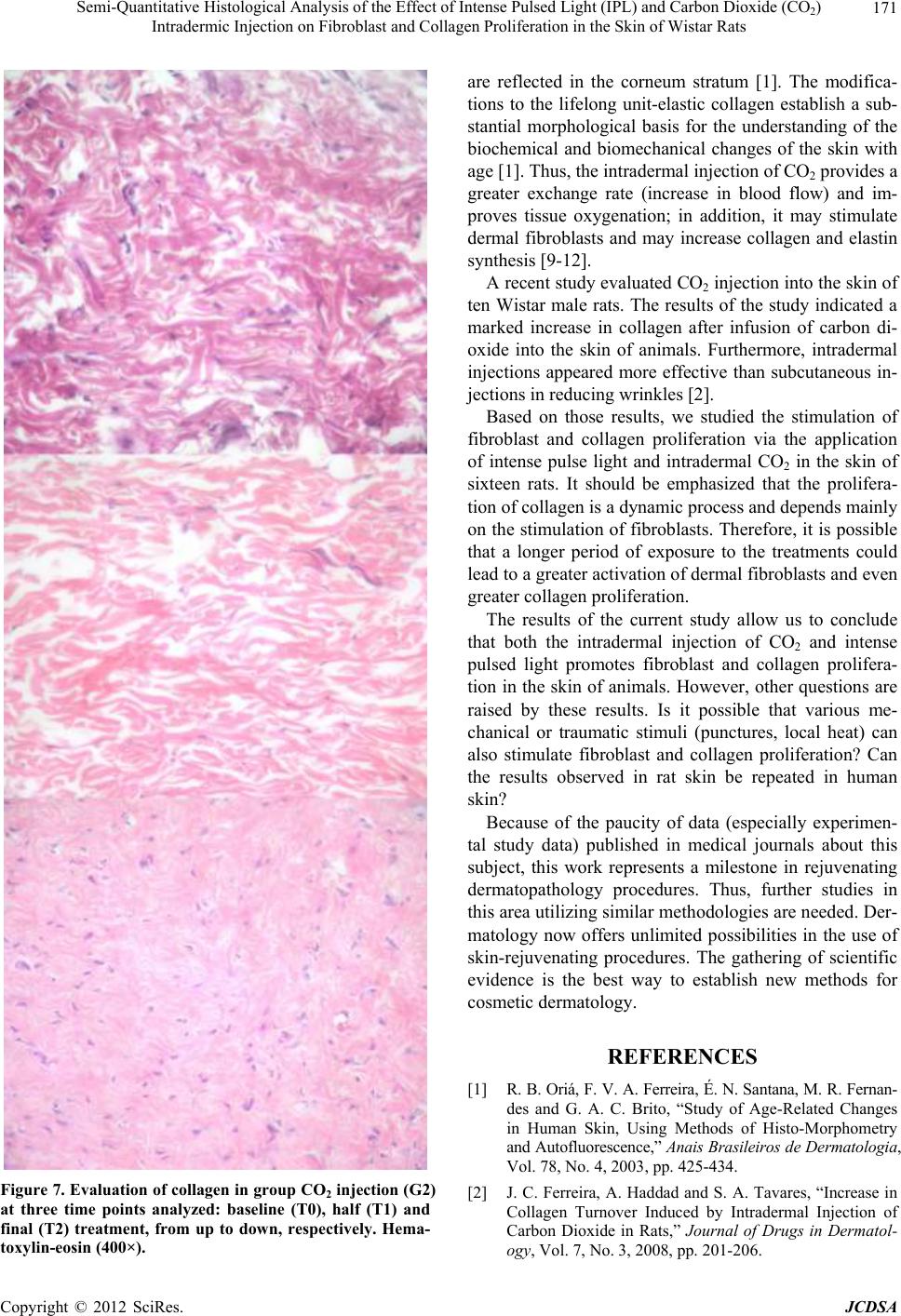 Semi-Quantitative Histological Analysis of the Effect of Intense Pulsed Light (IPL) and Carbon Dioxide (CO2) Intradermic Injection on Fibroblast and Collagen Proliferation in the Skin of Wistar Rats 171 Figure 7. Evaluation of collagen in group CO2 injection (G2) at three time points analyzed: baseline (T0), half (T1) and final (T2) treatment, from up to down, respectively. Hema- toxylin-eosin (400×). are reflected in the corneum stratum [1]. The modifica- tions to the lifelong unit-elastic collagen establish a sub- stantial morphological basis for the understanding of the biochemical and biomechanical changes of the skin with age [1]. Thus, the intradermal injection of CO2 provides a greater exchange rate (increase in blood flow) and im- proves tissue oxygenation; in addition, it may stimulate dermal fibroblasts and may increase collagen and elastin synthesis [9-12]. A recent study evaluated CO2 injection into the skin of ten Wistar male rats. The results of the study indicated a marked increase in collagen after infusion of carbon di- oxide into the skin of animals. Furthermore, intradermal injections appeared more effective than subcutaneous in- jections in reducing wrinkles [2]. Based on those results, we studied the stimulation of fibroblast and collagen proliferation via the application of intense pulse light and intradermal CO2 in the skin of sixteen rats. It should be emphasized that the prolifera- tion of collagen is a dynamic process and depends mainly on the stimulation of fibroblasts. Therefore, it is possible that a longer period of exposure to the treatments could lead to a greater activation of dermal fibroblasts and even greater collagen proliferation. The results of the current study allow us to conclude that both the intradermal injection of CO2 and intense pulsed light promotes fibroblast and collagen prolifera- tion in the skin of animals. However, other questions are raised by these results. Is it possible that various me- chanical or traumatic stimuli (punctures, local heat) can also stimulate fibroblast and collagen proliferation? Can the results observed in rat skin be repeated in human skin? Because of the paucity of data (especially experimen- tal study data) published in medical journals about this subject, this work represents a milestone in rejuvenating dermatopathology procedures. Thus, further studies in this area utilizing similar methodologies are needed. Der- matology now offers unlimited possibilities in the use of skin-rejuvenating procedures. The gathering of scientific evidence is the best way to establish new methods for cosmetic dermatology. REFERENCES [1] R. B. Oriá, F. V. A. Ferreira, É. N. Santana, M. R. Fernan- des and G. A. C. Brito, “Study of Age-Related Changes in Human Skin, Using Methods of Histo-Morphometry and Autofluorescence,” Anais Brasileiros de Dermatologia, Vol. 78, No. 4, 2003, pp. 425-434. [2] J. C. Ferreira, A. Haddad and S. A. Tavares, “Increase in Collagen Turnover Induced by Intradermal Injection of Carbon Dioxide in Rats,” Journal of Drugs in Dermatol- ogy, Vol. 7, No. 3, 2008, pp. 201-206. Copyright © 2012 SciRes. JCDSA  Semi-Quantitative Histological Analysis of the Effect of Intense Pulsed Light (IPL) and Carbon Dioxide (CO2) Intradermic Injection on Fibroblast and Collagen Proliferation in the Skin of Wistar Rats 172 [3] M. G. Catorze, “Laser: Bases and Use in Dermatology,” Medicina Cutánea Ibero-Latino-Americana, Vol. 37, No. 1, 2009, pp. 5-27. [4] V. G. Prieto, A. H. Diwan, C. R. Shea, P. Zhang and N. S. Sadick, “Effects of Intense Pulsed Light and the 1064 nm Nd:YAG Laser on Sun-Damaged Human Skin: Histolo- gic and Immunohistochemical Analysis,” Dermatologic Surgery, Vol. 31, No. 5, 2005, pp. 522-525. doi:10.1111/j.1524-4725.2005.31154 [5] R. C. R. Patriota, C. J. Rodrigues and L. C. Cucé, “Intense Pulsed Light in Photoaging: Clinical, Histopathological and Immunohistochemical Evaluation,” Anais Brasileiros de Dermatologia, Vol. 86, No. 6, 2011, pp. 1129-1133. doi:10.1590/S0365-05962011000600010 [6] D. J. Goldberg, “New Collagen Formation after Dermal Remodeling with an Intense Pulsed Light Source,” Jour- nal of Cutaneous Laser Therapy, Vol. 2, No. 2, 2000, pp. 59-61. [7] D. J. Goldberg and K. B. Cutler, “Nonablative Treatment of Rhytids with Intense Pulsed Light,” Lasers in Surgery and Medicine, Vol. 26, No. 2, 2000, pp. 196-200. doi:10.1002/(SICI)1096-9101(2000)26:2<196::AID-LSM 10>3.0.CO;2-9 [8] V. G. Prieto, P. S. Zhang and N. S. Sadick, “Evaluation of Pulsed Light and Radiofrequency Combined for the Treat- ment of Acne Vulgaris with Histologic Analysis of Facial Skin Biopsies,” Journal of Cosmetic and Laser Therapy, Vol. 7, No. 2, 2005, pp. 63-68. [9] C. Brandi, C. D’Aniello, L. Grimaldi, B. Bosi, I. Dei, P. Lattarulo, et al., “Carbon Dioxide Therapy in the Treat- ment of Localized Adiposities: Clinical Study and Histo- pathological Correlations,” Aesthetic Plastic Surgery, Vol. 25, No. 3, 2001, pp. 170-174. doi:10.1007/s002660010116 [10] C. Brandi, L. Grimaldi, G. Nisi, A. Brafa, A. Campa, M. Calabrò, et al., “The Role of Carbon Dioxide Therapy in the Treatment of Chronic Wounds,” In Vivo, Vol. 24, No. 2, 2010, pp. 223-226. [11] T. Brockow, A. Dillner, A. Franke and K. L. Resch, “An- algesic Effectiveness of Subcutaneous Carbon-Dioxide Insufflations as an Adjunct Treatment in Patients with Non-Specific Neck or Low Back Pain,” Complementary Therapies in Medicine, Vol. 9, No. 2, 2001, pp. 68-76. doi:10.1054/ctim.2001.0434 [12] A. Sönmez, M. Yaman, O. Yalçin, B. Ersoy, M. Serin and A. Sav, “Carbon Dioxide Therapy Increases Capillary Formation on Random Pedicled Skin Flaps in the Rat,” Journal of Plastic, Reconstructive & Aesthetic Surgery, Vol. 62, No. 7, 2009, pp. e236-e237. doi:10.1016/j.bjps.2009.01.067 [13] D. Kummer-Kloess, W. Kloess, N. Marienhoff, R. M. Schütz, M. Zwaan and H. D. Weiss, “Angiography during Interventional Procedures with Carbon Dioxide (CO2) (Carbo-Angiography) in Patients with Increased Contrast Media Risk,” Zentralblatt für Chirurgie, Vol. 122, 1997, pp. 725-729. [14] M. Zwaan, W. Kloess, C. Kagel, D. Kummer-Kloess, S. Matthies-Zwaan, R. M. Schütz, et al., “Carbon Dioxide as an Alternative Contrast Medium in Peripheral Angiogra- phy,” Rofo, Vol. 164, No. 5, 1996, pp. 445-448. doi:10.1055/s-2007-1015687 [15] M. Zwaan, J. Steinhoff, L. Fricke, C. Kagel, H. Lorch and H. D. Weiss, “The Angiography of Kidney Transplant Arteries Using Carbon Dioxide,” Dtsch Med Wochenschr, Vol. 122, 1997, pp. 1133-1136. doi:10.1055/s-2008-1047738 [16] B. R. Hartmann, E. Bassenge and M. Pittler, “Effect of Carbon Dioxide-Enriched Water and Fresh Water on the Cutaneous Microcirculation and Oxygen Tension in the Skin of the Foot,” Angiology, Vol. 48, No. 4, 1997, pp. 337-343. doi:10.1177/000331979704800406 [17] B. R. Hartmann, E. Bassenge and M. Hartmann, “Effects of Serial Percutaneous Application of Carbon Dioxide in Intermittent Claudication: Results of a Controlled Trial,” Angiology, Vol. 48, No. 11, 1997, pp. 957-963. doi:10.1177/000331979704801104 [18] K. Field, M. Bailey, L. L. Foresman, R. L. Harris, S. L. Motzel, R. A. Rockar, et al., “Medical Records for Ani- mals Used in Research, Teaching, and Testing: Public Statement from the American College of Laboratory Ani- mal Medicine,” Institute of Laboratory Animal Resources Journal, Vol. 48, 2007, pp. 37-41. [19] D. F. Kohn, T. E. Martin, P. L. Foley, T. H. Morris, M. M. Swindle, G. A. Vogler, et al., “Public Statement: Guide- lines for the Assessment and Management of Pain in Ro- dents and Rabbits,” Journal of American Association for Laboratory Animal Science, Vol. 46, No. 2, 2007, pp. 97- 108. [20] ACLAM, “ACLAM Position Statement on Animal Ex- perimentation,” Comparative Medicine, Vol. 53, No. 5, 2003, p. 472. [21] J. Artwohl, P. Brown, B. Corning, S. Stein and A. T. Force, “Report of the ACLAM Task Force on Rodent Euthanasia,” Journal of American Association for Labo- ratory Animal Science, Vol. 45, No. 1, 2006, pp. 98-105. [22] A. L. Domingos, S. Tucci, S. B. Garcia, J. de Bessa, A. J. Cologna and A. C. Martins, “Use of a Latex Biomem- brane for Bladder Augmentation in a Rabbit Model: Bio- compatibility, Clinical and Histological Outcomes,” In- ternational Journal of the Brazilian Society of Urology, Vol. 35, No. 2, 2009, pp. 217-224. [23] J. J. S. Gonçalves, L. E. V. Leão, R. G. Ferreira, R. Olivei- ra, L. H. Ota and R. S. D. Santos, “Semiquantitative Ana- lysis of Surgical Biopsies of Different Lung Lobes of Pa- tients with Usual Interstitial Pneumonia/Idiopathic Pul- monary Fibrosis,” Jornal Brasileiro de Pneumologia, Vol. 35, No. 7, 2009, pp. 676-682. [24] D. M. Hyde, T. E. King, T. McDermott, J. A. Waldron, T. V. Colby, W. M. Thurlbeck, et al., “Idiopathic Pulmonary Fibrosis. Quantitative Assessment of Lung Pathology. Com- parison of a Semiquantitative and a Morphometric Histo- pathologic Scoring System,” American Review of Respi- ratory Disease, Vol. 146, No. 4, 1992, pp. 1042-1047. [25] M. F. Y. Maeda, C. D. Silva, L. S. Harima, L. F. F. Silva, B. Ctenas and V. A. F. Alves, “Vascularization in Hepatic Cirrhosisbased on Autopsies,” Archives of Gastroentero- Copyright © 2012 SciRes. JCDSA  Semi-Quantitative Histological Analysis of the Effect of Intense Pulsed Light (IPL) and Carbon Dioxide (CO2) Intradermic Injection on Fibroblast and Collagen Proliferation in the Skin of Wistar Rats Copyright © 2012 SciRes. JCDSA 173 logy, Vol. 45, 2008, pp. 38-45. [26] D. Araf, M. Moura, C. J. Rodrigues and L. A. Figueiredo, “Homologous Scleral and Dura Mater Grafts in Rabbit Eyes,” Arquivos Brasileiros de Oftalmologia, Vol. 70, No. 6, 2007, pp. 981-987. doi:10.1590/S0004-27492007000600018 [27] W. C. Cavazana, M. L. P. B. de Simões, S. O. Yoshii, C. A. B. Amado and R. K. N. Cuman, “Sugar (Sucrose) and Medium Chain Triglycerides with Essential Fatty Acids in the Treatment of Wounds: An Experimental Study in Rats,” Brazilian Annals of Dermatology, Vol. 84, No. 3, 2009, pp. 229-236. doi:10.1590/S0365-05962009000300003 [28] D. Laury, “Intense Pulsed Light Technology and Its Im- provement on Skin Aging from the Patients’ Perspective Using Photorejuvenation Parameters,” Dermatology Online Journal, Vol. 9, No. 1, 2003, p. 5. [29] D. J. Goldberg and S. Silapunt, “Q-Switched Nd:YAG La- ser: Rhytid Improvement by Non-Ablative Dermal Re- modeling,” Journal of Cutaneous Laser Therapy, Vol. 2, No. 3, 2000, pp. 157-160. |