

Paper Menu >>
Journal Menu >>
 Surgical Science, 2012, 3, 460-462 http://dx.doi.org/10.4236/ss.2012.39091 Published Online September 2012 (http://www.SciRP.org/journal/ss) A Calcified Artery of Drummond, Could It Be a Sentinel Sign of Further Complications? Shahe Boghossian, Arpan K. Banerjee Heart of England NHS Trust, Birmingham, UK Email: [email protected] Received March 24, 2012; revised April 30, 2012; accepted May 18, 2012 ABSTRACT Objective: To und erstand the role of the artery of Drummo nd in the elderly population. The artery plays a crucial role in the anastomosis between the inferior mesenteric artery and sup erior mesenteric artery. Its maintenance is particularly important for the survivability of the bowel in events of stenosis of either one of the native arteries; SMA or IMA. Method: A 94 year old lady presents with post-prandial abdominal pain and significant emaciation. The patient under- goes clinical and radiological investigations to find out the cause of her ailments. Results: Abdominal X-ray revealed a serpentine structure that was later defined as the artery of Drummond, CT scan revealed a calcified and enlarged artery of Drummond in addition to an occluded origin of SMA and celiac artery as well as a severe stenosis origin of IMA. Conclusion: The artery of Drummond was immensely hypertrophied and was supplying the entire bowel through the native IMA; subsequent occlusion of the IMA was the main cause that caused the demise of the patient. Keywords: Artery of Drummond; Mesenteric Ischemia; Refeeding Syndrome 1. Case Report A 94 year old lady was admitted to Accident and Emer- gency complaining of six week history of abdominal pain, nausea and significant emaciation. She was alert with a GCS of 15. Examination of the abdomen revealed two well healed midline incisions. Palpation of the abdomen revealed fullness on the left side of her abdomen as well as a hard palpable tube like structure that rolled under- neath the examiner’s fingers. Auscultation of her abdo- men showed faint yet audible bowel sounds. Rectal ex- amination showed an empty rectum. Chest X-ray showed bibasal collapse but no obstruction of the bronchi and no signs of any pulmonary malignancy. On admission, her initial blood test showed the fol- lowing: Na: 132, K: 4.2, Creatin ine: 82 mmo l/lit, Alb: 40, Bilirubin: 16, Phosphate: 1.02, Hemoglobin: 14.4, Plate- lets: 252, White cell count: 11.32. Her observation chart was normal however the patient looked dehydrated and hence the patient was put on a slow IV drip. The patient looked generally emaciated and was put on IV rehydra- tion, oral rehydration, as well as fortified oral protein sachets. The patient was not able to tolerate the oral feed and hence a provisional diagnosis of Refeeding Syn- drome was put forward. Three days later, a random arte- rial blood gas showed an elevation of serum lactate to 6.5 mmol/lit with a base excess of –3.0 mmol/lit, pCO2: 4.58 kPa, pO2: 12.0 kPa, pH: 7.4 and a CRP of 161. Her white cell count was elevated at 19.38/ml. Unfortunately, the patient expired a week later. 2. Findings The abdominal and chest X-ray showed a calcified trans- versely oriented tube like structure the crossed the full spectrum of the abdomen (Figure 1). This was initially thought to be a foreign body but later confirmed to be a calcified vascular structure. Contrast enhanced Computer Tomography confirmed the structure to be a calcified artery of Drummond (Figure 2) with a stenosed origin of the Celiac, Superior Mesenteric Artery (Figure 3) and the Inferior Mesenteric Artery (Figure 4). The diagnosis eventually confirmed mesenteric ischemia in the elderly. 3. Discussion Chronic mesenteric ischemia is an indolent condition af- fecting mainly the vascular supply to the small bowel. It is mainly manifested by postprandial pain and subse- quent weight loss. The most common artery involved is the Superior Mesenter ic Artery (SMA) which supplies th e duodenum, the jejunum, the ileum as well as the right si de of the large bowel [1]. Occasionally, the coeliac art er y may be involved as well, either as a solitary condition or in association with widespread atherosclerosis of the A or ta . Congenital conditions such as acute angulation of the S MA with the Aorta or fibromuscular dysplasia have been C opyright © 2012 SciRes. SS 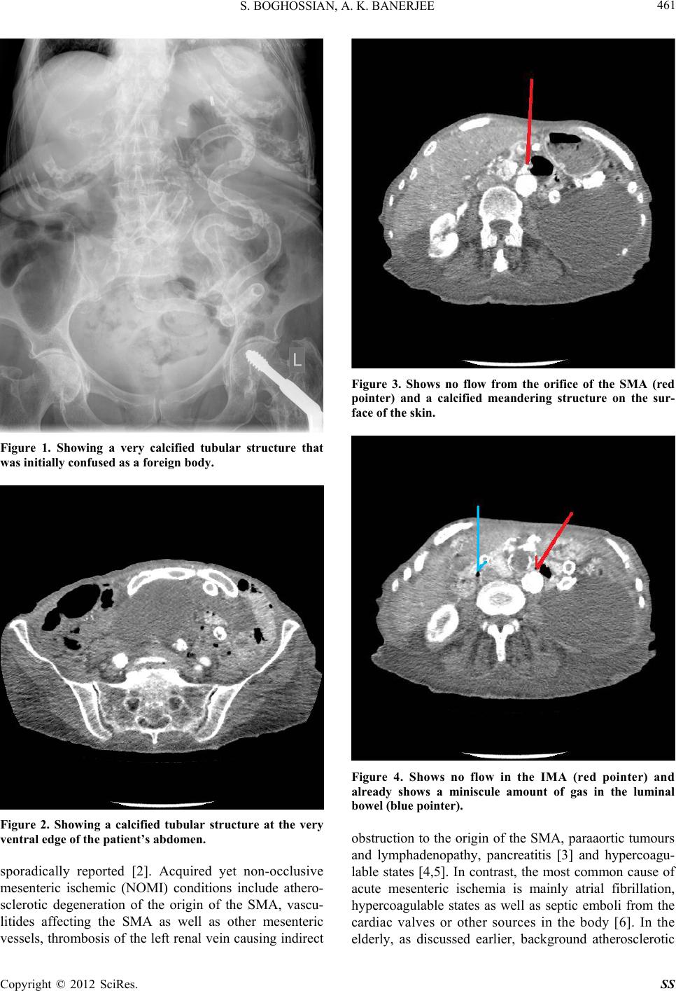 S. BOGHOSSIAN, A. K. BANERJEE 461 Figure 1. Showing a very calcified tubular structure that was initially confused as a foreign body. Figure 2. Showing a calcified tubular structure at the very ventral edge of the patient’s abdomen. sporadically reported [2]. Acquired yet non-occlusive mesenteric ischemic (NOMI) conditions include athero- sclerotic degeneration of the origin of the SMA, vascu- litides affecting the SMA as well as other mesenteric vessels, thrombosis of the left renal vein causing indirect Figure 3. Shows no flow from the orifice of the SMA (red pointer) and a calcified meandering structure on the sur- face of the skin. Figure 4. Shows no flow in the IMA (red pointer) and already shows a miniscule amount of gas in the luminal bowel (blue pointer). obstruction to the origin of the SMA, paraaortic tumours and lymphadenopathy, pancreatitis [3] and hypercoagu- lable states [4,5]. In contrast, the most common cause of acute mesenteric ischemia is mainly atrial fibrillation, hypercoagulable states as well as septic emboli from the cardiac valves or other sources in the body [6]. In the elderly, as discussed earlier, background atherosclerotic Copyright © 2012 SciRes. SS  S. BOGHOSSIAN, A. K. BANERJEE Copyright © 2012 SciRes. SS 462 disease is a common finding on non-invasive imaging studies, as demonstrated by the images shown under- neath (CT and AXR). However, in an acute ischemic event, the anastamotic feeder vessel would thrombose rendering the bowel non-viable and causing irreversible ischemic injury. This case highlights a variant whereby the coeliac axis territory and the SMA territory are sup- plied by a dominant artery of Drummond which in turn is supplied by the IMA as demonstrated by abdominal im- aging. The artery of Drummond is a crucial anastomosis between the superior and inferior mesenteric artery. Ad- ditionally the viability of this arc is crucial for the sur- vivability of Endovascular Aortic Aneurysm repairs whereby the origin of the inferior mesenteric artery is iatrogenically occluded [7,8]. Our assessment of the pa- tient’s condition certainly demonstrated th at she had long standing atherosclerotic disease as described from her weight loss, but we can certainly say that acute embolisa- tion of the inferior mesenteric artery (IMA) was a key factor in her rapid deterioration. The case highlights a case of hypertrophy of the artery of Drummond that was crucial in maintaining nutrient supply to the entire fore- gut and hindgut through the IMA. REFERENCES [1] P. R. Kvietys, “The Gastrointestinal Circulation,” Morgan & Claypool Life Sciences, San Rafael, 2010. [2] R. M. Hamed, and K. Ghandour, “Abdominal Angina and Intestinal Gangrene—A Catastrophic Presentation of Ar- terial Fibromuscular Dysplasia: Case Report and Review of the Literature,” Journal of Pediatric Surgery, Vol. 32, No. 9, 1997, pp. 1379-1380. doi:10.1016/S0022-3468(97)90328-6 [3] M. Hirota, K. Inoue, Y. Kimura, et al., “Non-Occlusive Mesenteric Ischemia and Its Associated Intestinal Gan- grene in Acute Pancreatitis,” Pancreatology, Vol. 3, No. 4, 2003, pp. 316-322. doi:10.1159/000071770 [4] N. Scally, M. E. O’Donnell, C. McClintock, M. Allen, S. J. Kirk and S. Dolan, “Mesenteric Artery Occlusion Se- condary to Activated Protein C Resistance: A Life- Threatening Combination,” Angiology, Vol. 58, No. 6, 2008, pp. 768-771. [5] N. Agaoglu, S. Turkyilmaz, C. Ozlu, M. K. Arslan and E. Colak, “Significance of Antithrombin III, Protein C and Protein S in Acute Mesenteric Ischemia Patients,” Acta Chirurgica Belgica, Vol. 104, No. 2, 2004, pp. 184-186. [6] D. Hussain, S. L. Sarfraz, S. K. Baliga and R. Hartung, “Acute Mesenteric Ischemia: Experience in a Tertiary Care Hospital,” Journal of Ayub Medical College Abbot- tabad, Vol. 21, No. 4, 2009, pp. 70-72. [7] J. Kulhanek, R. Chiu, S. Wann, “Multislice CT An- giographic Reconstruction of Marginal Artery of Drum- mond as Collateral to Occluded Superior Mesenteric Ar- tery,” American Heart Hospital Journal, Vol. 4, No. 4, 2006, pp. 290-291. doi:10.1111/j.1541-9215.2006.04931.x [8] D. L. Bhatt, “Guide to Peripheral and Cerebrovascular Intervention,” Remedica, London, 2004. |