

Paper Menu >>
Journal Menu >>
 Surgical Science, 2012, 3, 425-429 http://dx.doi.org/10.4236/ss.2012.39085 Published Online September 2012 (http://www.SciRP.org/journal/ss) Arthroscopic Classification of Suprapatellar Plica and Medial Synovial Plica Mitsuru Hanada1, Masaaki Takahashi2, Hiroshi Koyama1, Yukihiro Matsuyama1 1Department of Orthopaedic Surgery, Hamamatsu University School of Medicine, Hamamatsu, Japan 2Department of Joint Center, Juzen Hospital, Hamamatsu, Japan Email: [email protected] Received July 24, 2012; revised August 23, 2012; accepted September 6, 2012 ABSTRACT In the previous arthroscopic studies, the ratio of presence and type of plica was somewhat different. We arthroscopically investigated and classified suprapatellar plica and medial synovial plica in a Japanese population. Subjects and Metho d s: The anatomy of suprapatellar plica and medial synovial plica was studied arthroscopically in 130 knees. Original diagnosis of patients included in this study were 53 meniscal injuries, 51 ACL injuries, 17 osteoarthritis, 5 popliteal cysts, 3 osteochondritis dissecans, and 1 synovial osteochondromatosis. Results: The suprapatellar plica was present 73.8% and classified into 6 types which were arch type, medial type, lateral type, perforated type, pillar type and complete type. The medial synovial plica was present 62.3% and classified into 4 types which were narrow type, medium type, broad type and perforated type. No relationship between age and the pattern of the suprapatellar plica or medial synovial plica could be found. There was no trend to be correlation between the type of suprapatellar plica and medial synovial plica. Conclusion: We classified suprapatellar plica by only location and shape of plica and medial synovial plica by the size of plica. Keywords: Suprapatellar Plica; Medial Synovial Plica; Arthroscopy 1. Introduction The synovial plica in the knee joint is recognized as a normal structure that represents remnants of synovial mem- brane in embryonic development of the knee. However, sometimes the synovial plica may be involved in patho- logic processes [1-7]. In the fetal stage at 10 weeks, the developing knee joint consists of a single cavity with a synovial lining. At certain sites, such as at the medial part of the patella-femoral and infrapatellar regions, mes- enchymal tissue remains. These tissue strands may become plica. In the fetal stage from 11 to 20 weeks, an infrapa- tellar plica can be found in 50% of specimens, suprapatel- lar plica in 33%, and a mediopatellar plica in 37% [8]. Three types of synovial plica are generally known; the suprapatellar plica, medial patellar plica, and infrapatellar plica. Several studies have been attempted to classify the plica, but the findings have been conflicting. There were classifications by arthroscopy [9,10]. But before them, because suprapatellar plica and medial synovial plica were classified by the open dissection, they were uncer- tain. In the arthroscopic studies, the ratio of presence and type of plica was somewhat different. We arthroscopically investigated and classified plica in a Japanese population. 2. Subjects and Methods 2.1. Subjects From December 2002 to December 2007, we evaluated 125 patients (130 knees) who were undergone arthroscopic surgeries excluding the patients with a history of opera- tion of a knee and the patients who were plica syndrome. Only one surgeon performed arthroscopy. The original diagnoses of 130 knees with injuries or diseases, which were needed arthroscopic surgery, involved 53 meniscal injuries, 51 ACL injuries, 17 osteoarthritis, 5 popliteal cysts, 3 osteochondritis dissecans, and 1 synovial osteochodro- matosis (Table 1). The patient sample included 65 males (4 yrs to 80 years with an average age of 39.4 yrs) and 65 females (14 yrs to 80 yrs with an average of 47.9 yrs). There were 5 patients who were performed arthroscopic surgeries on both knees. However, there were not patients who were undergone arthroscopic surgery of both knees at the same time. 2.2. Classification of Suprapatellar Plica We modified and simplified the classification by Dandy [10] into our classification system described as below. For suprapatellar plica. C opyright © 2012 SciRes. SS 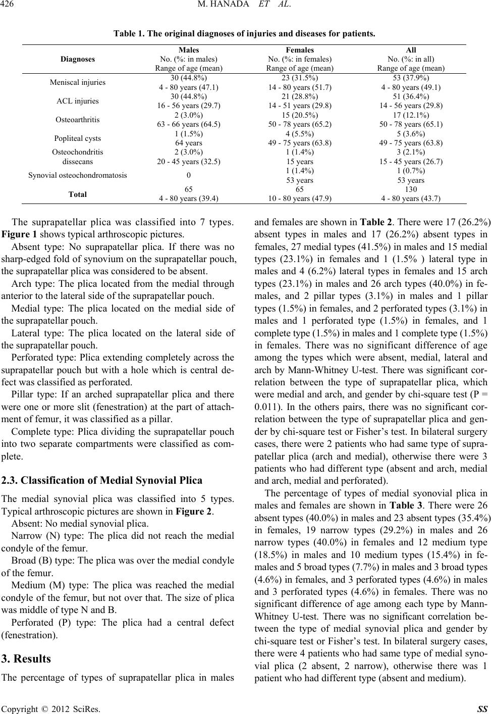 M. HANADA ET AL. 426 Table 1. The original diagnoses of injuries and diseases for patients. Diagnoses Males No. (%: in males) Range of age (mean) Females No. (%: in females) Range of age (mean) All No. (%: in all) Range of age (mean) Meniscal injuries 30 (44.8%) 4 - 80 years (47.1) 23 (31.5%) 14 - 80 years (51.7) 53 (37.9%) 4 - 80 years (49.1) ACL injuries 30 (44.8%) 16 - 56 years (29.7) 21 (28.8%) 14 - 51 years (29.8) 51 (36.4%) 14 - 56 years (29.8) Osteoarthritis 2 (3.0%) 63 - 66 years (64.5) 15 (20.5%) 50 - 78 years (65.2) 17 (12.1%) 50 - 78 years (65.1) Popliteal cysts 1 (1.5%) 64 years 4 (5.5%) 49 - 75 years (63.8) 5 (3.6%) 49 - 75 years (63.8) Osteochondritis dissecans 2 (3.0%) 20 - 45 years (32.5) 1 (1.4%) 15 years 3 (2.1%) 15 - 45 years (26.7) Synovial osteochondromatosis0 1 (1.4%) 53 years 1 (0.7%) 53 years Total 65 4 - 80 years (39.4) 65 10 - 80 years (47.9) 130 4 - 80 years (43.7) The suprapatellar plica was classified into 7 types. Figure 1 shows typical arthroscopic pictures. Absent type: No suprapatellar plica. If there was no sharp-edged fold of synovium on the suprapatellar pouch, the suprapatellar plica was considered to be absent. Arch type: The plica located from the medial through anterior to the lateral side of the suprapatellar pouch. Medial type: The plica located on the medial side of the suprapatellar pouch. Lateral type: The plica located on the lateral side of the suprapatellar pouch. Perforated type: Plica extending completely across the suprapatellar pouch but with a hole which is central de- fect was classified as perforated. Pillar type: If an arched suprapatellar plica and there were one or more slit (fenestration) at the part of attach- ment of femur, it was classified as a pillar. Complete type: Plica dividing the suprapatellar pouch into two separate compartments were classified as com- plete. 2.3. Classification of Medial Synovial Plica The medial synovial plica was classified into 5 types. Typical arthroscopic pictures are shown in Figure 2. Absent: No medial synovial plica. Narrow (N) type: The plica did not reach the medial condyle of the femur. Broad (B) type: The plica was over the medial condyle of the femur. Medium (M) type: The plica was reached the medial condyle of the femur, but not over that. The size of plica was middle of type N and B. Perforated (P) type: The plica had a central defect (fenestration). 3. Results The percentage of types of suprapatellar plica in males and females are shown in Table 2. There were 17 (26.2%) absent types in males and 17 (26.2%) absent types in females, 27 medial types (41.5%) in males and 15 medial types (23.1%) in females and 1 (1.5% ) lateral type in males and 4 (6.2%) lateral types in females and 15 arch types (23.1%) in males and 26 arch types (40.0%) in fe- males, and 2 pillar types (3.1%) in males and 1 pillar types (1.5%) in females, and 2 perforated types (3.1%) in males and 1 perforated type (1.5%) in females, and 1 complete type (1.5%) in males and 1 complete type (1.5%) in females. There was no significant difference of age among the types which were absent, medial, lateral and arch by Mann-Whitney U-test. There was significant cor- relation between the type of suprapatellar plica, which were medial and arch, and gender by chi-square test (P = 0.011). In the others pairs, there was no significant cor- relation between the type of suprapatellar plica and gen- der by chi-square test or Fisher’s test. In bilateral surgery cases, there were 2 patients who had same type of supra- patellar plica (arch and medial), otherwise there were 3 patients who had different type (absent and arch, medial and arch, medial and perforated). The percentage of types of medial syonovial plica in males and females are shown in Table 3. There were 26 absent types (40.0%) in males and 23 absent types (35.4%) in females, 19 narrow types (29.2%) in males and 26 narrow types (40.0%) in females and 12 medium type (18.5%) in males and 10 medium types (15.4%) in fe- males and 5 broad types (7.7%) in males and 3 broad types (4.6%) in females, and 3 perforated types (4.6%) in males and 3 perforated types (4.6%) in females. There was no significant difference of age among each type by Mann- Whitney U-test. There was no significant correlation be- tween the type of medial synovial plica and gender by chi-square test or Fisher’s test. In bilateral surgery cases, there were 4 patients who had same type of medial syno- vial plica (2 absent, 2 narrow), otherwise there was 1 patient who had different type (absent and medium). Copyright © 2012 SciRes. SS 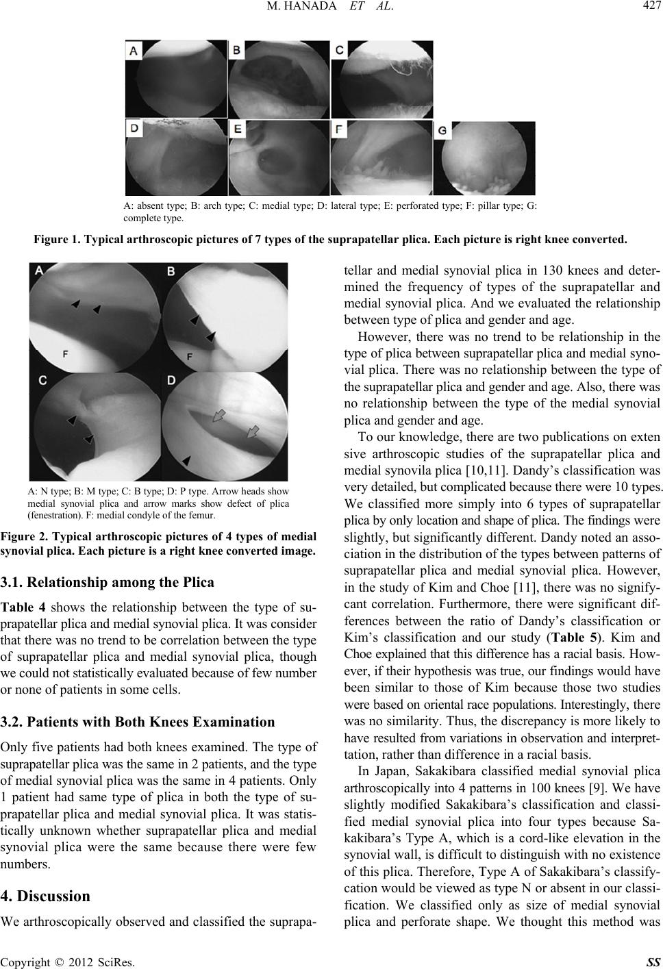 M. HANADA ET AL. 427 A: absent type; B: arch type; C: medial type; D: lateral type; E: perforated type; F: pillar type; G: complete type. Figure 1. Typical arthroscopic pictures of 7 types of the suprapatellar plica. Each picture is right knee converted. A: N type; B: M type; C: B type; D: P type. Arrow heads show medial synovial plica and arrow marks show defect of plica (fenestration). F: medial condyle of the femur. Figure 2. Typical arthroscopic pictures of 4 types of medial synovial plica. Each picture is a right knee converted image. 3.1. Relationship among the Plica Table 4 shows the relationship between the type of su- prapatellar plica and medial synovial plica. It was consider that there was no trend to be correlation between the type of suprapatellar plica and medial synovial plica, though we could not statistically evaluated because of few number or none of patients in some cells. 3.2. Patients with Both Knees Examination Only five patients had both knees examined. The type of suprapatellar plica was the same in 2 patients, and the type of medial synovial plica was the same in 4 patients. Only 1 patient had same type of plica in both the type of su- prapatellar plica and medial synovial plica. It was statis- tically unknown whether suprapatellar plica and medial synovial plica were the same because there were few numbers. 4. Discussion We arthroscopically observed and classified the suprapa- tellar and medial synovial plica in 130 knees and deter- mined the frequency of types of the suprapatellar and medial synovial plica. And we evaluated the relationship between type of plica and gender and age. However, there was no trend to be relationship in the type of plica between suprapatellar plica and medial syno- vial plica. There was no relationship between the type of the suprapatellar plica and gender and age. Also, there was no relationship between the type of the medial synovial plica and gender and age. To our knowledge, there are two publications on exten sive arthroscopic studies of the suprapatellar plica and medial synovila plica [10,11]. Dandy’s classification was very detailed, but complicated because there were 10 types. We classified more simply into 6 types of suprapatellar plica by only location and shape of plica. The findings were slightly, but significantly different. Dandy noted an asso- ciation in the distribution of the types between patterns of suprapatellar plica and medial synovial plica. However, in the study of Kim and Choe [11], there was no signify- cant correlation. Furthermore, there were significant dif- ferences between the ratio of Dandy’s classification or Kim’s classification and our study (Table 5). Kim and Choe explained that this difference has a racial basis. How- ever, if their hypothesis was true, our findings would have been similar to those of Kim because those two studies were based on oriental race populations. Interestingly, there was no similarity. Thus, the discrepancy is more likely to have resulted from variations in observation and interpret- tation, rather than difference in a racial basis. In Japan, Sakakibara classified medial synovial plica arthroscopically into 4 patterns in 100 knees [9]. We have slightly modified Sakakibara’s classification and classi- fied medial synovial plica into four types because Sa- kakibara’s Type A, which is a cord-like elevation in the synovial wall, is difficult to distinguish with no existence of this plica. Therefore, Type A of Sakakibara’s classify- cation would be viewed as type N or absent in our classi- fication. We classified only as size of medial synovial plica and perforate shape. We thought this method was Copyright © 2012 SciRes. SS 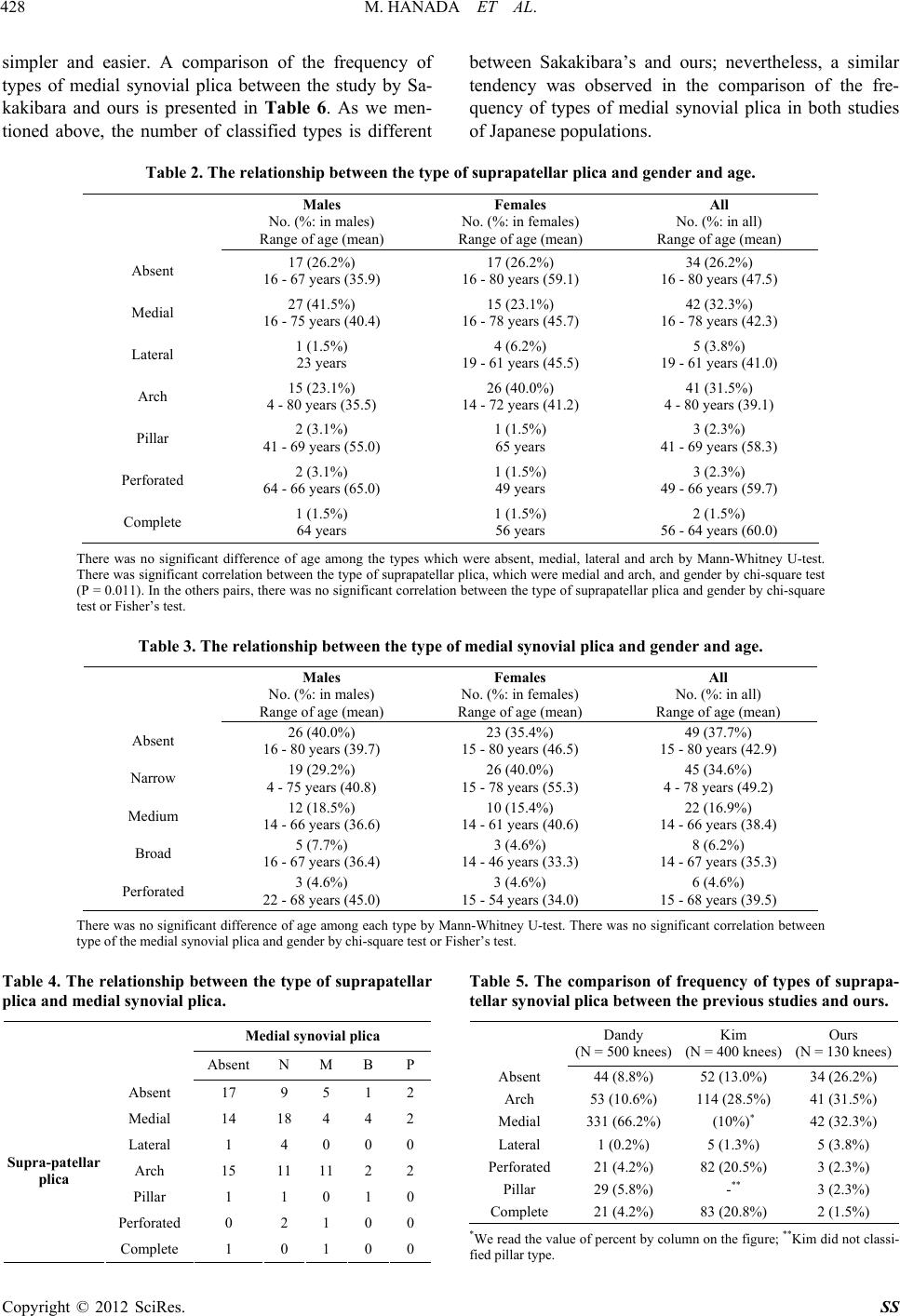 M. HANADA ET AL. 428 simpler and easier. A comparison of the frequency of types of medial synovial plica between the study by Sa- kakibara and ours is presented in Table 6. As we men- tioned above, the number of classified types is different between Sakakibara’s and ours; nevertheless, a similar tendency was observed in the comparison of the fre- quency of types of medial synovial plica in both studies of Japanese populations. Table 2. The relationship between the type of suprapatellar plica and gende r and age . Males No. (%: in males) Range of age (mean) Females No. (%: in females) Range of age (mean) All No. (%: in all) Range of age (mean) Absent 17 (26.2%) 16 - 67 years (35.9) 17 (26.2%) 16 - 80 years (59.1) 34 (26.2%) 16 - 80 years (47.5) Medial 27 (41.5%) 16 - 75 years (40.4) 15 (23.1%) 16 - 78 years (45.7) 42 (32.3%) 16 - 78 years (42.3) Lateral 1 (1.5%) 23 years 4 (6.2%) 19 - 61 years (45.5) 5 (3.8%) 19 - 61 years (41.0) Arch 15 (23.1%) 4 - 80 years (35.5) 26 (40.0%) 14 - 72 years (41.2) 41 (31.5%) 4 - 80 years (39.1) Pillar 2 (3.1%) 41 - 69 years (55.0) 1 (1.5%) 65 years 3 (2.3%) 41 - 69 years (58.3) Perforated 2 (3.1%) 64 - 66 years (65.0) 1 (1.5%) 49 years 3 (2.3%) 49 - 66 years (59.7) Complete 1 (1.5%) 64 years 1 (1.5%) 56 years 2 (1.5%) 56 - 64 years (60.0) There was no significant difference of age among the types which were absent, medial, lateral and arch by Mann-Whitney U-test. There was significant correlation between the type of suprapatellar plica, which were medial and arch, and gender by chi-square test (P = 0.011). In the others pairs, there was no significant correlation between the type of suprapatellar plica and gender by chi-square test or Fisher’s test. Table 3. The relationship between the type of medial synovial plica and gender and age. Males No. (%: in males) Range of age (mean) Females No. (%: in females) Range of age (mean) All No. (%: in all) Range of age (mean) Absent 26 (40.0%) 16 - 80 years (39.7) 23 (35.4%) 15 - 80 years (46.5) 49 (37.7%) 15 - 80 years (42.9) Narrow 19 (29.2%) 4 - 75 years (40.8) 26 (40.0%) 15 - 78 years (55.3) 45 (34.6%) 4 - 78 years (49.2) Medium 12 (18.5%) 14 - 66 years (36.6) 10 (15.4%) 14 - 61 years (40.6) 22 (16.9%) 14 - 66 years (38.4) Broad 5 (7.7%) 16 - 67 years (36.4) 3 (4.6%) 14 - 46 years (33.3) 8 (6.2%) 14 - 67 years (35.3) Perforated 3 (4.6%) 22 - 68 years (45.0) 3 (4.6%) 15 - 54 years (34.0) 6 (4.6%) 15 - 68 years (39.5) There was no significant difference of age among each type by Mann-Whitney U-test. There was no significant correlation between type of the medial synovial plica and gender by chi-square test or Fisher’s test. Table 4. The relationship between the type of suprapatellar plica and medial synovial plica. Medial synovial plica Absent N M B P Absent 17 9 5 1 2 Medial 14 18 4 4 2 Lateral 1 4 0 0 0 Arch 15 11 11 2 2 Pillar 1 1 0 1 0 Perforated 0 2 1 0 0 Supra-patellar plica Complete 1 0 1 0 0 Table 5. The comparison of frequency of types of suprapa- tellar synovial plica between the pre v ious studie s and our s. Dandy (N = 500 knees) Kim (N = 400 knees) Ours (N = 130 knees) Absent 44 (8.8%) 52 (13.0%) 34 (26.2%) Arch 53 (10.6%) 114 (28.5%) 41 (31.5%) Medial 331 (66.2%) (10%)* 42 (32.3%) Lateral 1 (0.2%) 5 (1.3%) 5 (3.8%) Perforated 21 (4.2%) 82 (20.5%) 3 (2.3%) Pillar 29 (5.8%) -** 3 (2.3%) Complete 21 (4.2%) 83 (20.8%) 2 (1.5%) *We read the value of percent by column on the figure; **Kim did not classi- fied pillar type. Copyright © 2012 SciRes. SS 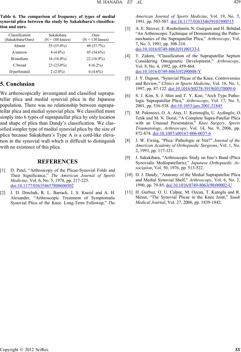 M. HANADA ET AL. 429 Table 6. The comparison of frequency of types of medial synovial plica betwe en the study by Sakakibara’s classifica - tion and ours. Classification (Sakakibara/Ours) Sakakibara (N = 100 knees) Ours (N = 130 knees) Absent 55 (55.0%) 49 (37.7%) A/narrow 4 (4.0%) 45 (34.6%) B/medium 16 (16.0%) 22 (16.9%) C/broad 23 (23.0%) 8 (6.2%) D/perforated 2 (2.0%) 6 (4.6%) 5. Conclusion We arthroscopically investigated and classified suprapa- tellar plica and medial synovial plica in the Japanese population. There was no relationship between suprapa- tellar plica and medial synovial plica. We classified more simply into 6 types of suprapatellar plica by only location and shape of plica than Dandy’s classification. We clas- sified simpler type of medial synovial plica by the size of plica because Sakakibara’s Type A is a cord-like eleva- tion in the synovial wall which is difficult to distinguish with no existence of this plica. REFERENCES [1] D. Patel, “Arthroscopy of the Plicae-Synovial Folds and Their Significance,” The American Journal of Sports Medicine, Vol. 6, No. 5, 1978, pp. 217-225. doi:10.1177/036354657800600502 [2] J. D. Dorchak, R. L. Barrack, J. S. Kneisl and A. H. Alexander, “Arthroscopic Treatment of Symptomatic Synovial Plica of the Knee. Long-Term Followup,” The American Journal of Sports Medicine, Vol. 19, No. 5, 1991, pp. 503-507. doi:10.1177/036354659101900515 [3] A. E. Strover, E. Rouholamin, N. Guirguis and H. Behdad. “An Arthroscopic Technique of Demonstrating the Patho- mechanics of the Suprapatellar Plica,” Arthroscopy, Vol. 7, No. 3, 1991, pp. 308-310. doi:10.1016/0749-8063(91)90133-I [4] T. Zidorn, “Classification of the Suprapatellar Septum Considering Ontogenetic Development,” Arthroscopy, Vol. 8, No. 4, 1992, pp. 459-464. doi:10.1016/0749-8063(92)90008-Y [5] J. Y. Dupont, “Synovial Plicae of the Knee. Controversies and Review,” Clinics in Sports Medicine, Vol. 16, No. 1, 1997, pp. 87-122. doi:10.1016/S0278-5919(05)70009-0 [6] S. J. Kim, S. J. Shin and T. Y. Koo, “Arch Type Patho- logic Suprapatellar Plica,” Arthroscopy, Vol. 17, No. 5, 2001, pp. 536-538. doi:10.1053/jars.2001.21845 [7] M. Pekmezci, O. A. Atay, U. Kerimoğlu, U. Aydingöz, O. Tetik and M. N. Doral, “A Complete Supra-Patellar Plica with an Unusual Presentation,” Knee Surgery, Sports Traumatology, Arthroscopy, Vol. 14, No. 9, 2006, pp. 872-874. doi:10.1007/s00167-006-0037-6 [8] J. W. Ewing, “Plica: Pathologic or Not?” Journal of the American Academy of Orthopaedic Surgeons, Vol. 1, No. 2, 1993, pp. 117-121. [9] J. Sakakibara, “Arthroscopic Study on Iino’s Band (Plica Synovialis Mediopatellaris),” Japanese Orthopaedic As- sociation, Vol. 50, 1976, pp. 513-522. [10] D. J. Dandy, “Anatomy of the Medial Suprapatellar Plica and Medial Synovial Shelf,” Arthroscopy, Vol. 6, No. 2, 1990, pp. 79-85. doi:10.1016/0749-8063(90)90002-U [11] H. Gurbuz, O. U. Calpur, M. Ozcan, T. Kutoglu and R. Mesut, “The Synovial Plicae in the Knee Joint,” Saudi Medical Journal, Vol. 27, 2006, pp. 1839-1842. Copyright © 2012 SciRes. SS |