

Paper Menu >>
Journal Menu >>
 Journal of Cancer Therapy, 2012, 3, 397-405 http://dx.doi.org/10.4236/jct.2012.324052 Published Online September 2012 (http://www.SciRP.org/journal/jct) 397 Baculovirus Mediated Experimental Research on Targeted Egr1-Kringle 5 Gene Radiotherapy in Lung Adenocarcinoma Haoping Xu, Rui Guo, Biao Li* Department of Nuclear Medicine, Rui Jin Hospital, Shanghai Jiao Tong University School of Medicine, Shanghai, China. Email: *[email protected] Received April 15th, 2012; revised May 27th, 2012; accepted June 15th, 2012 ABSTRACT Objective: To investigate the feasibility of temporally and spatially restricted Kringle5 expression induced by radiation, as well as the dual effect of radiotherapy and antiangiogenic therapy in lung adenocarcinoma in vitro. Methods: We first constructed recombinant baculovirus vectors containing Egr1 promoter and human plasminogen Kringle5 gene (rhK5), then transfected them into lung adenocarcinoma cells (A549). Transfect efficiency of the baculovirus for gene transfer in A549 cells and the activity of Egr1 promoter induced by X-radiation were detected by fluorescence micros- copy. The rhK5 mRNA transcription and rhK5 protein expression were detected by Real-time PCR and Western blot assay, respectively. The apoptosis asssay of human umbilical veins endothelial cells (HUVEC) was analyzed by flow cytometry. Results: The recombinant baculovirus were successfully transfected into A549 and HUVEC cells. As for the temporal regulation, the rhK5 mRNA transcription and rhK5 protein expression were elevated with the irradiation time significantly. And the HUVEC apoptotic percentage increased in relation to the irradiation time as well. As for the spa- tial regulation, rhK5 mRNA transcription level of A549 cell lines transfected with recombinant baculovirus Egr1-K5 was significantly higher than that of control groups after the same dose of X-radiation. When we analyzed the dose and frequency of X-radiation, no difference was observed among each dose after continuously three-times of irradiation. Conclusion: Baculovirus-mediated Egr1-K5 can be used in gene radiotherapy for its temporary and spatial controllable rhK5 expression by X-radiation and the consequent HUVEC apoptosis in vitro study. And low dose and more times of irradiation might be more effective. It would provide a promising way for the tumor treatment. Keywords: Radiotherapy; Lung Adenocarcinoma; Egr1 Promoter; Kringle 5; Baculovirus 1. Introduction Radiotherapy is widely used in the treatment of various types of cancer. However, its application is limited by the side-effects such as the radiation-induced damages of the normal tissues nearby and the radiation-resistance of certain tumors. As for the gene therapy, the results are less encouraging due to the inability to optimally localize the activity of therapeutic agents to tumor cells and limit damage to normal tissue. Also, there is a lack of effective therapeutic gene. Angiogenesis plays a key role in tumor progression. It was hypothesized that inhibition of angiogenesis would be an effective strategy to treat human cancer, and an active search for angiogenesis inducers and inhibitors began in 1971 [1]. Antiangiogenic therapy could destroy tumor vasculature and inhibit tumor growth. And it’s the- rapeutic benefit may even be greater when used in com- bination with established treatment modalities, such as surgery, chemotherapy, and radiation therapy [2]. Plas- minogen contains 5 kringles, and kringle 1 - 4 consists of angiostatin. The kringle 5 (K5) domain of human plas- minogen with low molecular weight of 14kDa and low immunogenicity is a specific inhibitor for endothelial cell proliferation and displays powerful antiangiogenic acti- vity [3]. Study also showed that kringle 5 could directly lead to apoptosis of anoxic tumor cells in vitro [4]. These features make K5 an appealing antitumor biopharmaceu- tical with combined antitumoral and antiangiogenic pro- perties. Although angiogenesis inhibition offers several advantages, it is expected to induce a cytostatic effect resulting in tumor stabilization not eradication. Further- more, single-agent antiangiogenic therapy may lead to a compensatory increase in the production of other angio- genic factors, which may then sustain angiogenesis [5]. In a recent study, the therapeutic effect of radiotherapy combined with K5 is investigated in Lewis lung carci- *Corresponding author. Copyright © 2012 SciRes. JCT 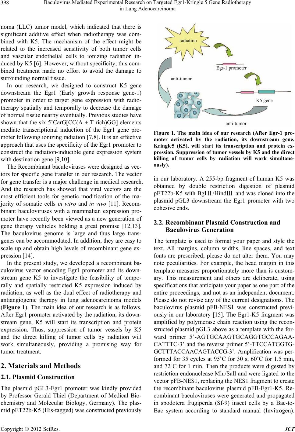 Baculovirus Mediated Experimental Research on Targeted Egr1-Kringle 5 Gene Radiotherapy in Lung Adenocarcinoma 398 noma (LLC) tumor model, which indicated that there is significant additive effect when radiotherapy was com- bined with K5. The mechanism of the effect might be related to the increased sensitivity of both tumor cells and vascular endothelial cells to ionizing radiation in- duced by K5 [6]. However, without specificity, this com- bined treatment made no effort to avoid the damage to surrounding normal tissue. In our research, we designed to construct K5 gene downstream the Egr1 (Early growth response gene-1) promoter in order to target gene expression with radio- therapy spatially and temporally to decrease the damage of normal tissue nearby eventually. Previous studies have shown that the six 5’CarG[CC(A + T rich)GG] elements mediate transcriptional induction of the Egr1 gene pro- moter following ionizing radiation [7,8]. It is an effective approach that uses the specificity of the Egr1 promoter to construct the radiation-inducible gene expression system with destination gene [9,10]. The Recombinant baculoviruses were designed as vec- tors for specific gene transfer in our research. The vector for gene transfer is a major challenge in medical research. And the research has showed that viral vectors are the most efficient tools for genetic modification of the ma- jority of somatic cells in vitro and in vivo [11]. Recom- binant baculoviruses with a mammalian expression pro- moter have recently been viewed as a new generation of gene therapy vehicles holding a great promise [12,13]. The baculovirus genome is large and thus large trans- genes can be accommodated. In addition, they are easy to scale up and obtain high levels of recombinant gene ex- pression [14]. In the present study, we developed a recombinant ba- culovirus vector encoding Egr1 promoter and its down- stream gene K5 to investigate the feasibility of tempo- rally and spatially restricted K5 expression induced by radiation, as well as the dual effect of radiotherapy and antiangiogenic therapy in lung adenocarcinoma models (Figure 1). The main idea of our research is as follows. After Egr1 promoter activated by the radiation, its down- stream gene, K5 will start its transcription and protein expression. Thus, suppression of tumor vessels by K5 and the direct killing of tumor cells by radiation will work simultaneously, providing a promising way for tumor treatment. 2. Materials and Methods 2.1. Plasmid Construction The plasmid pGL3-Egr1 promoter was kindly provided by Professor Gerald Thiel (Department of Medical Bio- chemistry and Molecular Biology, Germany). The plas- mid pET22b-K5 (His-tagged) was constructed previously Figure 1. The main idea of our research (After Egr-1 pro- moter activated by the radiation, its downstream gene, Kringle5 (K5), will start its transcription and protein ex- pression. Suppression of tumor vessels by K5 and the direct killing of tumor cells by radiation will work simultane- ously). in our laboratory. A 255-bp fragment of human K5 was obtained by double restriction digestion of plasmid pET22b-K5 with BgIⅡ/HindⅢ and was cloned into the plasmid pGL3 downstream the Egr1 promoter with two cohesive ends. 2.2. Recombinant Plasmid Construction and Baculovirus Generation The template is used to format your paper and style the text. All margins, column widths, line spaces, and text fonts are prescribed; please do not alter them. You may note peculiarities. For example, the head margin in this template measures proportionately more than is custom- ary. This measurement and others are deliberate, using specifications that anticipate your paper as one part of the entire proceedings, and not as an independent document. Please do not revise any of the current designations. The baculovirus plasmid pFB-NES1 was constructed previ- ously in our laboratory [15]. The Egr1-K5 fragment was amplified by polymerase chain reaction using the recon- structed plasmid pGL3 above as a template with the for- ward primer 5’-AGTGCAAGTGCAGGTGCCAGAA- CATTTC-3’ and the reverse primer 5’-TTCCATGGTG- GCTTTACCAACAGTACCG-3’. Amplification was per- formed for 35 cycles at 95˚C for 30 s, 60˚C for 1.5 min, and 72˚C for 1 min. Then the products were digested by restriction endonuclease Mlu/SalI and were ligated to the vector pFB-NES1, replacing the NES1 fragment to create the recombinant baculovirus plasmid pFB-Egr1-K5. Re- combinant baculoviruses were generated and propagated in spodotera frugiperda (Sf-9) insect cells by a Bac-to- Bac system according to standard manual (Invitrogen). Copyright © 2012 SciRes. JCT 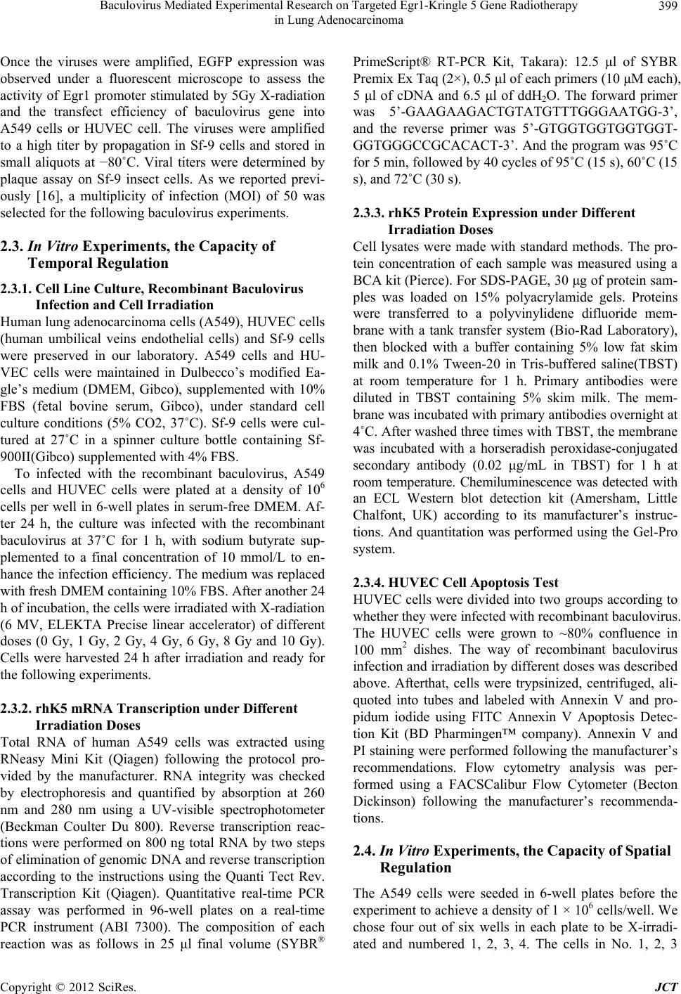 Baculovirus Mediated Experimental Research on Targeted Egr1-Kringle 5 Gene Radiotherapy in Lung Adenocarcinoma 399 Once the viruses were amplified, EGFP expression was observed under a fluorescent microscope to assess the activity of Egr1 promoter stimulated by 5Gy X-radiation and the transfect efficiency of baculovirus gene into A549 cells or HUVEC cell. The viruses were amplified to a high titer by propagation in Sf-9 cells and stored in small aliquots at −80˚C. Viral titers were determined by plaque assay on Sf-9 insect cells. As we reported previ- ously [16], a multiplicity of infection (MOI) of 50 was selected for the following baculovirus experiments. 2.3. In Vitro Experiments, the Capacity of Temporal Regulation 2.3.1. Cell Line Culture, Recombinant Baculovirus Infection and Cell Irradiation Human lung adenocarcinoma cells (A549), HUVEC cells (human umbilical veins endothelial cells) and Sf-9 cells were preserved in our laboratory. A549 cells and HU- VEC cells were maintained in Dulbecco’s modified Ea- gle’s medium (DMEM, Gibco), supplemented with 10% FBS (fetal bovine serum, Gibco), under standard cell culture conditions (5% CO2, 37˚C). Sf-9 cells were cul- tured at 27˚C in a spinner culture bottle containing Sf- 900II(Gibco) supplemented with 4% FBS. To infected with the recombinant baculovirus, A549 cells and HUVEC cells were plated at a density of 106 cells per well in 6-well plates in serum-free DMEM. Af- ter 24 h, the culture was infected with the recombinant baculovirus at 37˚C for 1 h, with sodium butyrate sup- plemented to a final concentration of 10 mmol/L to en- hance the infection efficiency. The medium was replaced with fresh DMEM containing 10% FBS. After another 24 h of incubation, the cells were irradiated with X-radiation (6 MV, ELEKTA Precise linear accelerator) of different doses (0 Gy, 1 Gy, 2 Gy, 4 Gy, 6 Gy, 8 Gy and 10 Gy). Cells were harvested 24 h after irradiation and ready for the following experiments. 2.3.2. rhK5 mRNA Transcription under Different Irradiation Doses Total RNA of human A549 cells was extracted using RNeasy Mini Kit (Qiagen) following the protocol pro- vided by the manufacturer. RNA integrity was checked by electrophoresis and quantified by absorption at 260 nm and 280 nm using a UV-visible spectrophotometer (Beckman Coulter Du 800). Reverse transcription reac- tions were performed on 800 ng total RNA by two steps of elimination of genomic DNA and reverse transcription according to the instructions using the Quanti Tect Rev. Transcription Kit (Qiagen). Quantitative real-time PCR assay was performed in 96-well plates on a real-time PCR instrument (ABI 7300). The composition of each reaction was as follows in 25 μl final volume (SYBR® PrimeScript® RT-PCR Kit, Takara): 12.5 μl of SYBR Premix Ex Taq (2×), 0.5 μl of each primers (10 μM each), 5 μl of cDNA and 6.5 μl of ddH2O. The forward primer was 5’-GAAGAAGACTGTATGTTTGGGAATGG-3’, and the reverse primer was 5’-GTGGTGGTGGTGGT- GGTGGGCCGCACACT-3’. And the program was 95˚C for 5 min, followed by 40 cycles of 95˚C (15 s), 60˚C (15 s), and 72˚C (30 s). 2.3.3. rhK5 Protein Expre ssion under Different Irradiation Doses Cell lysates were made with standard methods. The pro- tein concentration of each sample was measured using a BCA kit (Pierce). For SDS-PAGE, 30 μg of protein sam- ples was loaded on 15% polyacrylamide gels. Proteins were transferred to a polyvinylidene difluoride mem- brane with a tank transfer system (Bio-Rad Laboratory), then blocked with a buffer containing 5% low fat skim milk and 0.1% Tween-20 in Tris-buffered saline(TBST) at room temperature for 1 h. Primary antibodies were diluted in TBST containing 5% skim milk. The mem- brane was incubated with primary antibodies overnight at 4˚C. After washed three times with TBST, the membrane was incubated with a horseradish peroxidase-conjugated secondary antibody (0.02 μg/mL in TBST) for 1 h at room temperature. Chemiluminescence was detected with an ECL Western blot detection kit (Amersham, Little Chalfont, UK) according to its manufacturer’s instruc- tions. And quantitation was performed using the Gel-Pro system. 2.3.4. HUVEC Cell Apoptosis Test HUVEC cells were divided into two groups according to whether they were infected with recombinant baculovirus. The HUVEC cells were grown to ~80% confluence in 100 mm2 dishes. The way of recombinant baculovirus infection and irradiation by different doses was described above. Afterthat, cells were trypsinized, centrifuged, ali- quoted into tubes and labeled with Annexin V and pro- pidum iodide using FITC Annexin V Apoptosis Detec- tion Kit (BD Pharmingen™ company). Annexin V and PI staining were performed following the manufacturer’s recommendations. Flow cytometry analysis was per- formed using a FACSCalibur Flow Cytometer (Becton Dickinson) following the manufacturer’s recommenda- tions. 2.4. In Vitro Experiments, the Capacity of Spatial Regulation The A549 cells were seeded in 6-well plates before the experiment to achieve a density of 1 × 10⁶ cells/well. We chose four out of six wells in each plate to be X-irradi- ated and numbered 1, 2, 3, 4. The cells in No. 1, 2, 3 Copyright © 2012 SciRes. JCT 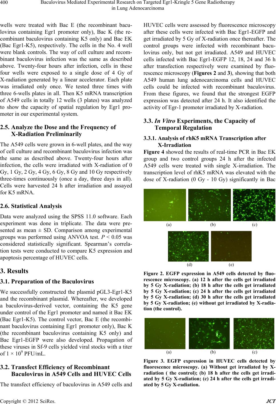 Baculovirus Mediated Experimental Research on Targeted Egr1-Kringle 5 Gene Radiotherapy in Lung Adenocarcinoma 400 wells were treated with Bac E (the recombinant bacu- lovirus containing Egr1 promoter only), Bac K (the re- combinant baculovirus containing K5 only) and Bac EK (Bac Egr1-K5), respectively. The cells in the No. 4 well were blank controls. The way of cell culture and recom- binant baculovirus infection was the same as described above. Twenty-four hours after infection, cells in these four wells were exposed to a single dose of 4 Gy of X-radiation generated by a linear accelerator. Each plate was irradiated only once. We tested three times with three 6-wells plates in all. Then K5 mRNA transcription of A549 cells in totally 12 wells (3 plates) was analyzed to show the capacity of spatial regulation by Egr1 pro- moter in our experimental system. 2.5. Analyze the Dose and the Frequency of X-Radiation Preliminarily The A549 cells were grown in 6-well plates, and the way of cell culture and recombinant baculovirus infection was the same as described above. Twenty-four hours after infection, the cells were irradiated with X-radiation of 0 Gy, 1 Gy, 2 Gy, 4 Gy, 6 Gy, 8 Gy and 10 Gy respectively three-times continuously (once a day, three days in all). Cells were harvested 24 h after irradiation and assayed for K5 mRNA. 2.6. Statistical Analysis Data were analyzed using the SPSS 11.0 software. Each experiment was done in triplicate. The data were pre- sented as mean ± SD. Comparison among experimental groups was performed using ANVOA test. P < 0.05 was considered statistically significant. Spearman’s correla- tion tests were conducted to compare K5 expression and apoptosis percentage of HUVEC cells. 3. Results 3.1. Preparation of the Baculovirus We successfully constructed the plasmid pGL3-Egr1-K5 and the recombinant plasmid. Whereafter, we developed a baculovirus-derived vector, containing the K5 gene under control of the Egr1 promoter and named it Bac EK (Bac Egr1-K5). The control vector, Bac E (the recombi- nant baculovirus containing Egr1 promoter only), Bac K (the recombinant baculovirus containing K5 only) and Bac Egr1-EGFP were also developed. Propagation of these viruses in Sf-9 cells yielded viral stocks with a titer of 1 × 10⁹ PFU/mL. 3.2. Transfect Efficiency of Recombinant Baculovirus in A549 Cells and HUVEC Cells The transfect efficiency of baculovirus in A549 cells and HUVEC cells were assessed by fluorescence microscopy after these cells were infected with Bac Egr1-EGFP and get irradiated by 5 Gy of X-radiation once thereafter. The control groups were infected with recombinant bacu- lovirus only, but not get irradiated. A549 and HUVEC cells infected with Bac Egr1-EGFP 12, 18, 24 and 36 h after transfection respectively were examined by fluo- rescence microscopy (Figures 2 and 3), showing that both A549 human lung adenocarcinoma cells and HUVEC cells could be infected with recombinant baculovirus. From these figures, we found that the strongest EGFP expression was detected after 24 h. It also identified the activity of Egr-1 promoter irradiated by X-radiation. 3.3. In Vitro Experiments, the Capacity of Temporal Regulation 3.3.1. Analysis of rhK5 mRNA Transcription after X-Irradiation Figure 4 showed the results of real-time PCR in Bac EK group and two control groups 24 h after the infected A549 cells were treated with single X-irradiation. The transcription level of rhK5 mRNA was elevated with the dose of X-radiation (0 Gy - 10 Gy) significantly in Bac (a) (b) (c) (d) (e) Figure 2. EGFP expression in A549 cells detected by fluo- rescence microscopy. (a) 12 h after the cells get irradiated by 5 Gy X-radiation; (b) 18 h after the cells get irradiated by 5 Gy X-radiation; (c) 24 h after the cells get irradiated by 5 Gy X-radiation; (d) 30 h after the cells get irradiated by 5 Gy X-radiation; (e) without get irradiated by X-radia- tion (the control). (a) (b) (c) Figure 3. EGFP expression in HUVEC cells detected by fluorescence microscopy. (a) Without get irradiated by X- radiation ( the control); (b) 18 h after the cells get irradi- ated by 5 Gy X-radiation; (c) 24 h after the cells get irradi- ated by 5 Gy X-radiation. Copyright © 2012 SciRes. JCT  Baculovirus Mediated Experimental Research on Targeted Egr1-Kringle 5 Gene Radiotherapy in Lung Adenocarcinoma 401 Figure 4. Kringle5 mRNA transcription in the infected A549 cells analyzed by real-time PCR (with single X-irra- diation). From top to bottom: in Bac EK group, in Bac K group and in Bac E group. EK group (P < 0.05), but not in the two control groups (P > 0.05). 3.3.2. rhK5 Expression in the Infected A549 Cells after X-Irradiati on Western Blot analysis was performed using antibodies to 6-histidine (His-tagged K5) for the expression of rhK5 in A549 cells infected with recombinant baculoviruses (Bac EK). A polyclonal antibody to 6-histidine detected a sin- gle protein band at 14kDa in the Bac EK group (Figure 5). Quantitative evaluation showed (Figure 6) that the rhK5 expression increased with the dose of radiation significantly (P < 0.05). 3.3.3. Cell Apoptosis Assay Res ul ts of Inf ec ted HUVEC Cells after X-Radiation The dual parameter fluorescent dot plots (Figure 7) shows the viable cell population in the lower left quad- rant (negative annexin-FITC and negative PI), the cells at (a) (b) (c) Figure 5. Kringle 5 expression in the infected A549 cells detected by Western blot 24 h after X-radiation. (a) In EK group, a single protein band at 14 kDa (Kringle5); (b) In Bac K group, no kringle5 expression; (c) In Bac E group, no Kringle5 expression. Figure 6. The quantitative evaluation of K5 expression: In- tegrated optical density (IOD) was measured and analyzed by software Gel-pro 4.0. The value of IODK5/IODGAPDH was showed in 0 Gy, 1 Gy, 2 Gy, 4 Gy, 6 Gy, 8 Gy and 10 Gy. Dose dependent K5 expr ession was observed. the early apoptosis are in the lower right quadrant (posi- tive annexin-FITC and negative PI) while the ones at the late apoptosis are in the upper right quadrant (positive annexin-FITC and positive-PI). As seen in Table 1, the early apoptotic cells percent- age increased in relation to the dose of X-radiation (P < 0.05) both in Bac EK plus radiotherapy group and the control radiotherapy alone group. Moreover, of the same dose of X-radiation, the apoptotic cells percentage of Bac EK plus radiotherapy group was significantly higher than that of radiotherapy alone group (P < 0.05). However, in Bac E and Bac K groups, no significant differences of the early apoptotic cells percentage was detected between Bac E/Bac K plus radiotherapy group and radiotherapy alone group (P > 0.05). These data suggest that Bac EK plus radiotherapy do best increase apoptosis of HUVEC cells compared with radiotherapy alone as the Egr-1 promoter was activated by radiotherapy and K5 played a Copyright © 2012 SciRes. JCT  Baculovirus Mediated Experimental Research on Targeted Egr1-Kringle 5 Gene Radiotherapy in Lung Adenocarcinoma Copyright © 2012 SciRes. JCT 402 Figure 7. Apoptotic effect of HUVEC cells 24 h after radiotherapy with or without Bac EK infection were determined by flow cytometry analysis using annexin V-FITC and propidium iodide. Cell population in bottom left, bottom right, top right and top left quadrants represented the proportion of viable cells, early apoptotic cells, late apoptotic cells and necrotic cells, re- spectively. Radi otherapy alo ne: (1) 0 Gy; (2) 1 Gy; ( 3) 2 Gy; (4) 4 Gy ; (5) 6 Gy; (6) 8 Gy; (7) 1 0 Gy; Radiotherapy w ith Bac EK: (8) 0 Gy; (9) 1 Gy; (10) 2 Gy; (11) 4 Gy; (12) 6 Gy; (13) 8 Gy; (14)10 Gy.  Baculovirus Mediated Experimental Research on Targeted Egr1-Kringle 5 Gene Radiotherapy in Lung Adenocarcinoma 403 Table 1. The early apoptotic cells percentage detected by fluorescence microscopy. Bac Egr-1-K5 with R (%) R alone (%) 0 Gy 10.67 ± 1.36 8.01 ± 1.85 1 Gy 32.16 ± 3.73 13.38 ± 2.51 2 Gy 34.66 ± 3.22 14.4 ± 4.17 4 Gy 41.52 ± 5.26 21.43 ± 1.39 6 Gy 44.42 ± 4.38 23.92 ± 5.76 8 Gy 49.06 ± 5.91 29.93 ± 3.17 10 Gy 52.55 ± 3.24 36.49 ± 4.62 R: radiotherapy. part in accelerating the cell apoptosis. 3.4. In Vitro Experiments, the Capacity of Spatial Regulation The real-time PCR results showed that the transcription of rhK5 mRNA in No.4 wells was significantly higher than those in other three wells (P < 0.05). The spatial regulation could be realized through the control of rhK5 expression. 3.5. Analyze the Dose and the Frequency of X-Radiation Preliminarily When we analyzed the K5 mRNA transcription in Bac EK group after the infected A549 cells were treated with continuously three-times of X-radiation (the same frac- tionated dose as single irradiation, once a day, 3 days in all), no significant difference was seen among these groups of different fractionated doses (Figure 8). 4. Discussion Most nonsmall cell lung cancer (NSCLC) patients were diagnosed at advanced stage and radiotherapy remains the treatment choice in such patients. Chemotherapy in combination with radiotherapy significantly improves the survival rate. However, the complete response rates are low and long-term survival remains poor, and the cyto- toxicity of chemotherapy is severe. All these indicate that exploration of more potent and safe therapeutic modali- ties is needed. The combination of gene therapy and ra- diotherapy has the potential to overcome many of the limitations of adverse tumor biology on cancer treatment and appears to be a promising approach to increase the therapeutic ratio of cancer therapy. In this study, the vector containing Egr1 promoter and its downstream K5 gene, an antiangiogenic agent gene, was constructed and tested by fluorescence microscopy for the ability of Egr1 promoter to regulate heterologous gene expression. The results of what the fluorescence microscopy showed also Figure 8. Kringle5 mRNA transcription in the infected A549 cells analyzed by real-time PCR (with continuously three-times of X-radiation) of Bac EK group. identified that radiation can induce the transcription of Egr1 as reported previously [9,17], and laid the ground- work for the next research. There are several potential advantages in using radia- tion-inducible genetic constructs with therapeutic radia- tion over other gene therapy delivery systems for cancer treatment. One advantage is that the already well-deve- loped technology of radiation targeting might add to the localization of toxin production provided by genetic therapy. As our study showed, when infected HUVEC cells received the same dose of X-radiation, the apoptotic cells percentage of “Bac EK plus radiotherapy” group was significantly higher than that of radiotherapy alone group (P < 0.05). A number of preclinical studies have indicated that antiangiogenic agents can enhance the tu- mor response to radiation [18,19]. Under X-radiation- induced cell stress condition, K5 could produce additive effect on endothelial cell apoptosis. Another advantage is that it controls gene expression spatially and temporally. The system we describe exploits the benefits of a tumor- specific vector in combination with radiotherapy which triggers and increases gene expression. Since therapeutic agents such as K5 might bring sideeffects to normal tis- sues, spatial control of protein expression can be very beneficial to limit K5 expressing locally in the tumor. There are two challenges in this method when it is pro- jected as a gene therapy strategy against cancer cells. First, introduction of the Egr1 promoter-driven antian- giogenic gene to the tumor site would require an appro- priate vector. The therapeutic gene must be present in the cancer cells when the tumor is irradiated by the X-radia- tion. However, specific irradiation of the tumor site with radiotherapy may give us specific activation of Egr1 promoter in the tumor cells. Autograps californica multi- ple nucleopolyhedrovirus (AcMNPV)-based vector, tra- ditionally used as a biopesticide to kill infected inserts, is recently tested as a new type of delivery vehicle for transgene expression in mammalian cells [20]. These viruses can enter but not replicate in mammalian cells. With mammalian expression promoters, recombinant Copyright © 2012 SciRes. JCT  Baculovirus Mediated Experimental Research on Targeted Egr1-Kringle 5 Gene Radiotherapy in Lung Adenocarcinoma 404 baculoviruses provide a high transduction efficiency in different cells and tissues, including several tumor cell lines [21]. One of the attractive advantages of using AcMNPV as a cancer gene therapy vector is the large cloning capacity conferred by its 130-kb viral genome, which may be used to deliver a large functional gene or multiple genes from a single vector [22]. Other empirical advantages of baculovirus vectors include easy construc- tion of a recombinant viral vector and simple procedure of purifying large quantities of viruses with high titers. It would be possible to scale up the less labor-intensive process to pharmaceutical levels [23]. A second problem is the dose and the frequency of X-radiation to activate Egr1 promoter and work as radiotherapy at the same time. We found that the K5 expression activated by Egr1 pro- moter dose-dependently increased in this vitro study, that is, the most K5 expression was achieved using 10 Gy, the highest dose in our study. Since a dose of 10 Gy is not used in a curative treatment setting, and patients are not treated with a single dose of irradiation but with fraction- ated radiotherapy, we tried using continuously three- times of X-radiation. To our surprise, the study demon- strated that there’s no significant difference of K5 ex- pression between high-dose groups and low-dose groups after three times of irradiation. On the basis of these re- sults we deduce this daily repeated small irradiation dose results in the same activation efficiency of Egr1 promoter. The mechanism has not been available yet. Moreover, irradiated by 2 Gy daily is suitable for clinical treatment and may decrease normal tissue toxicity compared with a single large dose. In summary, we report one therapeutic system with radiation-inducible promoter, Egr1, which made it have the capacity of temporal regulation as well as spatial re- gulation. In this system, radiotherapy and gene therapy worked simultaneously, and their effects were greater than those of either modality alone. Low dose and more times might be the most efficient therapeutic model of this combined system. On the basis of the results above, further study in vivo has been carried on to observe the effect of this therapeutic system. And the potential bene- fits of exogenous gene induction by radiotherapy include increased local tumor control as well as the potential for treatment of metastatic disease. 5. Acknowledgements This work was supported by Shanghai Leading Aca- demic Discipline Project S30203. REFERENCES [1] J. Folkman, E. Merler, C. Abernathy and G. Williams, “Isolation of a Tumor Factor Responsible for Angiogene- sis,” Journal of Experimental Medicine, Vol. 133, No. 2, 1971, pp. 275-288. doi:10.1084/jem.133.2.275 [2] P. Wachsberger, R. Burd and A. P. Dicker, “Tumor Re- sponse to Ionizing Radiation Combined with Antiangio- genesis or Vascular Targeting Agents: Exploring Mecha- nisms of Interaction,” Clinical Cancer Research, Vol. 9, No. 6, 2003, pp. 1957-1971. [3] Y. Cao, R. Cao and N. Veitonmaki, “Kringle Structures and Antiangiogenesis,” Current Medicinal Chemistry— Anti-Cancer Agents, Vol. 2, No. 6, 2002, pp. 667-681. doi:10.2174/1568011023353705 [4] D. J. Davidson, C. Haskell, S. Majest, A. Kherzai, D. A. Egan, K. A. Walter, et al., “Kringle 5 of Human Plasmi- nogen Induces Apoptosis of Endothelial and Tumor Cells through Surface-Expressed Glucose-Regulated Protein 78,” Cancer Research, Vol. 65, No. 11, 2005, pp. 4663- 4672. doi:10.1158/0008-5472.CAN-04-3426 [5] L. Bello, C. Giussani, G. Carrabba, M. Pluderi, F. Costa and A. Bikfalvi, “Angiogenesis and Invasion in Glio- mas,” Cancer Treatment and Research, Vol. 117, 2004, pp. 263-284. doi:10.1007/978-1-4419-8871-3_16 [6] G.-H. Jin, D.-Y. Ma, N. Wu, F. M. M. T. Marikar, S.-Z. Jin, W.-W. Jiang, et al., “Combination of Human Plas- minogen Kringle 5 with Ionizing Radiation Significantly Enhances the Efficacy of Antitumor Effect,” Interna- tional Journal of Cancer, Vol. 121, No. 11, 2007, pp. 2539-2546. doi:10.1002/ijc.22708 [7] M. M. Ahmed, “Regulation of Radiation-Induced Apop- tosis by Early Growth Response-1 Gene in Solid Tu- mors,” Current Cancer Drug Targets, Vol. 4, No. 1, 2004, pp. 43-52. doi:10.2174/1568009043481704 [8] O. Greco, B. Marples, G. U. Dachs, K. J. Williams, A. V. Patterson and S. D. Scott, “Novel Chimeric Gene Pro- moters Responsive to Hypoxia and Ionizing Radiation,” Gene Therapy, Vol. 9, No. 20, 2002, pp. 1403-1411. doi:10.1038/sj.gt.3301823 [9] M. Y. Wu, X. Y. Wu, Q. S. Li and R. M. Zheng, “Ex- pression of Egr-1 Gene and Its Correlation with the On- cogene Proteins in Non-Irradiated and Irradiated Eso- phageal Squamous Cell Carcinoma,” Diseases of the Esophagus, Vol. 19, No. 4, 2006, pp. 267-272. doi:10.1111/j.1442-2050.2006.00575.x [10] H. B. Ma, X. J. Wang, Z. L. Di, H. Xia, Z. Li, J. Liu, et al., “Construction of Targeted Plasmid Vector pcDNA3.1- Egr.1p-p16 and Its Expression in Pancreatic Cancer JF305 Cells Induced by Radiation in Vitro,” World Jour- nal of Gastroenterology, Vol. 13, No. 31, 2007, pp. 4214- 4218. [11] N. Khalighinejad, H. Hariri, O. Behnamfar, A. Yousefi and A. Momeni, “Adenoviral Gene Therapy in Gastric Cancer: A Review,” World Journal of Gastroenterology, Vol. 14, No. 2, 2008, pp. 180-184. doi:10.3748/wjg.14.180 [12] F. M. Boyce and N. L. Bucher, “Baculovirus-Mediated Gene Transfer into Mammalian Cells,” Proceedings of the National Academy of Sciences USA, Vol. 93, No. 6, 1996, pp. 2348-2352. doi:10.1073/pnas.93.6.2348 [13] H. Matilainen, J. Rinne, L. Gilbert, V. Marjomaki, H. Copyright © 2012 SciRes. JCT  Baculovirus Mediated Experimental Research on Targeted Egr1-Kringle 5 Gene Radiotherapy in Lung Adenocarcinoma Copyright © 2012 SciRes. JCT 405 Reunanen and C. Oker-Blom, “Baculovirus Entry into Human Hepatoma Cells,” Journal of Virology, Vol. 79, No. 24, 2005, pp. 15452-15459. doi:10.1128/JVI.79.24.15452-15459.2005 [14] D. L. Jarvis and A. Garcia Jr., “Long-Term Stability of Baculoviruses Stored under Various Conditions,” Bio- techniques, Vol. 16, 1994, pp. 508-513. [15] W. Huang, X. L. Tian, Y. L. Wu, J. Zhong, L. F. Yu, S. P. Hu, et al., “Suppression of Gastric Cancer Growth by Baculovirus Vectormediated Transfer of Normal Epithe- lial Cell Specific-1 Gene,” World Journal of Gastroen- terology, Vol. 14, No. 38, 2008, pp. 5810-5815. doi:10.3748/wjg.14.5810 [16] R. Guo, Y. F. Zhang, S. Liang, H. P. Xu, M. Zhang and B. Li, “Sodium Butyrate Enhances the Expression of Bacu- lovirus-Mediated Sodium/Iodide Symporter Gene in A549 Lung Adenocarcinoma Cells,” Nuclear Medicine Communications, Vol. 31, No. 10, 2010, pp. 916-921. [17] J. M. McLoughlin, T. M. McCarty, C. Cunningham, V. Clark, N. Senzer, J. Nemunaitis, et al., “TNFerade, an Adenovector Carrying the Transgene forHuman Tumor Necrosis Factor a, for Patients with Advanced Solid Tu- mors: Surgical Experience and Long-Term Follow-Up,” Annals of Surgical Oncology, Vol.12, No.10, 2005, pp. 825-830. doi:10.1245/ASO.2005.03.023 [18] R. P. Dings, B. W. Williams, C. W. Song, A. W. Grif- fioen, K. H. Mayo and R. J. Griffin, “Anginex Synergizes with Radiation Therapy to Inhibit Tumor Growth by Ra- diosensitizing Endothelial Cells,” International Journal of Cancer, Vol. 115, No. 2, 2005, pp. 312-319. doi:10.1002/ijc.20850 [19] S. Nasu, K. K. Ang, Z. Fan and L. Milas, “C225 Antiepi- dermal Growth Factor Receptor Antibody Enhances Tu- mor Radiocurability,” International Journal of Radiation Oncology•Biology•Physics, Vol. 51, No. 2, 2001, pp. 474- 477. [20] J. P. Condreay and T. A. Kost, “Baculovirus Expression Vectors for Insect and Mammalian Cells,” Current Drug Targets, Vol. 8, No. 10, 2007, pp. 1126-1131. doi:10.2174/138945007782151351 [21] C. Hofmann, V. Sandig, G. Jennings, M. Rudolph, P. Schlag and M. Strauss, “Efficient Gene Transfer into Human Hepatocytes by Baculovirus Vectors,” Proceed- ings of the National Academy of Sciences of the USA, Vol. 92, 1995, pp. 10099-10103. doi:10.1073/pnas.92.22.10099 [22] T. A. Kost and J. P. Condreay, “Recombinant Baculovi- ruses as Mammalian Cell Gene-Delivery Vectors,” Trends in Biotechnology, Vol. 20, No. 4, 2002, pp. 173- 180. doi:10.1016/S0167-7799(01)01911-4 [23] S. Ghosh, M. K. Parvez, K. Banerjee, S. K. Sarin and S. E. Hasnain, “Baculovirus as Mammalian Cell Expression Vector for Gene Therapy: An Emerging Strategy,” Mo- lecular Therapy, Vol. 6, No. 1, 2002, pp. 5-11. doi:10.1006/mthe.2000.0643 |